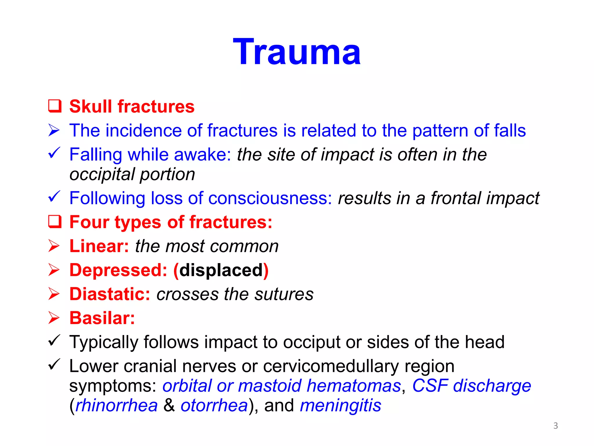
Trauma Brain Injury
Head injuries by physical forces may result
in:
Skull fractures
Parenchymal injuries
Vascular injuries
All three can coexist
Trauma may produce closed or open head
injuries
Injuries may be penetrating or blunt
1
Trauma
The consequences of CNS trauma depend
on
Anatomic location of the lesion
Limited capacity of the brain for functional
repair
Injury of several cubic cm of brain
parenchyma may be:
Clinically silent: e.g. in the frontal lobe
Severely disabling: in the spinal cord
Fatal: in the brainstem
2
Trauma
 Skull fractures
 The incidence of fractures is related to the pattern of falls
 Falling while awake: the site of impact is often in the
occipital portion
 Following loss of consciousness: results in a frontal impact
 Four types of fractures:
 Linear: the most common
 Depressed: (displaced)
 Diastatic: crosses the sutures
 Basilar:
 Typically follows impact to occiput or sides of the head
 Lower cranial nerves or cervicomedullary region
symptoms: orbital or mastoid hematomas, CSF discharge
(rhinorrhea & otorrhea), and meningitis
3
“LINEAR” “DEPRESSED
”, aka
“DISPLACED”
4
Trauma
Parenchymal injuries
 Concussion
 Mild traumatic brain injury commonly due to
change in the momentum of the head
 Characterized by a transient neurologic
dysfunction that includes: loss of
consciousness, temporary respiratory arrest and
loss of reflexes
 The neurologic recovery is complete, but
amnesia for the event persists
 Concussion usually does not produce
parenchymal damage
5
Trauma
Parenchymal injuries
 Contusion
 Bruise in the brain caused by blunt trauma
 Occurs when brain contacts rough skull surfaces
 Frontal and temporal lobes are the most
common locations
 Coup and contrecoup contusions
 Symptoms include: drowsiness, confusion,
agitation, hemiparesis, unequal pupil size
Laceration
 Caused by penetration of an object and tearing
of tissue
6
Trauma
Parenchymal injuries
Diffuse axonal injury
Injury to axons at nodes of Ranvier with
impairment of axoplasmic flow
Is due to acceleration /deceleration even
in absence of impact
Results in coma after trauma without
evidence of direct parenchymal injuries
7
Contusion
Morphologic changes
Acute: wedge-shaped hemorrhage
Subacute: necrosis / liquefaction
Chronic: plaque jaune lesions; depressed,
retracted, yellowish brown patches
Stages of changes:
Early: edema and hemorrhage
Next few hours: blood extravasation into
the white matter and subarachnoid space
By 24 hours: neuronal reaction to damage
Old contusions: gliosis and hemosiderin
laden macrophages 8
9
Trauma
Traumatic vascular injury
Results from direct trauma and disruption of
the vessel wall and leads to hemorrhage
Hemorrhage may occur in
Epidural
Subdural
Subarachnoid
Intraparenchymal compartments
10
11
Epidural hematoma
Associated with skull fractures
Does not cross suture lines
Due to tearing of dural arteries, mostly the
middle meningeal artery
Extravasation of blood separates the dura
from the inner surface of the skull
Blood accumulates slowly (“lucid interval” -
talk and die syndrome)
may expand rapidly and becomes a
neurosurgical emergency requiring prompt
drainage
12
13
EPIDURAL
HEMATOMA
14
Subdural hematoma
Due to rupture of bridging veins
May cross suture lines
Predisposing conditions:
Abnormal hemostasis
Elderly: with brain atrophy due to
stretching of veins
Infants: because of their thin-walled veins
15
16
Subdural hematoma
Clinical features:
Hematomas manifest within 48 hours of injury
Most commonly over the lateral aspects of
cerebral hemispheres
Slowly progressive neurological deterioration
headache, and confusion are the most
common features, but drowsiness, focal
neurological deficits, and sometimes
dementia may also occur
17
Subdural hematoma
 Morphology – Acute
 Freshly clotted blood along the brain surface
 The underlying brain is flattened
 Venous bleeding is self-limited → breakdown and
organization:
 Lysis of the clot (about 1 week)
 Growth of fibroblasts from the dural surface (2
weeks)
 Early development of hyalinized connective tissue
(1 to 3 months)
 Retraction of the lesion as granulation tissue
matures until there is only a thin layer of
connective tissue (subdural membranes)
18
SUBDURAL
HEMATOMA
19
20
Subarachnoid hemorrhage
 Is often a manifestation of cerebrovascular
disease
 Most common cause is rupture of saccular
(berry) aneurysm
 May also result from extension of a traumatic
hematoma, vascular malformation, hematologic
disturbances, and tumors
 Blood accumulates in ventricles, sulci, and
cisterns- chiasmatic cistern via A.Cerebral
artery.
 Clinical features include: sudden headache,
nuchal rigidity, neurological dificits on one side,
and stupor
21
22
SUBARACHNOID 23
Intracerebral hemorrhage
Is often a manifestation of cerebrovascular
disease
Occurs most commonly in middle to late
adult life (peak at 60 years)
Hypertension is the most common cause
Other causes include: cerebral amyloid
angiopathy, coagulation disorders,
neoplasms, vasculitis, aneurysms, and
vascular malformations
24
INTRAPARENCHYMAL
26
27
28
Sequelae of brain trauma
Broad neurologic syndromes may manifest
months or years after trauma:
Post-traumatic hydrocephalus
Post-traumatic dementia
Punch-drunk syndrome
Post-traumatic epilepsy
Risk of infections
Psychiatric disorders
29
30
Spinal cord trauma
31
Associated with transient or
permanent displacement of
vertebral column
The level of injury determines
the extent of manifestation:
Thoracic segments or below –
paraplegia
Cervical segments –
tetraplegia
Above C4 – respiratory arrest

Traumatic brain injury

  • 1.
    Trauma Brain Injury Headinjuries by physical forces may result in: Skull fractures Parenchymal injuries Vascular injuries All three can coexist Trauma may produce closed or open head injuries Injuries may be penetrating or blunt 1
  • 2.
    Trauma The consequences ofCNS trauma depend on Anatomic location of the lesion Limited capacity of the brain for functional repair Injury of several cubic cm of brain parenchyma may be: Clinically silent: e.g. in the frontal lobe Severely disabling: in the spinal cord Fatal: in the brainstem 2
  • 3.
    Trauma  Skull fractures The incidence of fractures is related to the pattern of falls  Falling while awake: the site of impact is often in the occipital portion  Following loss of consciousness: results in a frontal impact  Four types of fractures:  Linear: the most common  Depressed: (displaced)  Diastatic: crosses the sutures  Basilar:  Typically follows impact to occiput or sides of the head  Lower cranial nerves or cervicomedullary region symptoms: orbital or mastoid hematomas, CSF discharge (rhinorrhea & otorrhea), and meningitis 3
  • 4.
  • 5.
    Trauma Parenchymal injuries  Concussion Mild traumatic brain injury commonly due to change in the momentum of the head  Characterized by a transient neurologic dysfunction that includes: loss of consciousness, temporary respiratory arrest and loss of reflexes  The neurologic recovery is complete, but amnesia for the event persists  Concussion usually does not produce parenchymal damage 5
  • 6.
    Trauma Parenchymal injuries  Contusion Bruise in the brain caused by blunt trauma  Occurs when brain contacts rough skull surfaces  Frontal and temporal lobes are the most common locations  Coup and contrecoup contusions  Symptoms include: drowsiness, confusion, agitation, hemiparesis, unequal pupil size Laceration  Caused by penetration of an object and tearing of tissue 6
  • 7.
    Trauma Parenchymal injuries Diffuse axonalinjury Injury to axons at nodes of Ranvier with impairment of axoplasmic flow Is due to acceleration /deceleration even in absence of impact Results in coma after trauma without evidence of direct parenchymal injuries 7
  • 8.
    Contusion Morphologic changes Acute: wedge-shapedhemorrhage Subacute: necrosis / liquefaction Chronic: plaque jaune lesions; depressed, retracted, yellowish brown patches Stages of changes: Early: edema and hemorrhage Next few hours: blood extravasation into the white matter and subarachnoid space By 24 hours: neuronal reaction to damage Old contusions: gliosis and hemosiderin laden macrophages 8
  • 9.
  • 10.
    Trauma Traumatic vascular injury Resultsfrom direct trauma and disruption of the vessel wall and leads to hemorrhage Hemorrhage may occur in Epidural Subdural Subarachnoid Intraparenchymal compartments 10
  • 11.
  • 12.
    Epidural hematoma Associated withskull fractures Does not cross suture lines Due to tearing of dural arteries, mostly the middle meningeal artery Extravasation of blood separates the dura from the inner surface of the skull Blood accumulates slowly (“lucid interval” - talk and die syndrome) may expand rapidly and becomes a neurosurgical emergency requiring prompt drainage 12
  • 13.
  • 14.
  • 15.
    Subdural hematoma Due torupture of bridging veins May cross suture lines Predisposing conditions: Abnormal hemostasis Elderly: with brain atrophy due to stretching of veins Infants: because of their thin-walled veins 15
  • 16.
  • 17.
    Subdural hematoma Clinical features: Hematomasmanifest within 48 hours of injury Most commonly over the lateral aspects of cerebral hemispheres Slowly progressive neurological deterioration headache, and confusion are the most common features, but drowsiness, focal neurological deficits, and sometimes dementia may also occur 17
  • 18.
    Subdural hematoma  Morphology– Acute  Freshly clotted blood along the brain surface  The underlying brain is flattened  Venous bleeding is self-limited → breakdown and organization:  Lysis of the clot (about 1 week)  Growth of fibroblasts from the dural surface (2 weeks)  Early development of hyalinized connective tissue (1 to 3 months)  Retraction of the lesion as granulation tissue matures until there is only a thin layer of connective tissue (subdural membranes) 18
  • 19.
  • 20.
  • 21.
    Subarachnoid hemorrhage  Isoften a manifestation of cerebrovascular disease  Most common cause is rupture of saccular (berry) aneurysm  May also result from extension of a traumatic hematoma, vascular malformation, hematologic disturbances, and tumors  Blood accumulates in ventricles, sulci, and cisterns- chiasmatic cistern via A.Cerebral artery.  Clinical features include: sudden headache, nuchal rigidity, neurological dificits on one side, and stupor 21
  • 22.
  • 23.
  • 24.
    Intracerebral hemorrhage Is oftena manifestation of cerebrovascular disease Occurs most commonly in middle to late adult life (peak at 60 years) Hypertension is the most common cause Other causes include: cerebral amyloid angiopathy, coagulation disorders, neoplasms, vasculitis, aneurysms, and vascular malformations 24
  • 26.
  • 27.
  • 28.
  • 29.
    Sequelae of braintrauma Broad neurologic syndromes may manifest months or years after trauma: Post-traumatic hydrocephalus Post-traumatic dementia Punch-drunk syndrome Post-traumatic epilepsy Risk of infections Psychiatric disorders 29
  • 30.
  • 31.
    Spinal cord trauma 31 Associatedwith transient or permanent displacement of vertebral column The level of injury determines the extent of manifestation: Thoracic segments or below – paraplegia Cervical segments – tetraplegia Above C4 – respiratory arrest