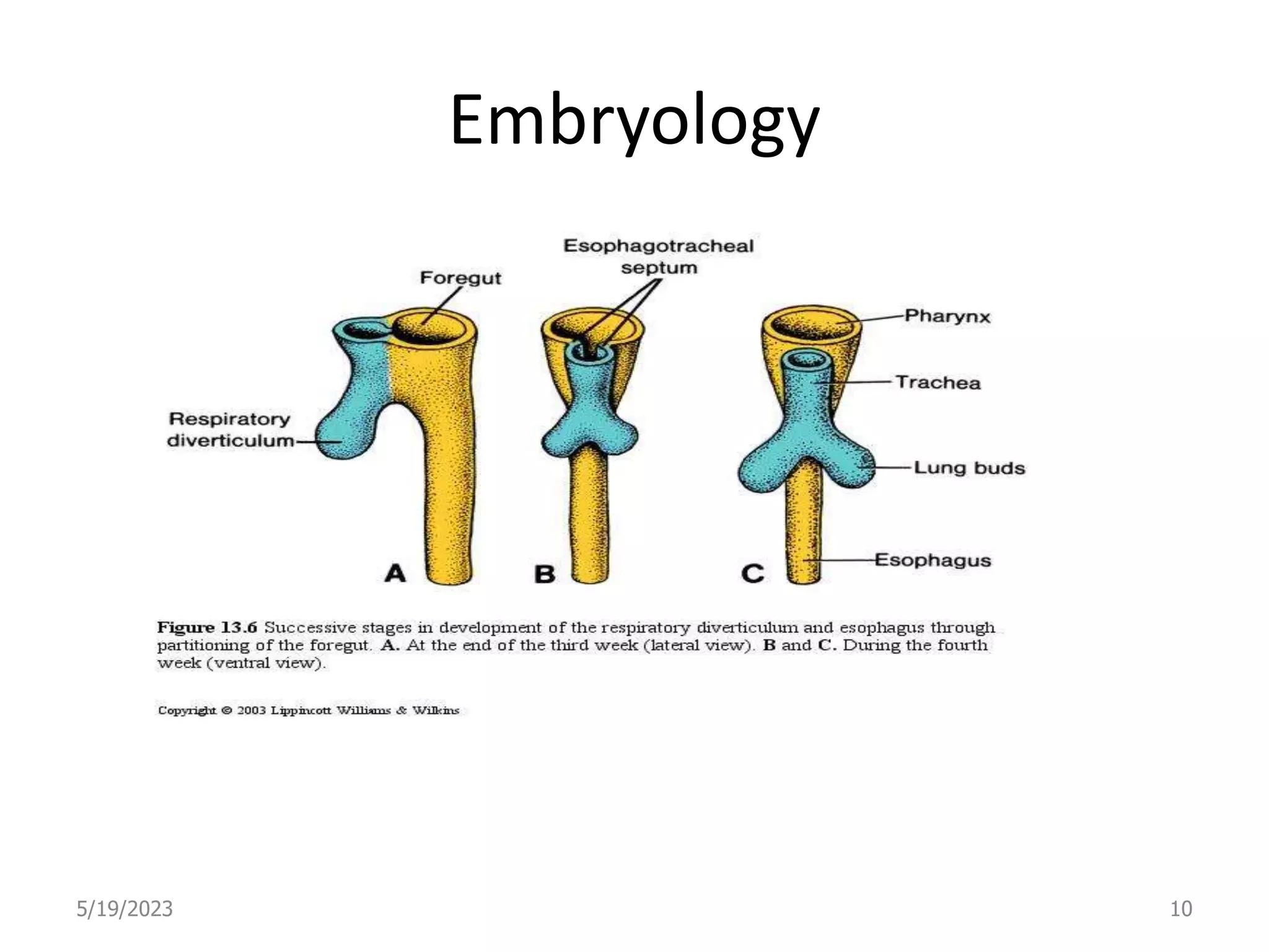
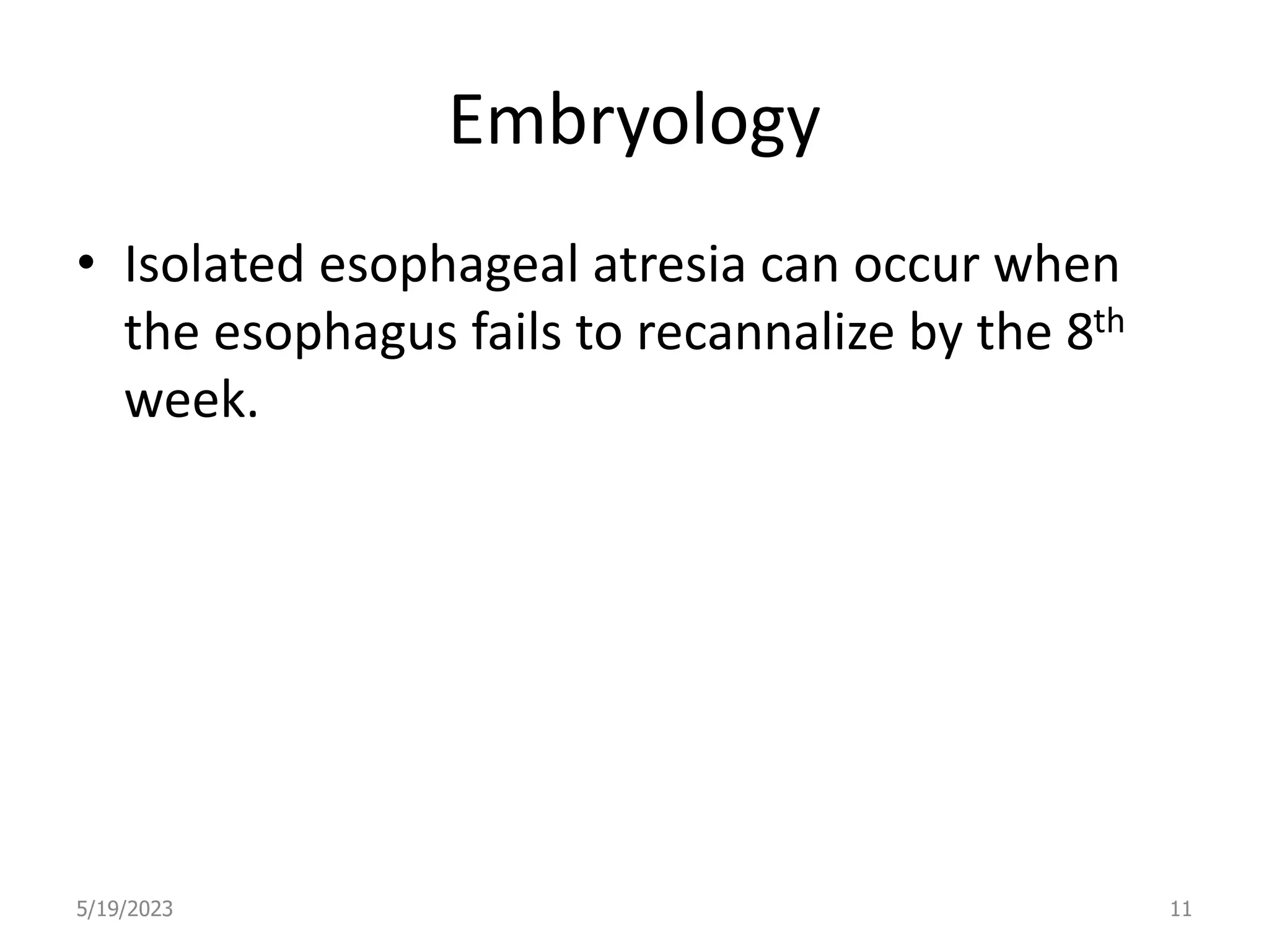
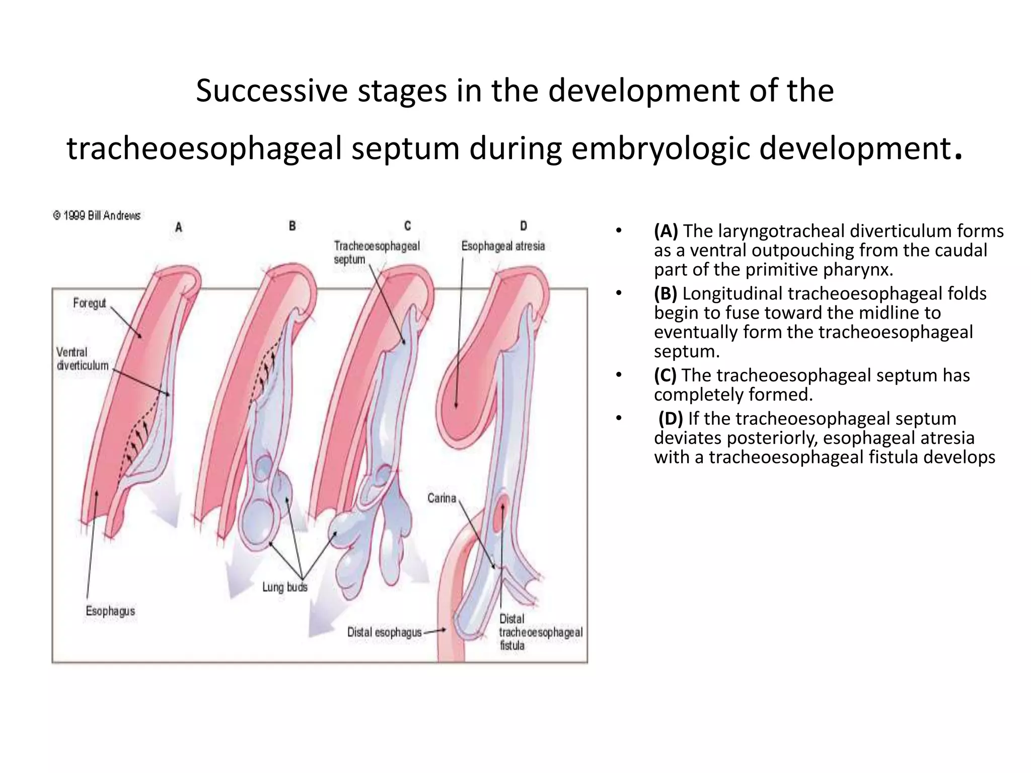
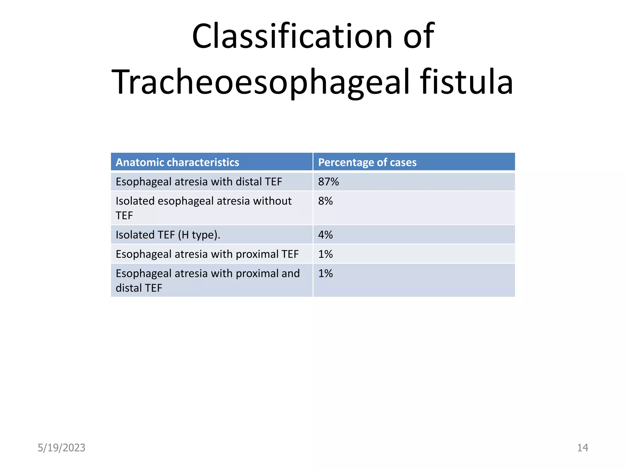
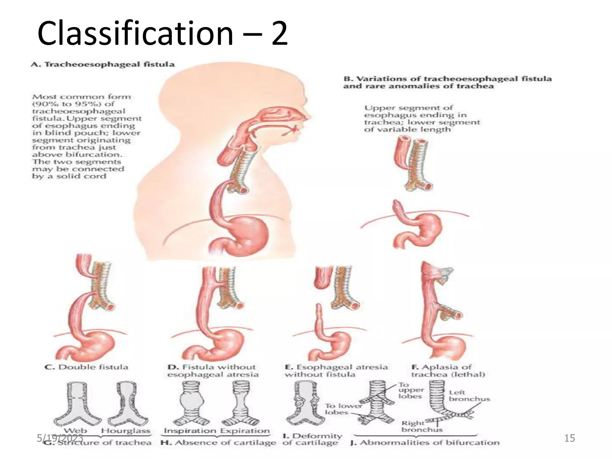
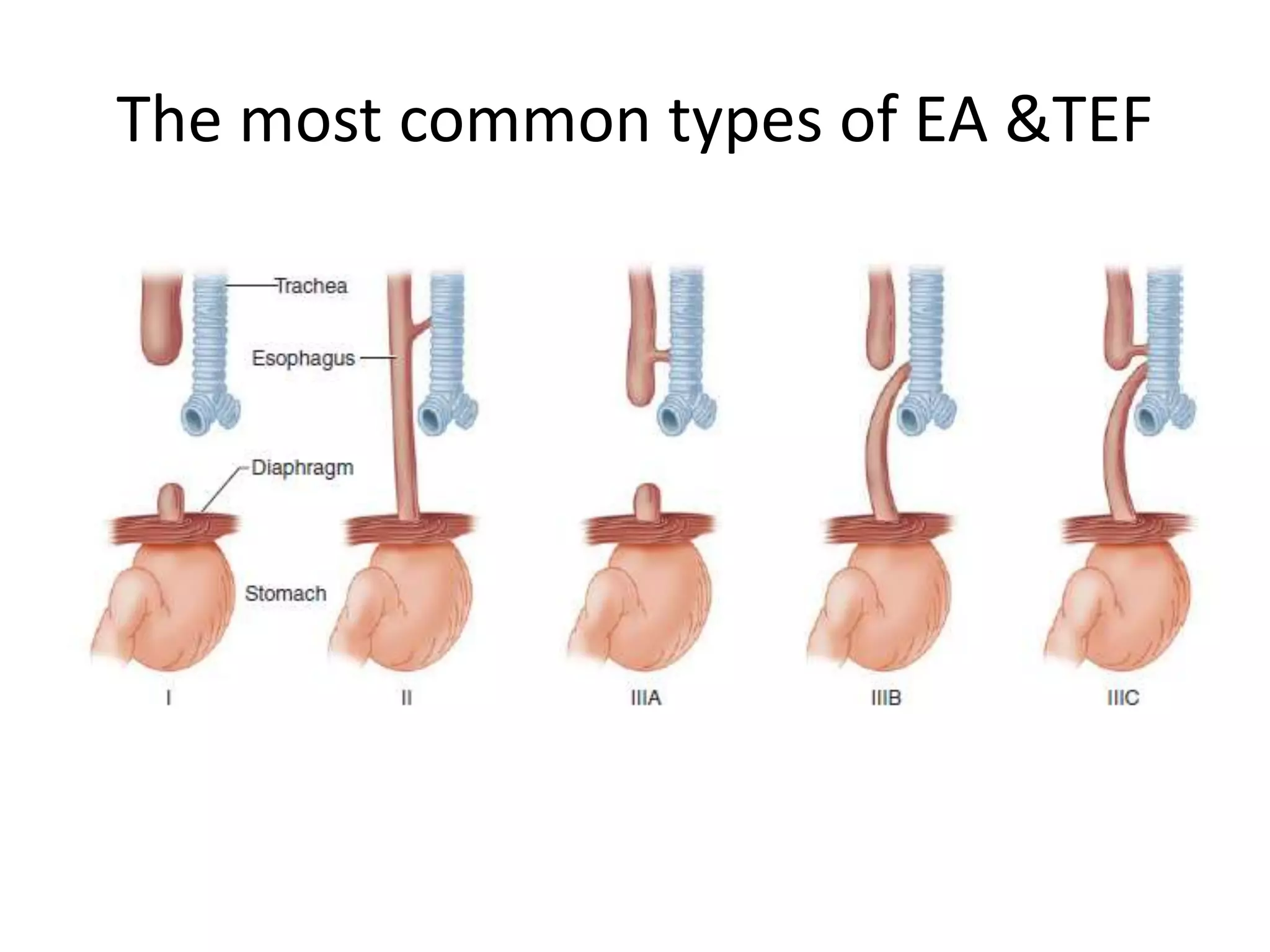
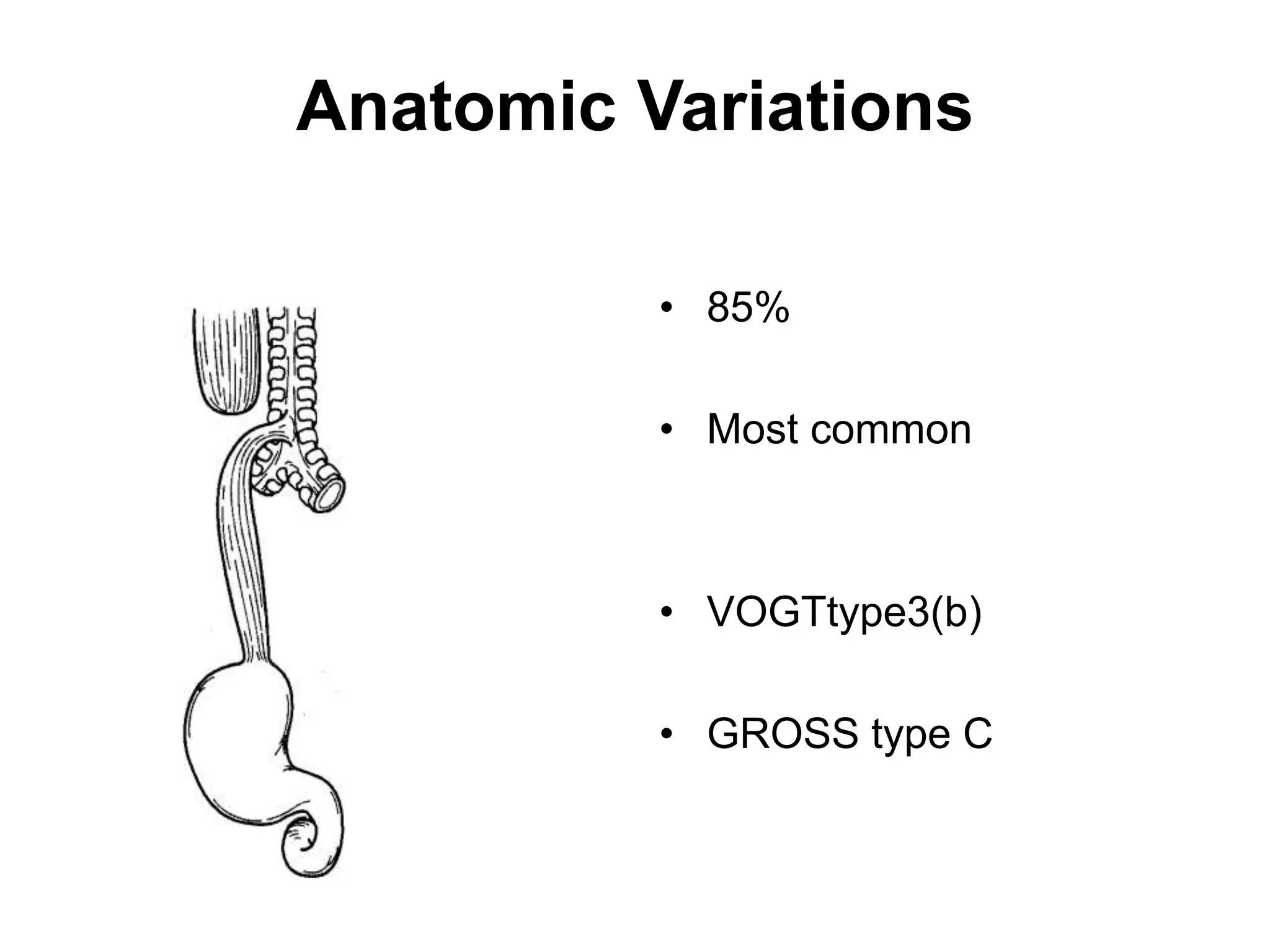
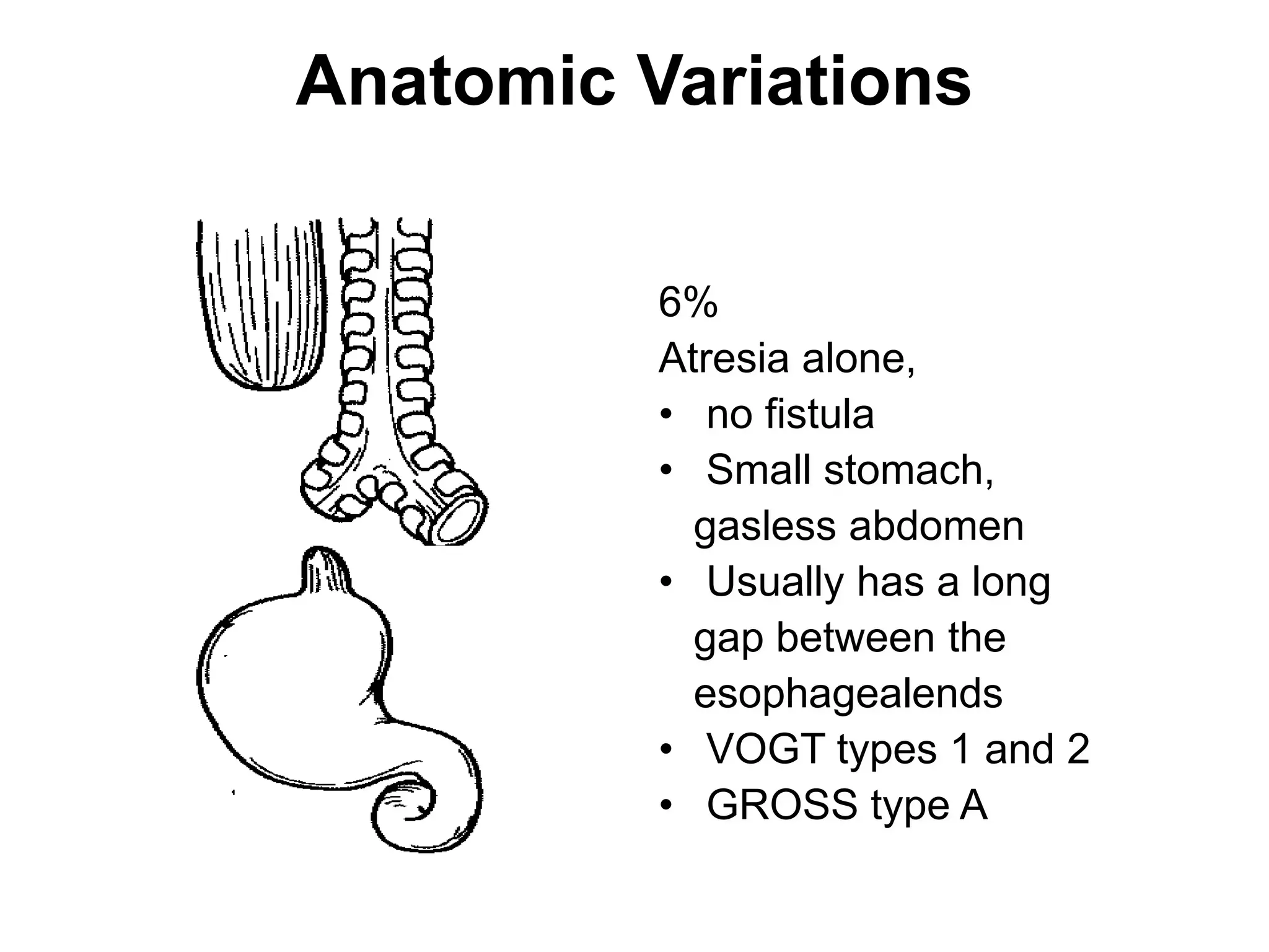
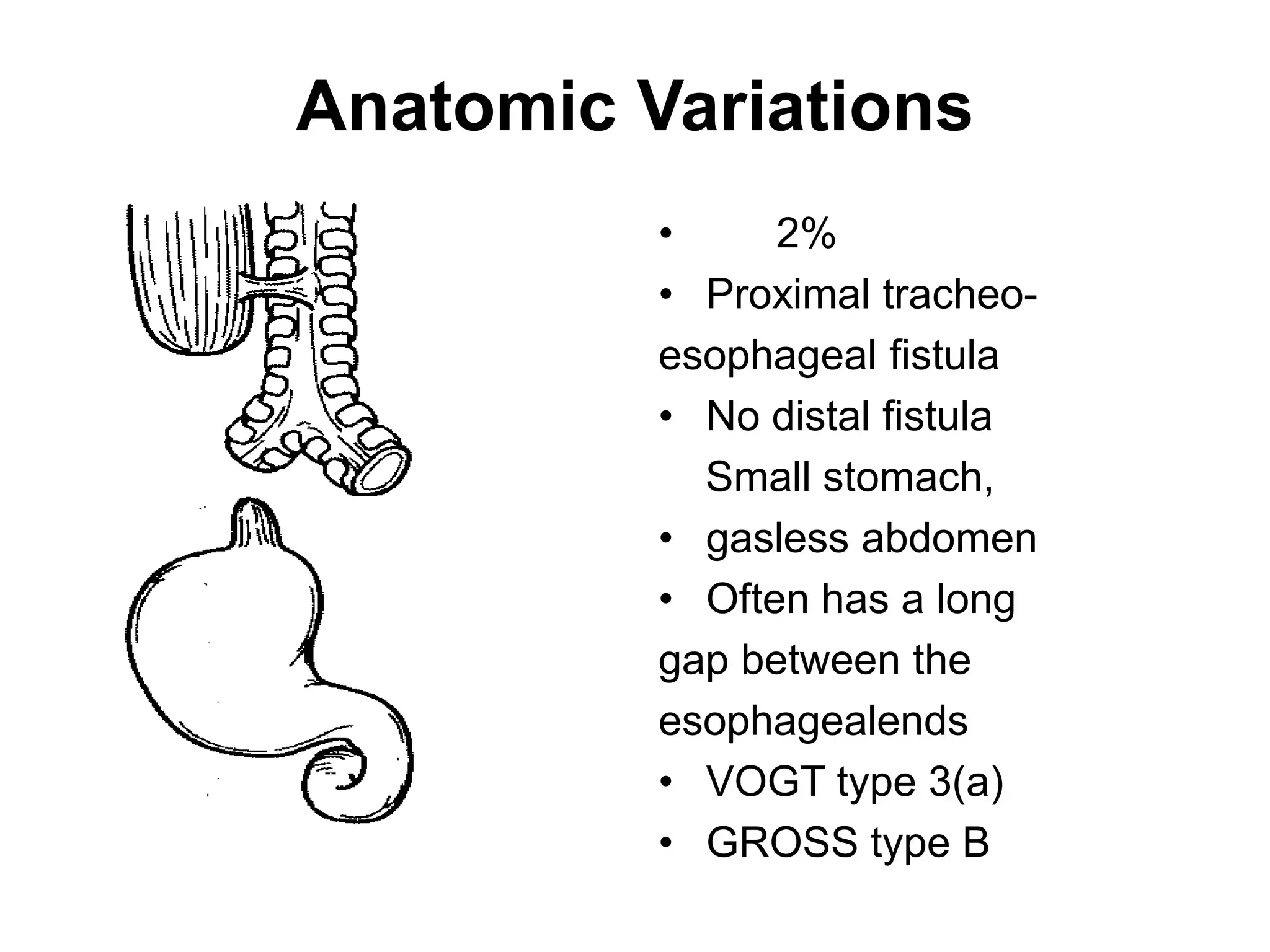
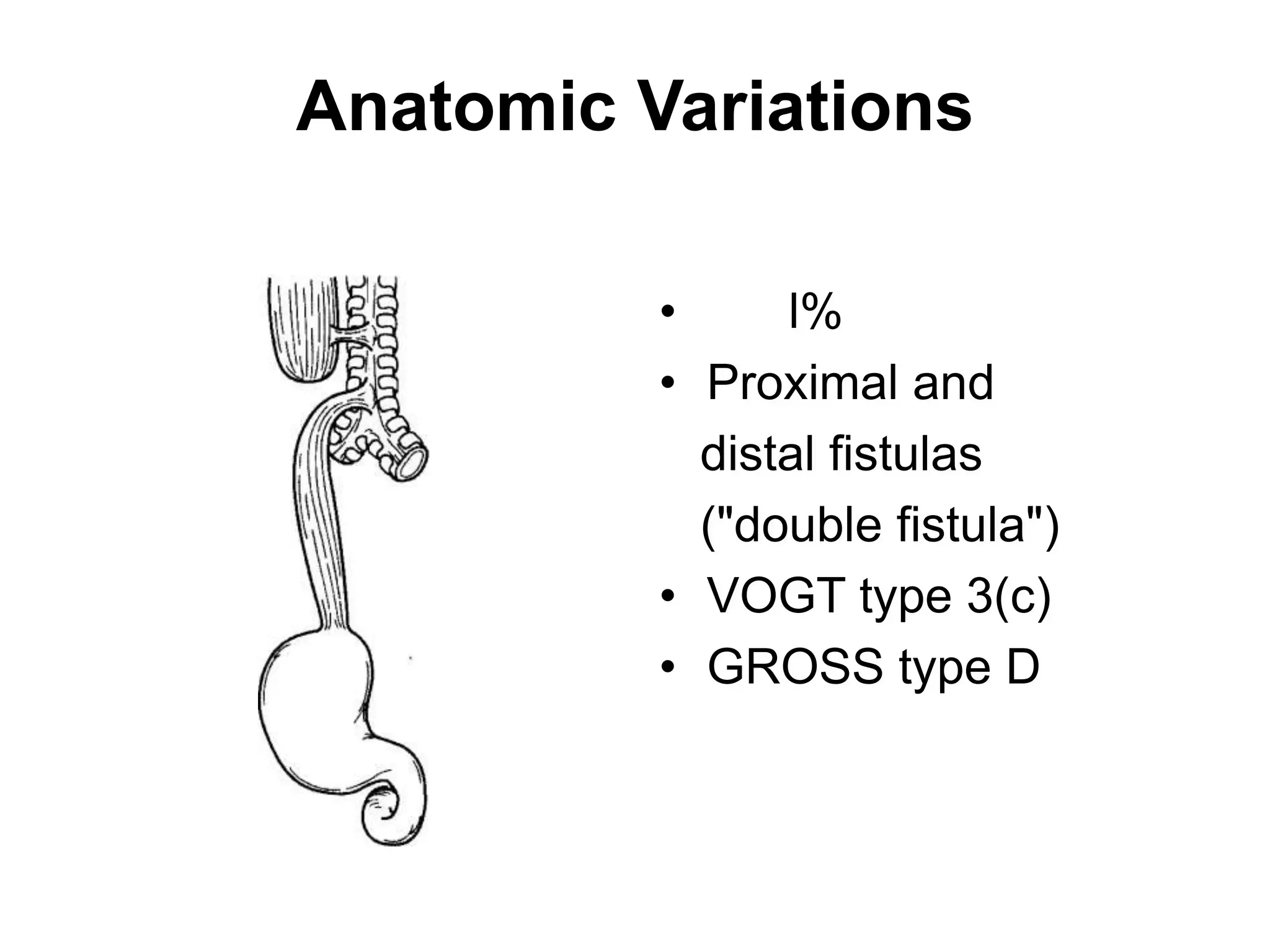
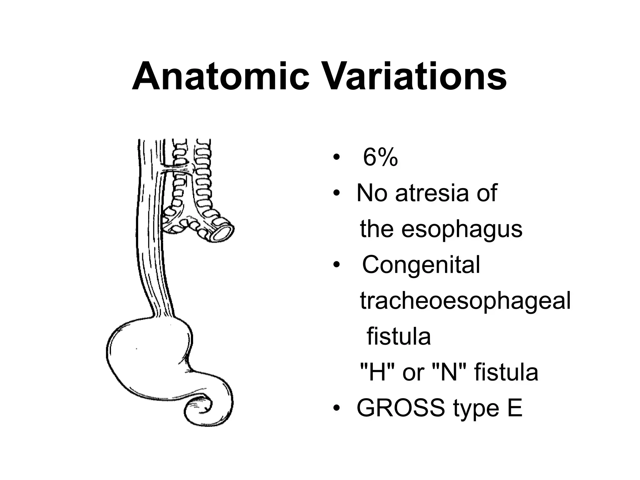
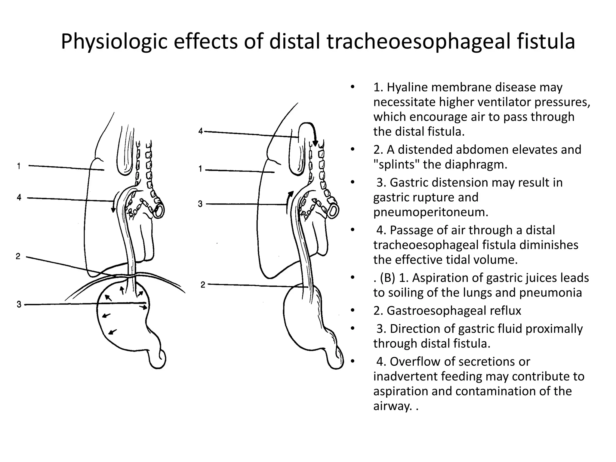
Eyayalem M.
Esophageal Atresia and
Tracheoesophageal Fistula
INTRODUCTION
• Tracheoesophageal fistula(TEF) is a congenital or
acquired communication between the trachea
and esophagus.
• Esophageal atresia is a congenital abnormality in
which the midportion of the esophagus is absent.
• Incidence is between 1 in 3,570 -1 in 4,500
• Esophageal atresia(EA) occurs in association with
TEF in about 90% of instances.
• TEF most likely leads to fatal pulmonary
complications.
5/19/2023 2
Introduction
• Most congenital TEFs are diagnosed
immediately following birth or in infancy.
• They are commonly associated with other
congenital anomalies.
• Acquired TEF occur secondary to malignant
disease, infection,, trauma, etc.
5/19/2023 3
Introduction
• Most congenital TEFs are diagnosed
immediately following birth or in infancy.
• They are commonly associated with other
congenital anomalies.
• Acquired TEF occur secondary to malignant
disease, infection,, trauma, etc.
5/19/2023 4
Etiology
• Exact aetiology of TEF is unclear.
• Embryologically believed to be due to
incomplete closure of laryngotracheal groove.
• Observed in mothers that used imidazole
containing decongestants in the first trimester
of pregnancy.
• Association with trisomies 18, 21, 13.
5/19/2023 5
Etiology
• Associated risk factors
• Advanced maternal age
• European ethnicity
• Obesity
• Low socioeconomic status
• Tobacco smoking
• Infants < 1500g ( highest mortality).
5/19/2023 6
Etiology
• Approximately 50% affected infants have
associated anomalies.
• Syndromes include VATER/VACTERL( vertebral,
anorectal, cardiac, tracheal, esophageal, renal,
radial).
• Some genetic factors have been implicated
with discrete mutations in syndromic cases.
5/19/2023 7
EMBRYOLOGY
• The esophagus and trachea both develop from
the primitive foregut.
• During 4 – 6 weeks of life the caudal part of
the foregut forms the ventral diverticular
which gives rise to the trachea.
5/19/2023 8
Embryology
• The longitudinal tracheoesophageal fold fuses
to form a septum, dividing the foregut into a
ventral laryngotracheal tube and a dorsal
esophagus.
• Posterior deviation of the septum causes
incomplete separation of the esophagus from
the laryngo tracheal tube resulting in TEF.
5/19/2023 9
Embryology
5/19/2023 10
Embryology
• Isolated esophageal atresia can occur when
the esophagus fails to recannalize by the 8th
week.
5/19/2023 11
Successive stages in the development of the
tracheoesophageal septum during embryologic development.
• (A) The laryngotracheal diverticulum forms
as a ventral outpouching from the caudal
part of the primitive pharynx.
• (B) Longitudinal tracheoesophageal folds
begin to fuse toward the midline to
eventually form the tracheoesophageal
septum.
• (C) The tracheoesophageal septum has
completely formed.
• (D) If the tracheoesophageal septum
deviates posteriorly, esophageal atresia
with a tracheoesophageal fistula develops
Introduction
Classification of
Tracheoesophageal fistula
Anatomic characteristics Percentage of cases
Esophageal atresia with distal TEF 87%
Isolated esophageal atresia without
TEF
8%
Isolated TEF (H type). 4%
Esophageal atresia with proximal TEF 1%
Esophageal atresia with proximal and
distal TEF
1%
5/19/2023 14
Classification – 2
5/19/2023 15
The most common types of EA &TEF
Anatomic Variations
• 85%
• Most common
• VOGTtype3(b)
• GROSS type C
Anatomic Variations
6%
Atresia alone,
• no fistula
• Small stomach,
gasless abdomen
• Usually has a long
gap between the
esophagealends
• VOGT types 1 and 2
• GROSS type A
Anatomic Variations
• 2%
• Proximal tracheo-
esophageal fistula
• No distal fistula
Small stomach,
• gasless abdomen
• Often has a long
gap between the
esophagealends
• VOGT type 3(a)
• GROSS type B
Anatomic Variations
• l%
• Proximal and
distal fistulas
("double fistula")
• VOGT type 3(c)
• GROSS type D
Anatomic Variations
• 6%
• No atresia of
the esophagus
• Congenital
tracheoesophageal
fistula
"H" or "N" fistula
• GROSS type E
Physiologic effects of distal tracheoesophageal fistula
• 1. Hyaline membrane disease may
necessitate higher ventilator pressures,
which encourage air to pass through
the distal fistula.
• 2. A distended abdomen elevates and
"splints" the diaphragm.
• 3. Gastric distension may result in
gastric rupture and
pneumoperitoneum.
• 4. Passage of air through a distal
tracheoesophageal fistula diminishes
the effective tidal volume.
• . (B) 1. Aspiration of gastric juices leads
to soiling of the lungs and pneumonia
• 2. Gastroesophageal reflux
• 3. Direction of gastric fluid proximally
through distal fistula.
• 4. Overflow of secretions or
inadvertent feeding may contribute to
aspiration and contamination of the
airway. .
Associated Abnormalities
Incidence of Associated Anomalies in Esophageal Atresia.
Anomaly Frequency (%)
• Congenital heart disease 25
• Urinary tract 22
• Orthopaedic (mostly vertebral and radial) 15
• Gastrointestinal (e.g., duodenal
• atresia,imperforate anus) 22
• Chromosomal (usually trisomy 18 or 21) 7
• Total with one or more associated 58
anomalies
Associated Congenital Anomalies Reported in Patients with Esophageal
Atresia
System affected
Musculoskeletal
Gastrointestinal
Cardiac
Genitourinary.
Potential anomalies
Hemivertebrae, radial dysplasia or
amelia, polydactyly, syndactyly, rib
malformations, scoliosis, lower limb
defects
Imperforate anus, duodenal atresia,
malrotation, intestinal
malformations, Meckel's
diverticulum, annular pancreas
Ventricular septal defect, patent
ductus arteriosus, tetralogy of Fallot,
atrial septal defect, single umbilical
artery, right-sided aortic arch
Renal agenesis or dysplasia,
horseshoe kidney, polycystic kidney,
ureteral and urethral malformations,
hypospadias
DIAGNOSIS OF ESOPHAGEAL
ATRESIA
• Antenatal Diagnosis (maternal
polyhydramnios, a small stomach, a distended
upper esophageal pouch, or abnormal
swallowing)
• Diagnostic suspicion is increased when
abnormalities known to be associated with
esophageal atresia are identified.
Fetal MRI
• This 32 week
fetus had
esophageal
atresia and an
absent stomach,
resulting in
marked
polyhydramnios
Clinical Diagnosis
• Prematurity
• Any excessively drooling (copious, fine, white,
frothy bubbles of mucus in the mouth and,
sometimes, the nose).
Clinical Diagnosis
Diagnosis of esophageal
atresia is confirmed when a
10-gauge (French) catheter
cannot be passed beyond 10
cm from the gums
A smaller-caliber tube is not used
because it may curl up in the upper
esophageal segment, giving a false
impression of esophageal continuity.
The chest radiograph
• A plain radiograph will
confirm the tube has
not reached the
stomach
The Gasless Abdomen
• Absence of gas in the
abdomen suggests that
the patient has either
atresia without a fistula
or atresia with a
proximal fistula only
Contrast studies
• should be performed by an
experienced pediatric
radiologist, or after transfer
to the tertiary institution,
and with the use of a small
amount (0.5 to 1 mL) of
water-soluble contrast. Care
must be taken to avoid
aspiration.
Management
• Measures should be taken to reduce the risk of
aspiration(continuous suctioning of the upper pouch,
the infant's head should be elevated).
• In infants with respiratory failure, endotracheal
intubation should be performed.
• Transfer to a major tertiary pediatric institution is
best not delayed .
34
treatment
Goals of the initial treatment:
1)Attention to ventilation
2)↓ upper pouch pressure
3)Determine appropriate time for surgery
So:
For ↓ aspiration risk:
1)elevate neonate’s head at least 30º in infant warmer
2)Use “sump” catheter on34 continiouse suction
IV AB and electrolyte.

chapter 6-1 Esophageal atresia and TEF.ppt

  • 1.
    Eyayalem M. Esophageal Atresiaand Tracheoesophageal Fistula
  • 2.
    INTRODUCTION • Tracheoesophageal fistula(TEF)is a congenital or acquired communication between the trachea and esophagus. • Esophageal atresia is a congenital abnormality in which the midportion of the esophagus is absent. • Incidence is between 1 in 3,570 -1 in 4,500 • Esophageal atresia(EA) occurs in association with TEF in about 90% of instances. • TEF most likely leads to fatal pulmonary complications. 5/19/2023 2
  • 3.
    Introduction • Most congenitalTEFs are diagnosed immediately following birth or in infancy. • They are commonly associated with other congenital anomalies. • Acquired TEF occur secondary to malignant disease, infection,, trauma, etc. 5/19/2023 3
  • 4.
    Introduction • Most congenitalTEFs are diagnosed immediately following birth or in infancy. • They are commonly associated with other congenital anomalies. • Acquired TEF occur secondary to malignant disease, infection,, trauma, etc. 5/19/2023 4
  • 5.
    Etiology • Exact aetiologyof TEF is unclear. • Embryologically believed to be due to incomplete closure of laryngotracheal groove. • Observed in mothers that used imidazole containing decongestants in the first trimester of pregnancy. • Association with trisomies 18, 21, 13. 5/19/2023 5
  • 6.
    Etiology • Associated riskfactors • Advanced maternal age • European ethnicity • Obesity • Low socioeconomic status • Tobacco smoking • Infants < 1500g ( highest mortality). 5/19/2023 6
  • 7.
    Etiology • Approximately 50%affected infants have associated anomalies. • Syndromes include VATER/VACTERL( vertebral, anorectal, cardiac, tracheal, esophageal, renal, radial). • Some genetic factors have been implicated with discrete mutations in syndromic cases. 5/19/2023 7
  • 8.
    EMBRYOLOGY • The esophagusand trachea both develop from the primitive foregut. • During 4 – 6 weeks of life the caudal part of the foregut forms the ventral diverticular which gives rise to the trachea. 5/19/2023 8
  • 9.
    Embryology • The longitudinaltracheoesophageal fold fuses to form a septum, dividing the foregut into a ventral laryngotracheal tube and a dorsal esophagus. • Posterior deviation of the septum causes incomplete separation of the esophagus from the laryngo tracheal tube resulting in TEF. 5/19/2023 9
  • 10.
  • 11.
    Embryology • Isolated esophagealatresia can occur when the esophagus fails to recannalize by the 8th week. 5/19/2023 11
  • 12.
    Successive stages inthe development of the tracheoesophageal septum during embryologic development. • (A) The laryngotracheal diverticulum forms as a ventral outpouching from the caudal part of the primitive pharynx. • (B) Longitudinal tracheoesophageal folds begin to fuse toward the midline to eventually form the tracheoesophageal septum. • (C) The tracheoesophageal septum has completely formed. • (D) If the tracheoesophageal septum deviates posteriorly, esophageal atresia with a tracheoesophageal fistula develops
  • 13.
  • 14.
    Classification of Tracheoesophageal fistula Anatomiccharacteristics Percentage of cases Esophageal atresia with distal TEF 87% Isolated esophageal atresia without TEF 8% Isolated TEF (H type). 4% Esophageal atresia with proximal TEF 1% Esophageal atresia with proximal and distal TEF 1% 5/19/2023 14
  • 15.
  • 16.
    The most commontypes of EA &TEF
  • 17.
    Anatomic Variations • 85% •Most common • VOGTtype3(b) • GROSS type C
  • 18.
    Anatomic Variations 6% Atresia alone, •no fistula • Small stomach, gasless abdomen • Usually has a long gap between the esophagealends • VOGT types 1 and 2 • GROSS type A
  • 19.
    Anatomic Variations • 2% •Proximal tracheo- esophageal fistula • No distal fistula Small stomach, • gasless abdomen • Often has a long gap between the esophagealends • VOGT type 3(a) • GROSS type B
  • 20.
    Anatomic Variations • l% •Proximal and distal fistulas ("double fistula") • VOGT type 3(c) • GROSS type D
  • 21.
    Anatomic Variations • 6% •No atresia of the esophagus • Congenital tracheoesophageal fistula "H" or "N" fistula • GROSS type E
  • 22.
    Physiologic effects ofdistal tracheoesophageal fistula • 1. Hyaline membrane disease may necessitate higher ventilator pressures, which encourage air to pass through the distal fistula. • 2. A distended abdomen elevates and "splints" the diaphragm. • 3. Gastric distension may result in gastric rupture and pneumoperitoneum. • 4. Passage of air through a distal tracheoesophageal fistula diminishes the effective tidal volume. • . (B) 1. Aspiration of gastric juices leads to soiling of the lungs and pneumonia • 2. Gastroesophageal reflux • 3. Direction of gastric fluid proximally through distal fistula. • 4. Overflow of secretions or inadvertent feeding may contribute to aspiration and contamination of the airway. .
  • 23.
    Associated Abnormalities Incidence ofAssociated Anomalies in Esophageal Atresia. Anomaly Frequency (%) • Congenital heart disease 25 • Urinary tract 22 • Orthopaedic (mostly vertebral and radial) 15 • Gastrointestinal (e.g., duodenal • atresia,imperforate anus) 22 • Chromosomal (usually trisomy 18 or 21) 7 • Total with one or more associated 58 anomalies
  • 24.
    Associated Congenital AnomaliesReported in Patients with Esophageal Atresia System affected Musculoskeletal Gastrointestinal Cardiac Genitourinary. Potential anomalies Hemivertebrae, radial dysplasia or amelia, polydactyly, syndactyly, rib malformations, scoliosis, lower limb defects Imperforate anus, duodenal atresia, malrotation, intestinal malformations, Meckel's diverticulum, annular pancreas Ventricular septal defect, patent ductus arteriosus, tetralogy of Fallot, atrial septal defect, single umbilical artery, right-sided aortic arch Renal agenesis or dysplasia, horseshoe kidney, polycystic kidney, ureteral and urethral malformations, hypospadias
  • 25.
    DIAGNOSIS OF ESOPHAGEAL ATRESIA •Antenatal Diagnosis (maternal polyhydramnios, a small stomach, a distended upper esophageal pouch, or abnormal swallowing) • Diagnostic suspicion is increased when abnormalities known to be associated with esophageal atresia are identified.
  • 26.
    Fetal MRI • This32 week fetus had esophageal atresia and an absent stomach, resulting in marked polyhydramnios
  • 27.
    Clinical Diagnosis • Prematurity •Any excessively drooling (copious, fine, white, frothy bubbles of mucus in the mouth and, sometimes, the nose).
  • 28.
    Clinical Diagnosis Diagnosis ofesophageal atresia is confirmed when a 10-gauge (French) catheter cannot be passed beyond 10 cm from the gums A smaller-caliber tube is not used because it may curl up in the upper esophageal segment, giving a false impression of esophageal continuity.
  • 29.
    The chest radiograph •A plain radiograph will confirm the tube has not reached the stomach
  • 30.
    The Gasless Abdomen •Absence of gas in the abdomen suggests that the patient has either atresia without a fistula or atresia with a proximal fistula only
  • 31.
    Contrast studies • shouldbe performed by an experienced pediatric radiologist, or after transfer to the tertiary institution, and with the use of a small amount (0.5 to 1 mL) of water-soluble contrast. Care must be taken to avoid aspiration.
  • 32.
    Management • Measures shouldbe taken to reduce the risk of aspiration(continuous suctioning of the upper pouch, the infant's head should be elevated). • In infants with respiratory failure, endotracheal intubation should be performed. • Transfer to a major tertiary pediatric institution is best not delayed .
  • 34.
    34 treatment Goals of theinitial treatment: 1)Attention to ventilation 2)↓ upper pouch pressure 3)Determine appropriate time for surgery So: For ↓ aspiration risk: 1)elevate neonate’s head at least 30º in infant warmer 2)Use “sump” catheter on34 continiouse suction IV AB and electrolyte.