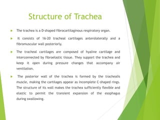
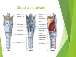
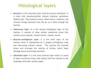
Anatomy of
Trachea
By- CHIRAG AJMERA
Assistant Professor, MJF Group of Colleges and Hospitals
Introduction
 The trachea, commonly known as the windpipe, is a
fibrocartilaginous tube about 10-11 cm long.
 The trachea begins just under the larynx (voice box) and
runs down behind the sternum. The trachea then divides
into two smaller tubes called bronchus: one bronchus for
each lung.
 The main function of the trachea is to transport air in
and out of the lungs during the act of breathing.
 It protects the respiratory tract by warming and
moistening the air and propelling foreign particles
upwards towards the pharynx for expulsion.
 The trachea helps defend against disease. The mucus in
the trachea helps capture microorganisms such as viruses
and harmful bacteria before they enter the lungs.
Relations of Trachea
 Anteriorly –
 Sternum
 Sternothyroid muscles
 Arch of aorta ,
 Brachiocephalic trunk
 left common carotid arteries
Relations of Trachea
 Posteriorly –
 Oesophagus
 Vertebral column
 Right side
 Right lung and pleura
 Right vagus
 Azygos vein
 Left side
 Arch of aorta
 left common carotid
arteries
 left subclavian arteries
Structure of Trachea
 The trachea is a D-shaped fibrocartilaginous respiratory organ.
 It consists of 16-20 tracheal cartilages anterolaterally and a
fibromuscular wall posteriorly.
 The tracheal cartilages are composed of hyaline cartilage and
interconnected by fibroelastic tissue. They support the trachea and
keep it open during pressure changes that accompany air
ventilation.
 The posterior wall of the trachea is formed by the trachealis
muscle, making the cartilages appear as incomplete C-shaped rings.
The structure of its wall makes the trachea sufficiently flexible and
elastic to permit the transient expansion of the esophagus
during swallowing.
Structure diagram
Histology
 The trachea consists of four histological layers.
 Mucosa
 Submucosal layer
 Musculo-cartilaginous layer
 Adventitia
Histological layers
Histological layers
 Mucosa-It is the innermost layer lined by mucous membrane. It
is lined with pseudostratified ciliated columnar epithelium
(Goblet cell). They produce mucus, which warms, moistens, and
removes foreign particles from the air as it flows through the
trachea.
 Submucosa layer- It is the second histological layer of the
trachea. It consists of loose areolar connective tissue that
contains mucus glands, smooth muscle, vessels, nerves.
 Musculo-cartilaginous layer- It is the third layer of the
trachea which is represented by C shaped cartilaginous rings
and intervening smooth muscle. This narrows the tracheal
lumen and increases the velocity of airflow, which helps
dislodge mucus and foreign particles.
 Adventitia layer- It is the most external layer which is a band
of loose connective tissue that loosely bind the trachea to the
esophagus and other nearby organs.
Blood supply
 Arterial supply- Inferior thyroid arteries
 Venous supply - Brachiocephalic veins
 Lymphatic drainage - pretracheal and paratracheal lymph nodes.
 Nerve supply –
 Parasympathetic nerves supply by the laryngeal nerve
 Sympathetic nerves supply the trachealis muscle
Function
 The main function is to transport oxygenated air from the upper
respiratory tract towards the alveoli for gas exchange. The resulting
carbon dioxide filled air is transported by the tracheobronchial system
towards the oral cavity and nose for excretion.
 The trachea also protects the respiratory tract via physical and
immunological methods. In addition to oxygen and carbon dioxide,
environmental air also contains many potentially harmful agents such as
pathogens, debris, gases and chemicals. The goblet cells lining the
trachea secrete mucus which traps any unwanted substances. Immune
cells and their secretions prevent harmful microorganisms from reaching
the lower airways. The trapped particles are then removed from the
trachea. This system pushes them upwards back into the pharynx and
mouth for excretion by the coughing reflex.
 The trachea also helps regulate the temperature of the air coming in
and out of the lungs.
 On cold days, the trachea helps warm and provide humidity to the air
before it reaches the lungs.
 On hot days, the trachea helps cool the air through evaporation.
Thank you
This slides covers about the Introduction, Relations,
Structure, blood supply, nerve supply and Function of the
Trachea

Tracheal anatomy

  • 1.
    Anatomy of Trachea By- CHIRAGAJMERA Assistant Professor, MJF Group of Colleges and Hospitals
  • 2.
    Introduction  The trachea,commonly known as the windpipe, is a fibrocartilaginous tube about 10-11 cm long.  The trachea begins just under the larynx (voice box) and runs down behind the sternum. The trachea then divides into two smaller tubes called bronchus: one bronchus for each lung.  The main function of the trachea is to transport air in and out of the lungs during the act of breathing.  It protects the respiratory tract by warming and moistening the air and propelling foreign particles upwards towards the pharynx for expulsion.  The trachea helps defend against disease. The mucus in the trachea helps capture microorganisms such as viruses and harmful bacteria before they enter the lungs.
  • 4.
    Relations of Trachea Anteriorly –  Sternum  Sternothyroid muscles  Arch of aorta ,  Brachiocephalic trunk  left common carotid arteries
  • 5.
    Relations of Trachea Posteriorly –  Oesophagus  Vertebral column  Right side  Right lung and pleura  Right vagus  Azygos vein  Left side  Arch of aorta  left common carotid arteries  left subclavian arteries
  • 6.
    Structure of Trachea The trachea is a D-shaped fibrocartilaginous respiratory organ.  It consists of 16-20 tracheal cartilages anterolaterally and a fibromuscular wall posteriorly.  The tracheal cartilages are composed of hyaline cartilage and interconnected by fibroelastic tissue. They support the trachea and keep it open during pressure changes that accompany air ventilation.  The posterior wall of the trachea is formed by the trachealis muscle, making the cartilages appear as incomplete C-shaped rings. The structure of its wall makes the trachea sufficiently flexible and elastic to permit the transient expansion of the esophagus during swallowing.
  • 7.
  • 8.
    Histology  The tracheaconsists of four histological layers.  Mucosa  Submucosal layer  Musculo-cartilaginous layer  Adventitia
  • 9.
  • 10.
    Histological layers  Mucosa-Itis the innermost layer lined by mucous membrane. It is lined with pseudostratified ciliated columnar epithelium (Goblet cell). They produce mucus, which warms, moistens, and removes foreign particles from the air as it flows through the trachea.  Submucosa layer- It is the second histological layer of the trachea. It consists of loose areolar connective tissue that contains mucus glands, smooth muscle, vessels, nerves.  Musculo-cartilaginous layer- It is the third layer of the trachea which is represented by C shaped cartilaginous rings and intervening smooth muscle. This narrows the tracheal lumen and increases the velocity of airflow, which helps dislodge mucus and foreign particles.  Adventitia layer- It is the most external layer which is a band of loose connective tissue that loosely bind the trachea to the esophagus and other nearby organs.
  • 11.
    Blood supply  Arterialsupply- Inferior thyroid arteries  Venous supply - Brachiocephalic veins  Lymphatic drainage - pretracheal and paratracheal lymph nodes.  Nerve supply –  Parasympathetic nerves supply by the laryngeal nerve  Sympathetic nerves supply the trachealis muscle
  • 12.
    Function  The mainfunction is to transport oxygenated air from the upper respiratory tract towards the alveoli for gas exchange. The resulting carbon dioxide filled air is transported by the tracheobronchial system towards the oral cavity and nose for excretion.  The trachea also protects the respiratory tract via physical and immunological methods. In addition to oxygen and carbon dioxide, environmental air also contains many potentially harmful agents such as pathogens, debris, gases and chemicals. The goblet cells lining the trachea secrete mucus which traps any unwanted substances. Immune cells and their secretions prevent harmful microorganisms from reaching the lower airways. The trapped particles are then removed from the trachea. This system pushes them upwards back into the pharynx and mouth for excretion by the coughing reflex.  The trachea also helps regulate the temperature of the air coming in and out of the lungs.  On cold days, the trachea helps warm and provide humidity to the air before it reaches the lungs.  On hot days, the trachea helps cool the air through evaporation.
  • 13.
    Thank you This slidescovers about the Introduction, Relations, Structure, blood supply, nerve supply and Function of the Trachea