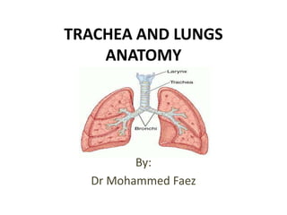
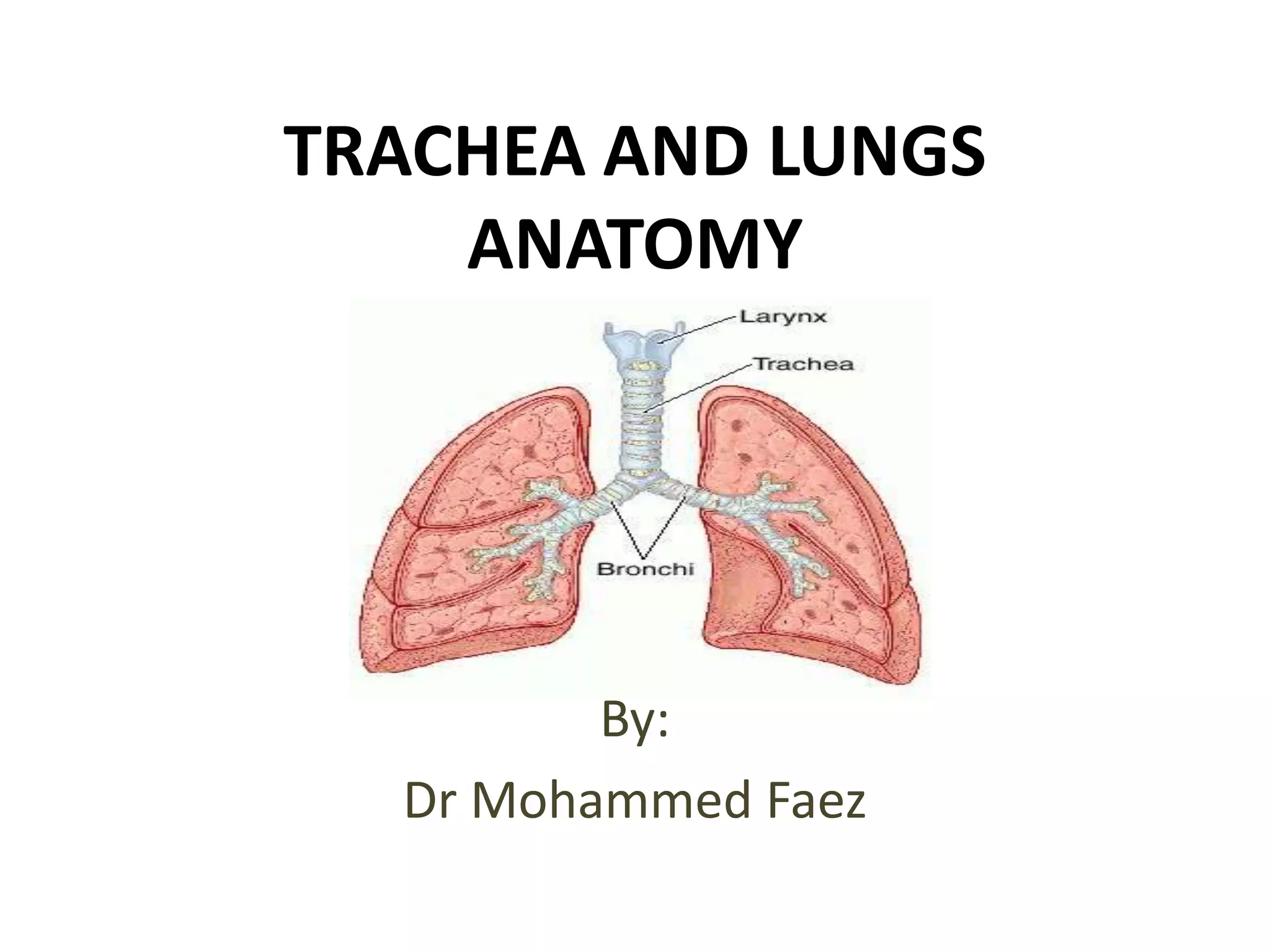
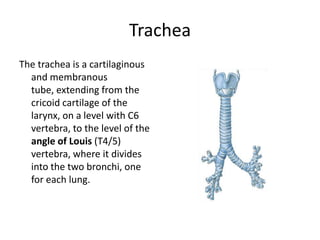
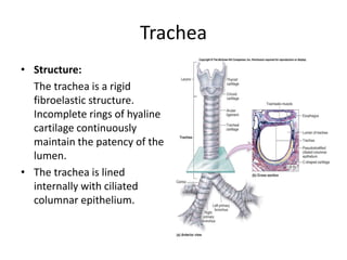
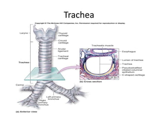
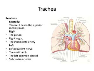
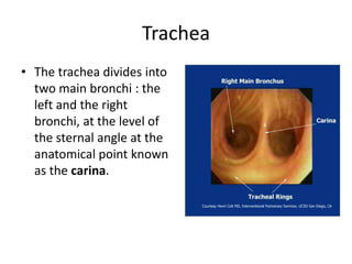
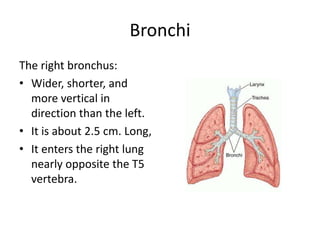
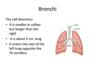
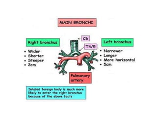
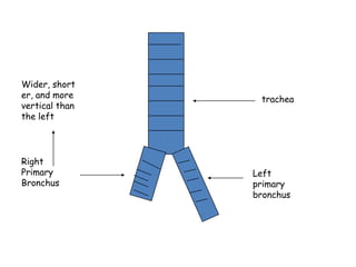
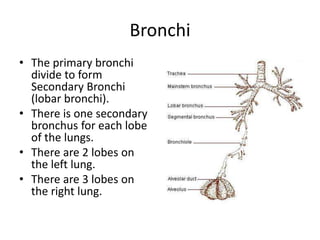
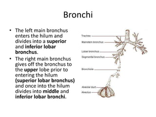
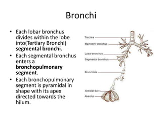
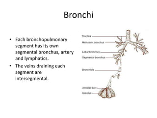
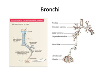
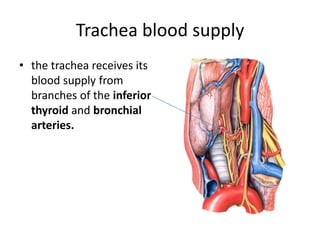
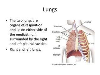
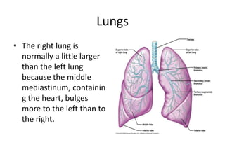
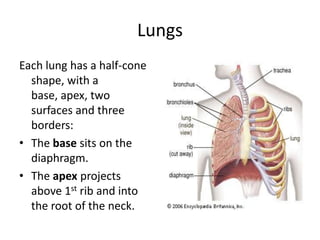
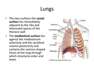
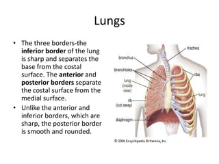
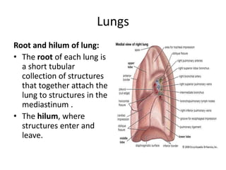
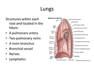
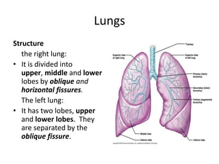
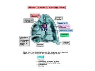
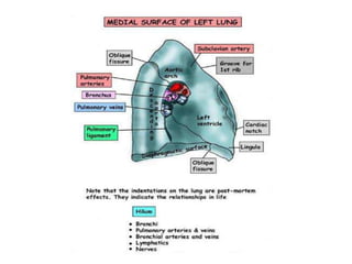
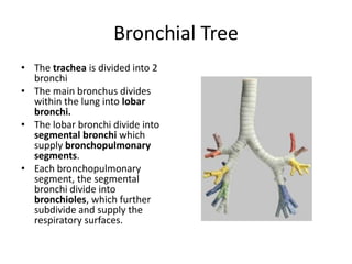
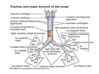
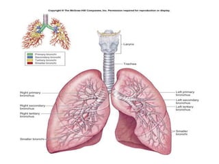
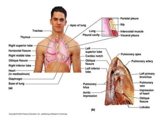
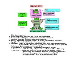
TRACHEA AND LUNGS ANATOMYBy:Dr Mohammed Faez
TracheaThe trachea is a cartilaginous and membranous tube, extending from the cricoid cartilageof the larynx, on a level with C6 vertebra, to the level of the angle of Louis (T4/5) vertebra, where it divides into the two bronchi, one for each lung.
TracheaStructure: The trachea is a rigid fibroelastic structure. Incomplete rings of hyaline cartilage continuously maintain the patency of the lumen. The trachea is lined internally with ciliated columnar epithelium.
Trachea
TracheaIt measures about 11 cm. In length. Its diameter, from side to side, is from 2 to 2.5 cm.It has 18-22 cartilaginous ringsIt is greater in the male than in the female.
TracheaRelations:Anteriorly:Neck:The isthmus of the thyroid gland (2nd, 3rd and 4th tracheal rings )The inferior thyroid veinsThe sternothyroid and sternohyoid musclesThe cervical fasciaThorax: The manubriumsterniThe remains of the thymusThe left innominate veinThe aortic archLeft common carotid arteriesThe deep cardiac plexus.
TracheaRelations:Posteriorly : the esophagusLaterally:Neck:Common carotid arteriesRight and left lobes of the thyroid glandInferior thyroid arteriesThe recurrent nerves
TracheaRelations:Laterally:Thorax: it lies in the superior mediastinum.	RightThe pleura Right vagus, The innominate artery	Left Left recurrent nerveThe aortic archThe left common carotid Subclavian arteries
Inferior thyroid a.Vagus n.Recurrent laryngeal n.Thoracic duct Sympathetic trunkTrachea
TracheaThe trachea divides into two main bronchi : the left and the right bronchi, at the level of the sternal angle at the anatomical point known as the carina.
BronchiThe right bronchus:Wider, shorter, and more vertical in direction than the left. It is about 2.5 cm. Long, It enters the right lung nearly opposite the T5 vertebra.
BronchiThe Left Bronchus It is smaller in caliber but longer than the rightIt is about 5 cm. long.It enters the root of the left lung opposite the T6 vertebra.
Wider, shorter, and more vertical than the lefttracheaRight Primary BronchusLeft primary bronchus
BronchiThe primary bronchi divide to form Secondary Bronchi (lobar bronchi).There is one secondary bronchus for each lobe of the lungs.There are 2 lobes on the left lung.There are 3 lobes on the right lung.
BronchiThe left main bronchus enters the hilum and divides into a superior and inferior lobar bronchus. The right main bronchus gives off the bronchus to the upper lobe prior to entering the hilum(superior lobar bronchus)and once into the hilumdivides into middle and inferiorlobar bronchi.
BronchiEach lobar bronchus divides within the lobe into(Tertiary Bronchi) segmental bronchi.Each segmental bronchus enters a bronchopulmonary segment.Each bronchopulmonary segment is pyramidal in shape with its apex directed towards the hilum.
BronchiEach bronchopulmonarysegment has its own segmental bronchus, artery and lymphatics.The veins draining each segment are intersegmental.
Bronchi
Trachea blood supplythe trachea receives its blood supply from branches of the inferior thyroid and bronchial arteries.
LungsThe two lungs are organs of respiration and lie on either side of the mediastinum surrounded by the right and left pleural cavities.Right and left lungs.
LungsThe right lung is normally a little larger than the left lung because the middle mediastinum, containing the heart, bulges more to the left than to the right.
LungsEach lung has a half-cone shape, with a base, apex, two surfaces and three borders:The base sits on the diaphragm. The apex projects above 1st rib and into the root of the neck.
LungsThe two surfaces-the costal surface lies immediately adjacent to the ribs and intercostal spaces of the thoracic wall. The mediastinal surface lies against the mediastinumanteriorly and the vertebral column posteriorly and contains the comma-shaped hilum of the lung through which structures enter and leave.
LungsThe three borders-the inferior border of the lung is sharp and separates the base from the costal surface. The anterior and posterior borders separate the costal surface from the medial surface. Unlike the anterior and inferior borders, which are sharp, the posterior border is smooth and rounded.
LungsRoot and hilum of lung:The root of each lung is a short tubular collection of structures that together attach the lung to structures in the mediastinum .The hilum, where structures enter and leave.
LungsStructures within each root and located in the hilum:A pulmonary artery Two pulmonary veinsA main bronchusBronchial vessel NervesLymphatics
LungsStructure	the right lung:It is divided into upper, middle and lower lobes by oblique and horizontal fissures.	The left lung:It has two lobes, upper and lower lobes.  They are separated by the oblique fissure.
LungsRead about right and left lungs relations and  lungs impressions.
Bronchial TreeThe trachea is divided into 2 bronchiThe main bronchus divides within the lung into lobar bronchi.The lobar bronchi divide into segmental bronchi which supply bronchopulmonary segments.Each bronchopulmonary segment, the segmental bronchi divide into bronchioles, which further subdivide and supply the respiratory surfaces.
Bronchopulmonary SegmentThere are ten bronchopulmonary segments in each lungEach bronchopulmonary segment is shaped like an irregular cone with the apex at the origin of the segmental bronchus and the base projected peripherally onto the surface of the lung.
Bronchopulmonary SegmentA bronchopulmonary segment is the smallest, functionally independent region of a lung and the smallest area of lung that can be isolated and removed without affecting adjacent regions.
Blood supply of the lungThe bronchi and parenchymal tissue of the lungs are supplied by bronchial arteries a branches of the descending thoracic aorta.Bronchial veins, which also communicate with pulmonary veins, drain into the azygos and hemiazygos.
Lymphatic drainage of the lungslymph returns from the periphery towards the hilartracheobronchial groups of nodes and from here to mediastinallymph trunks.
Nerve supply of the lungsA pulmonary plexus is located at the root of each lung. The plexus is composed of sympathetic fibres (from the sympathetic trunk) and parasympathetic fibres (from the vagus).
THANK YOU
Trachea + Lungs
Trachea + Lungs
Trachea + Lungs
Trachea + Lungs

Trachea + Lungs

  • 1.
    TRACHEA AND LUNGSANATOMYBy:Dr Mohammed Faez
  • 2.
    TracheaThe trachea isa cartilaginous and membranous tube, extending from the cricoid cartilageof the larynx, on a level with C6 vertebra, to the level of the angle of Louis (T4/5) vertebra, where it divides into the two bronchi, one for each lung.
  • 3.
    TracheaStructure: The tracheais a rigid fibroelastic structure. Incomplete rings of hyaline cartilage continuously maintain the patency of the lumen. The trachea is lined internally with ciliated columnar epithelium.
  • 4.
  • 5.
    TracheaIt measures about11 cm. In length. Its diameter, from side to side, is from 2 to 2.5 cm.It has 18-22 cartilaginous ringsIt is greater in the male than in the female.
  • 6.
    TracheaRelations:Anteriorly:Neck:The isthmus ofthe thyroid gland (2nd, 3rd and 4th tracheal rings )The inferior thyroid veinsThe sternothyroid and sternohyoid musclesThe cervical fasciaThorax: The manubriumsterniThe remains of the thymusThe left innominate veinThe aortic archLeft common carotid arteriesThe deep cardiac plexus.
  • 7.
    TracheaRelations:Posteriorly : theesophagusLaterally:Neck:Common carotid arteriesRight and left lobes of the thyroid glandInferior thyroid arteriesThe recurrent nerves
  • 8.
    TracheaRelations:Laterally:Thorax: it liesin the superior mediastinum. RightThe pleura Right vagus, The innominate artery Left Left recurrent nerveThe aortic archThe left common carotid Subclavian arteries
  • 9.
    Inferior thyroid a.Vagusn.Recurrent laryngeal n.Thoracic duct Sympathetic trunkTrachea
  • 10.
    TracheaThe trachea dividesinto two main bronchi : the left and the right bronchi, at the level of the sternal angle at the anatomical point known as the carina.
  • 11.
    BronchiThe right bronchus:Wider,shorter, and more vertical in direction than the left. It is about 2.5 cm. Long, It enters the right lung nearly opposite the T5 vertebra.
  • 12.
    BronchiThe Left BronchusIt is smaller in caliber but longer than the rightIt is about 5 cm. long.It enters the root of the left lung opposite the T6 vertebra.
  • 14.
    Wider, shorter, andmore vertical than the lefttracheaRight Primary BronchusLeft primary bronchus
  • 15.
    BronchiThe primary bronchidivide to form Secondary Bronchi (lobar bronchi).There is one secondary bronchus for each lobe of the lungs.There are 2 lobes on the left lung.There are 3 lobes on the right lung.
  • 16.
    BronchiThe left mainbronchus enters the hilum and divides into a superior and inferior lobar bronchus. The right main bronchus gives off the bronchus to the upper lobe prior to entering the hilum(superior lobar bronchus)and once into the hilumdivides into middle and inferiorlobar bronchi.
  • 17.
    BronchiEach lobar bronchusdivides within the lobe into(Tertiary Bronchi) segmental bronchi.Each segmental bronchus enters a bronchopulmonary segment.Each bronchopulmonary segment is pyramidal in shape with its apex directed towards the hilum.
  • 18.
    BronchiEach bronchopulmonarysegment hasits own segmental bronchus, artery and lymphatics.The veins draining each segment are intersegmental.
  • 19.
  • 20.
    Trachea blood supplythetrachea receives its blood supply from branches of the inferior thyroid and bronchial arteries.
  • 21.
    LungsThe two lungsare organs of respiration and lie on either side of the mediastinum surrounded by the right and left pleural cavities.Right and left lungs.
  • 22.
    LungsThe right lungis normally a little larger than the left lung because the middle mediastinum, containing the heart, bulges more to the left than to the right.
  • 23.
    LungsEach lung hasa half-cone shape, with a base, apex, two surfaces and three borders:The base sits on the diaphragm. The apex projects above 1st rib and into the root of the neck.
  • 24.
    LungsThe two surfaces-thecostal surface lies immediately adjacent to the ribs and intercostal spaces of the thoracic wall. The mediastinal surface lies against the mediastinumanteriorly and the vertebral column posteriorly and contains the comma-shaped hilum of the lung through which structures enter and leave.
  • 25.
    LungsThe three borders-theinferior border of the lung is sharp and separates the base from the costal surface. The anterior and posterior borders separate the costal surface from the medial surface. Unlike the anterior and inferior borders, which are sharp, the posterior border is smooth and rounded.
  • 26.
    LungsRoot and hilumof lung:The root of each lung is a short tubular collection of structures that together attach the lung to structures in the mediastinum .The hilum, where structures enter and leave.
  • 27.
    LungsStructures within eachroot and located in the hilum:A pulmonary artery Two pulmonary veinsA main bronchusBronchial vessel NervesLymphatics
  • 28.
    LungsStructure the right lung:Itis divided into upper, middle and lower lobes by oblique and horizontal fissures. The left lung:It has two lobes, upper and lower lobes. They are separated by the oblique fissure.
  • 29.
    LungsRead about rightand left lungs relations and lungs impressions.
  • 32.
    Bronchial TreeThe tracheais divided into 2 bronchiThe main bronchus divides within the lung into lobar bronchi.The lobar bronchi divide into segmental bronchi which supply bronchopulmonary segments.Each bronchopulmonary segment, the segmental bronchi divide into bronchioles, which further subdivide and supply the respiratory surfaces.
  • 33.
    Bronchopulmonary SegmentThere areten bronchopulmonary segments in each lungEach bronchopulmonary segment is shaped like an irregular cone with the apex at the origin of the segmental bronchus and the base projected peripherally onto the surface of the lung.
  • 34.
    Bronchopulmonary SegmentA bronchopulmonarysegment is the smallest, functionally independent region of a lung and the smallest area of lung that can be isolated and removed without affecting adjacent regions.
  • 38.
    Blood supply ofthe lungThe bronchi and parenchymal tissue of the lungs are supplied by bronchial arteries a branches of the descending thoracic aorta.Bronchial veins, which also communicate with pulmonary veins, drain into the azygos and hemiazygos.
  • 39.
    Lymphatic drainage ofthe lungslymph returns from the periphery towards the hilartracheobronchial groups of nodes and from here to mediastinallymph trunks.
  • 41.
    Nerve supply ofthe lungsA pulmonary plexus is located at the root of each lung. The plexus is composed of sympathetic fibres (from the sympathetic trunk) and parasympathetic fibres (from the vagus).
  • 42.