Downloaded 174 times
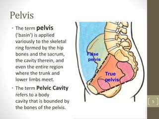
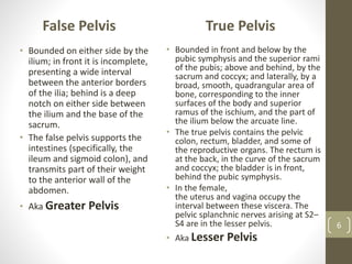
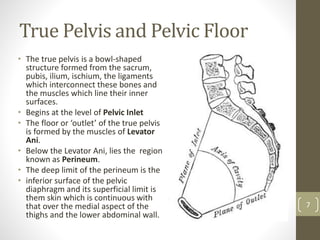

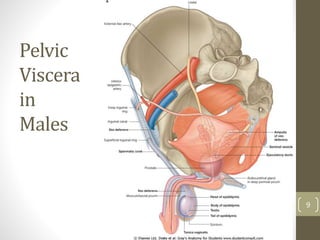


This 37 slide presentation provides an overview of the pelvis and pelvic floor anatomy. It discusses the bones that make up the pelvis, the true and false pelvis cavities, and the pelvic viscera in males and females. The muscles of the pelvic floor are described in detail, including the levator ani muscle and its components. Other structures covered include the pelvic fasciae, vasculature, nerves, pudendal canal, and related clinical terms. The presentation provides a comprehensive review of key anatomical structures and relationships in the pelvis region.


![ANATOMY OF THE LOWER URINARY TRACT AND MALE [Autosaved] [Autosaved].pptx](https://cdn.slidesharecdn.com/ss_thumbnails/anatomyofthelowerurinarytractandmaleautosavedautosaved-240526080531-9d6371e3-thumbnail.jpg?width=640&height=640&fit=bounds)


![Hypothalamus short ppt by Dr. Neha [PT].pptx](https://cdn.slidesharecdn.com/ss_thumbnails/hypothalamusbydr-260124145759-b9f94a93-thumbnail.jpg?width=640&height=640&fit=bounds)


