Introduction
• A tissue= a mass of similar cells and cell products that forms a
discrete region of an organ and performs a specific function
• The four basic tissue types in the body:
the epithelial, connective, muscular, and nervous tissue
o all these tissues exist and function in close association with one
another.
• The four basic tissues differ from each other in the:
Types and functions of their cells
Characteristics of the matrix (extracellular material) that surrounds
the cells
Relative amount of space occupied by cells versus matrix 3
4.
Introduction cont’d
• Epithelialtissue, or epithelium, consists of sheets of cells
that:
cover the external surfaces of the body
line the internal cavities
form various organs and glands, and line their ducts
• Characteristics of epithelial tissues
i. Continuous sheets of closely packed, tightly joined cells
ii. Cells are attached to basement membrane – 2 layers
Basal lamina - proteins and polysaccharides secreted by epithelial cells
Reticular lamina - protein fibres and glycoproteins secreted by
underlying connective tissue 4
5.
Introduction cont’d
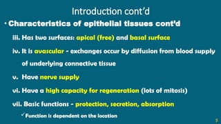
• Characteristicsof epithelial tissues cont’d
iii. Has two surfaces: apical (free) and basal surface
iv. It is avascular - exchanges occur by diffusion from blood supply
of underlying connective tissue
v. Have nerve supply
vi. Have a high capacity for regeneration (lots of mitosis)
vii. Basic functions - protection, secretion, absorption
Function is dependent on the location
5
6.
Classification of Epithelial
Tissues
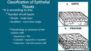
•Itis according to the:
Number of cell layers
oSimple – single layer
oStratified – more than single
Morphology or structure of the
surface cells
oSquamous = flat
oCuboidal = squarish or roundish
oColumnar = tall and narrow cells
6
7.
Types of Epithelialtissues
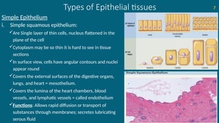
Simple Epithelium
i. Simple squamous epithelium:
Are Single layer of thin cells, nucleus flattened in the
plane of the cell
Cytoplasm may be so thin it is hard to see in tissue
sections
In surface view, cells have angular contours and nuclei
appear round
Covers the external surfaces of the digestive organs,
lungs, and heart = mesothelium.
Covers the lumina of the heart chambers, blood
vessels, and lymphatic vessels = called endothelium
Functions: Allows rapid diffusion or transport of
substances through membranes; secretes lubricating
serous fluid
7
8.
Types of Epithelialtissues cont’d
Simple Epithelium cont’d
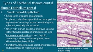
ii. Simple cuboidal epithelium
Single layer of square or round cells
In glands, cells often pyramidal and arranged like
segments of an orange around a central space;
spherical, centrally placed nuclei
Often with a brush border of microvilli in some
kidney tubules; ciliated in bronchioles of lung
Representative locations: Liver; thyroid,
mammary, salivary, and other glands; most
kidney tubules; bronchioles
Functions: Absorption and secretion; production
and movement of respiratory mucus
8
Simple Cuboidal Epithelium in Kidney
Tubules (×400).
9.
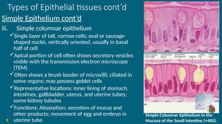
Types of Epithelialtissues cont’d
Simple Epithelium cont’d
iii. Simple columnar epithelium
Single layer of tall, narrow cells; oval or sausage-
shaped nuclei, vertically oriented, usually in basal
half of cell
Apical portion of cell often shows secretory vesicles
visible with the transmission electron microscope
(TEM)
Often shows a brush border of microvilli; ciliated in
some organs; may possess goblet cells
Representative locations: Inner lining of stomach,
intestines, gallbladder, uterus, and uterine tubes;
some kidney tubules
Functions: Absorption; secretion of mucus and
other products; movement of egg and embryo in
uterine tube
9
Simple Columnar Epithelium in the
Mucosa of the Small Intestine (×400).
10.
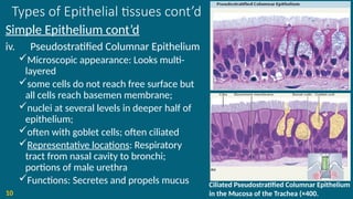
Types of Epithelialtissues cont’d
Simple Epithelium cont’d
iv. Pseudostratified Columnar Epithelium
Microscopic appearance: Looks multi-
layered
some cells do not reach free surface but
all cells reach basemen membrane;
nuclei at several levels in deeper half of
epithelium;
often with goblet cells; often ciliated
Representative locations: Respiratory
tract from nasal cavity to bronchi;
portions of male urethra
Functions: Secretes and propels mucus
10
Ciliated Pseudostratified Columnar Epithelium
in the Mucosa of the Trachea (×400.
11.
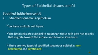
Types of Epithelialtissues cont’d
Stratified Epithelium cont’d
i. Stratified squamous epithelium
contains multiple cell layers.
The basal cells are cuboidal to columnar; these cells give rise to cells
that migrate toward the surface and become squamous.
There are two types of stratified squamous epithelia: non-
keratinized and keratinized.
11
12.
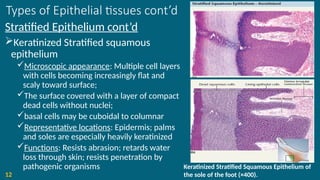
Types of Epithelialtissues cont’d
Stratified Epithelium cont’d
Keratinized Stratified squamous
epithelium
Microscopic appearance: Multiple cell layers
with cells becoming increasingly flat and
scaly toward surface;
The surface covered with a layer of compact
dead cells without nuclei;
basal cells may be cuboidal to columnar
Representative locations: Epidermis; palms
and soles are especially heavily keratinized
Functions: Resists abrasion; retards water
loss through skin; resists penetration by
pathogenic organisms
12
Keratinized Stratified Squamous Epithelium of
the sole of the foot (×400).
13.
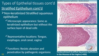
Types of Epithelialtissues cont’d
Stratified Epithelium cont’d
Non-keratinized Stratified squamous
epithelium
Microscopic appearance: Same as
keratinized epithelium but without the
surface layer of dead cells
Representative locations: Tongue,
esophagus, anal canal, vagina
Functions: Resists abrasion and
penetration by pathogenic organisms
13
Nonkeratinized Stratified Squamous Epithelium
in the Mucosa of the Vagina (×400).
14.
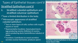
Types of Epithelialtissues cont’d
Stratified Epithelium cont’d
ii. Stratified cuboidal epithelium and
stratified columnar epithelium
have a limited distribution in the body.
Microscopic appearance of stratified
cuboidal epithelium:
Two or more layers of cells; surface cells roughly
square or round
Representative locations: Sweat gland ducts;
egg-producing vesicles (follicles) of ovaries;
sperm-producing ducts (seminiferous tubules) of
testes
Functions: Contributes to sweat secretion;
secretes ovarian hormones; produces sperm
14
Stratified Cuboidal Epithelium in the Duct of a
Sweat Gland (×400).
15.
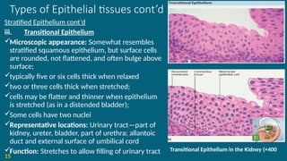
Types of Epithelialtissues cont’d
Stratified Epithelium cont’d
iii. Transitional Epithelium
Microscopic appearance: Somewhat resembles
stratified squamous epithelium, but surface cells
are rounded, not flattened, and often bulge above
surface;
typically five or six cells thick when relaxed
two or three cells thick when stretched;
cells may be flatter and thinner when epithelium
is stretched (as in a distended bladder);
Some cells have two nuclei
Representative locations: Urinary tract—part of
kidney, ureter, bladder, part of urethra; allantoic
duct and external surface of umbilical cord
Function: Stretches to allow filling of urinary tract
15
Transitional Epithelium in the Kidney (×400
16.
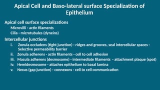
Apical Cell andBaso-lateral surface Specialization of
Epithelium
Apical cell surface specializations
Microvilli - actin filaments
Cilia - microtubules (dyneins)
Intercellular junctions
i. Zonula occludens (tight junction) - ridges and grooves, seal intercellular spaces -
Selective permeability barrier
ii. Zonula adherens - actin filaments - cell to cell adhesion
iii. Macula adherens (desmosome) - intermediate filaments - attachment plaque (spot)
iv. Hemidesmosome - attaches epithelium to basal lamina
v. Nexus (gap junction) - connexons - cell to cell communication
17.
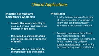
Clinical Applications
Immotile ciliasyndrome
(Kartegener’s syndrome):
• Disorder that causes infertility in
male and chronic respiratory tract
infection in both sexes.
• It is caused by immobility of cilia
and flagella induced by deficiency
of dynein.
• Dynein protein is responsible for
movements of cilia and flagella.
Metaplasia:
• It is the transformation of one type
of tissue to another in response to
injury. This condition is usually
reversible if the injury is removed.
• Example: pseudostratified ciliated
columnar epithelium of the
respiratory passages, e.g. trachea, of
heavy smokers may undergo
squamous metaplasia, transforming
into stratified squamous epithelium.
19
Introduction
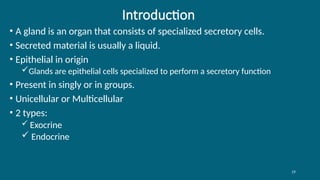
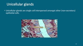
• A glandis an organ that consists of specialized secretory cells.
• Secreted material is usually a liquid.
• Epithelial in origin
Glands are epithelial cells specialized to perform a secretory function
• Present in singly or in groups.
• Unicellular or Multicellular
• 2 types:
Exocrine
Endocrine
21
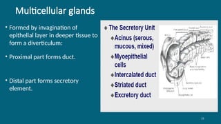
Multicellular glands
• Formedby invagination of
epithelial layer in deeper tissue to
form a diverticulum:
• Proximal part forms duct.
• Distal part forms secretory
element.
22.
22
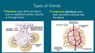
Types of Glands
Exocrine: pour their secretions
onto an epithelial surface, directly
or through ducts.
Endocrine (ductless): pour
their secretions directly into
the blood.
23.
23
Classification of exocrineglands
• Based on shape & branching pattern of duct
• Based on mode of release of their product
• Based on the nature of their secretion
24.
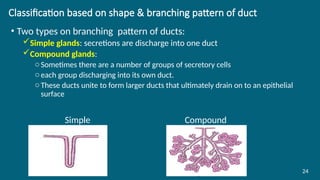
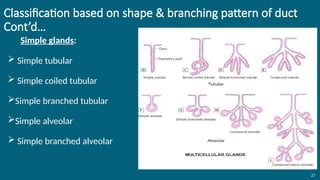
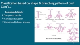
Classification based onshape & branching pattern of duct
• Two types on branching pattern of ducts:
Simple glands: secretions are discharge into one duct
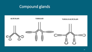
Compound glands:
oSometimes there are a number of groups of secretory cells
oeach group discharging into its own duct.
oThese ducts unite to form larger ducts that ultimately drain on to an epithelial
surface
Simple Compound
24
25.
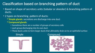
Classification based onbranching pattern of duct
• Based on shape of secretory units (tubular or alveolar) & branching pattern of
ducts.
• 2 types on branching pattern of ducts:
Simple glands: secretions are discharge into one duct
Compound glands:
o Sometimes there are a number of groups of secretory cells
o each group discharging into its own duct.
o These ducts unite to form larger ducts that ultimately drain on to an epithelial surface
Simple Compound
25
26.
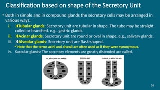
Classification based onshape of the Secretory Unit
• Both in simple and in compound glands the secretory cells may be arranged in
various ways:
i. ‰Tubular glands: Secretory unit are tubular in shape. The tube may be straight,
coiled or branched. e.g., gastric glands.
ii. Acinar glands
‰ : Secretory unit are round or oval in shape, e.g., salivary glands.
iii. ‰Alveolar glands: Secretory unit are flask-shaped.
Note that the terms acini and alveoli are often used as if they were synonymous.
iv. Saccular glands: The secretory elements are greatly distended are called.
26
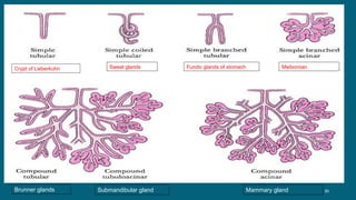
30
Crypt of LieberkuhnSweat glands Fundic glands of stomach Meibomian glands
Brunner glands Submandibular gland Mammary gland
31.
31
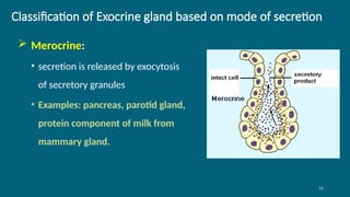
Classification of Exocrinegland based on mode of secretion
Merocrine:
• secretion is released by exocytosis
of secretory granules
• Examples: pancreas, parotid gland,
protein component of milk from
mammary gland.
32.
32
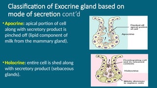
Classification of Exocrinegland based on
mode of secretion cont’d
•Apocrine: apical portion of cell
along with secretory product is
pinched off (lipid component of
milk from the mammary gland).
•Holocrine: entire cell is shed along
with secretory product (sebaceous
glands).
34
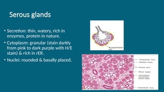
Serous glands
• Secretion:thin, watery, rich in
enzymes, protein in nature.
• Cytoplasm: granular (stain darkly
from pink to dark purple with H/E
stain) & rich in rER.
• Nuclei: rounded & basally placed.
35.
35
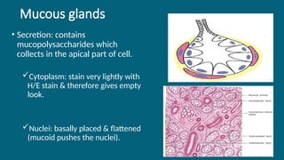
Mucous glands
• Secretion:contains
mucopolysaccharides which
collects in the apical part of cell.
Cytoplasm: stain very lightly with
H/E stain & therefore gives empty
look.
Nuclei: basally placed & flattened
(mucoid pushes the nuclei).
36.
36
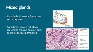
Mixed glands
• Containboth serous & mucous
secretory units.
• Sometimes serous cells form
crescentic caps on mucous acini
called as serous demilunes.
37.
37
Structural Organization ofExocrine Glands
• It consists of three components:
i. Parenchyma
The secretory cells of a gland constitute its parenchyma
ii. Stroma
The stroma is the connective tissue in which the parenchyma lies
It forms septa that divides the glandular tissue into lobules
Aggregations of lobules may form distinct lobes.
The connective tissue covering the entire gland forms a capsule for it
iii. Duct system
Could be within a lobule as intra-lobular, between a lobule as interlobular or lies
between adjacent lobes as inter-lobar .
Intra-lobular has the least diameter while interlobar has the greatest diameter


![Epithelium[1]](https://cdn.slidesharecdn.com/ss_thumbnails/epithelium1-200323141425-thumbnail.jpg?width=640&height=640&fit=bounds)


![Polymer [ बहुलक ] Chemistry Notes PDF - Irfanullah Mehar - JJ Sir Chemistry.pdf](https://cdn.slidesharecdn.com/ss_thumbnails/polymerchemistrynotespdf-irfanullahmehar-jjsirchemistry-260210172118-3f9b37f7-thumbnail.jpg?width=640&height=640&fit=bounds)
