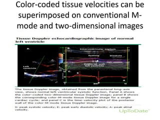
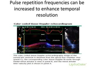
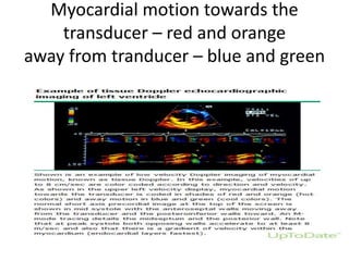
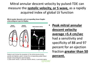
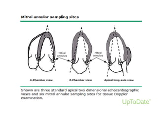
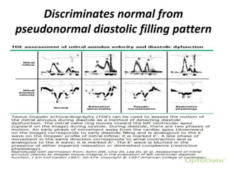
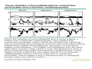
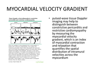
Tissue Doppler
Echocardiography(TDE)
S.R.Sruthi Meenaxshi MBBS,MD,PDF
• Tissue Doppler echocardiography (TDE) has
become an established component of the
diagnostic ultrasound examination; it permits
an assessment of myocardial motion using
Doppler ultrasound imaging.
• The technique uses frequency shifts of
ultrasound waves to calculate myocardial
velocity;
• focus on lower velocity frequency shifts
TECHNICAL ASPECTS
• Two techniques have been used to assess
myocardial function:
• pulsed-TDE
• color-coded TDE
• TDE modification of doppler of blood flow and
calculates velocity of frequency shifts in
similar manner
• A primary advantage of TDE is that Doppler
shifts of tissue motion are of high amplitude,
being approximately 40 dB higher than
Doppler signals from blood flow
• In instrumentation feature common to both
pulsed and color-coded TDE involves removal
of the high-pass filter used for routine
Doppler to assess blood flow
• This is to focus on the lower velocity values of
myocardial motion
Pulsed TDE
• similar to pulsed-Doppler of blood flow
• . The gate of the sample volume of pulsed-TDE
is usually opened to 1 cm and directed to
assess the region of interest
• Most commonly the mitral annulus at lateral
and medial sites from the apical four-chamber
view
Color-coded TDE
• instrumentation uses the autocorrelator
technique to calculate and display multigated
points of color-coded blood velocity along a
series of ultrasound scan lines within a two-
dimensional sector
• Color-coded blood velocity data are then
superimposed on conventional gray scale two-
dimensional images in real time.
Color-coded tissue velocities can be
superimposed on conventional M-
mode and two-dimensional images
Pulse repetition frequencies can be
increased to enhance temporal
resolution
Myocardial motion towards the
transducer – red and orange
away from tranducer – blue and green
CLINICAL APPLICATIONS
• In the assessment of left ventricular (LV)
systolic and diastolic function.
• Pulsed TDE is routinely used in clinical practice
• measures of mitral annular velocity have
established usefulness for assessment of LV
systolic and diastolic function, estimation of LV
filling pressures, and in the diagnosis of
hypertrophic cardiomyopathy, cardiac
amyloidosis, and the athletic heart
• Mitral annular velocity alone or in
combination with mitral inflow velocity (E) to
estimate LV diastolic function are the most
commonly used clinical applications.
• TDE assessment of mitral annular velocity (e’)
has been widely accepted as a component of
determining left ventricular (LV) diastolic
function.
• TDE also may quantify regional and global LV
function through the assessment of
myocardial velocity data.
Assessment of global and regional
systolic left venticular function
• color-coded TDE objectively quantified a wide
range of alterations in regional contractility
induced by inotropic modulation with
dobutamine and esmolol.
• Dobutamine produced significant increases in
peak systolic endocardial velocity, systolic time
velocity integral (TVI), and diastolic TVI;
• infusion of esmolol, there were significant
decreases in these indices of myocardial
contractility.
Strain and strain rate imaging
• To quantify global and regional LV function
• Strain is the ratio of change in length over the original
length or the fraction or percentage change from the
original or unstressed dimension
• Quantification of deformation is applied to describe
the contraction/relaxation pattern of the myocardium
• strain rate is the rate of this deformation and is
associated with LV contractility
Use in dobutamine stress
echocardiography
• Dobutamine stress echocardiography is a
technique for evaluating regional wall motion
abnormalities due to ischemia that is induced
by pharmacologic stress
• it is useful for the diagnosis of coronary heart
disease or determining the viability of
dysfunctional myocardium
To assess LV dyssynchrony for CRT
• TDE measures of the severity of LV
intraventricular dyssynchrony may provide
prognostic information to patients with heart
failure who typically have a delay in electrical
activation, such as left bundle branch block
(LBBB)
• TDE may also play a role for evaluating the effect
of CRT or biventricular pacing on LV function and
reverse remodeling.
Mitral annular velocity to assess LV
function
• Mitral annular motion assessed by M-mode
echocardiography has historically been used as
an index of global LV systolic function
• viewed from the apical windows
• Mitral annular descent reflects the longitudinal
shortening of the LV chamber and correlates with
other global measures of LV function, such as
stroke volume
Mitral annular descent velocity by pulsed-TDE can
measure the systolic velocity, or S wave, as a rapidly
acquired index of global LV function
• Peak mitral annular
descent velocity
average >5.4 cm/sec
had a sensitivity and
specificity of 88 and 97
percent for an ejection
fraction greater than 50
percent.
Use in evaluating chronic aortic
regurgitation
• TDE may be helpful for identifying subclinical
LV dysfunction in patients with chronic severe
aortic regurgitation who are asymptomatic
but may be candidates for surgery
• A systolic annular excursion <12 mm and a
resting mitral annular velocity <9.5 cm/sec
were the best indicators of subclinical LV
dysfunction
Assessment of diastolic function
• Peak negative
myocardial
velocity can
provide a
quantitative
assessment of
diastolic
dysfunction.
TDE IN DD
• Segmental and global
function can be measured.
For global function, the
region of interest is placed
at the septal and lateral
borders of the mitral
annulus.
• During systole, the annulus
descends towards the apex,
whereas it recoils back
toward the base in early (e')
and late (a') diastole
Discriminates normal from
pseudonormal diastolic filling pattern
An average E/e' ratio below 8 is associated with normal filling
pressures and ratio >14 is associated with elevated filling pressures
• 2016 American Society of Echocardiography and European Association of
Cardiovascular Imaging guidelines
• For Diastolic dysfunction
• -E/e’ >14; the E/e’ is the ratio of early mitral inflow velocity (E) to mitral
annular early diastolic velocity (e’)
• -Septal e’ velocity <7 cm/s or lateral e’ velocity <10 cm/s
• -TR velocity >2.8 m/s; this criterion should not be used in patients with
significant pulmonary disease.
• -LA maximum volume index >34mL/m2 (should not be applied in athletes,
patients with more than mild mitral valve stenosis or regurgitation, or
those in atrial fibrillation).
Prognostic utility in heart failure
• Mitral annular Ea (also called E’) has
important prognostic utility in heart failure
patients.
• In patients with impaired systolic function
poor prognostic indicators were
 S <3 cm/s
mitral deceleration time <140ms
E/E’ >15
Differentiating constrictive and
restrictive pericarditis
Differentiating restrictive cardiomyopathy and
constrictive pericarditis
• The early diastolic Doppler tissue velocity at the
mitral annulus (E') is decreased (<8 cm/sec) in
restrictive cardiomyopathy, due to an intrinsic
decrease in myocardial contraction and
relaxation.
• In contrast, the transmitral E' is frequently
increased (>12 cm/sec) in constrictive
pericarditis, since the longitudinal movement of
the myocardium is enhanced because of
constricted radial motion
MITRAL ANNULUS REVERSUS IN CP
• mitral lateral (and tricuspid) annular E'
velocities are often relatively reduced in
patients with constrictive pericarditis
("annular reversus")
• This reduction may be the result of lateral
adhesion of the pericardium while the
longitudinal movement of the septal annulus
is unimpeded
Annulus reversus in constrictive
pericarditis
MYOCARDIAL VELOCITY GRADIENT
• pulsed-wave tissue Doppler
imaging may help to
distinguish between
constrictive pericarditis and
restrictive cardiomyopathy
by measuring the
myocardial velocity
gradient, which is an index
of myocardial contraction
and relaxation that
quantifies the spatial
distribution of intramural
velocities across the
myocardium
TDE in differntiating constrictive pericarditis
and restrictive cardiomyopathy
• . Ea obtained by pulsed-TDE is useful to
distinguish patients with constrictive
pericarditis from restrictive cardiomyopathy
• Since restrictive cardiomyopathy is a disease
of the myocardium, e’ is reduced, usually
<6.0 cm/sec, whereas constrictive pericarditis
is a disease of the pericardium and e’ velocity
is preserved or elevated >10 cm/sec.
HOCM shows lower systolic and
diastolic velocity in TDE
Tissue doppler Echocardiography (TDE)

Tissue doppler Echocardiography (TDE)

  • 1.
  • 2.
    • Tissue Dopplerechocardiography (TDE) has become an established component of the diagnostic ultrasound examination; it permits an assessment of myocardial motion using Doppler ultrasound imaging. • The technique uses frequency shifts of ultrasound waves to calculate myocardial velocity; • focus on lower velocity frequency shifts
  • 3.
    TECHNICAL ASPECTS • Twotechniques have been used to assess myocardial function: • pulsed-TDE • color-coded TDE
  • 4.
    • TDE modificationof doppler of blood flow and calculates velocity of frequency shifts in similar manner • A primary advantage of TDE is that Doppler shifts of tissue motion are of high amplitude, being approximately 40 dB higher than Doppler signals from blood flow
  • 5.
    • In instrumentationfeature common to both pulsed and color-coded TDE involves removal of the high-pass filter used for routine Doppler to assess blood flow • This is to focus on the lower velocity values of myocardial motion
  • 6.
    Pulsed TDE • similarto pulsed-Doppler of blood flow • . The gate of the sample volume of pulsed-TDE is usually opened to 1 cm and directed to assess the region of interest • Most commonly the mitral annulus at lateral and medial sites from the apical four-chamber view
  • 7.
    Color-coded TDE • instrumentationuses the autocorrelator technique to calculate and display multigated points of color-coded blood velocity along a series of ultrasound scan lines within a two- dimensional sector • Color-coded blood velocity data are then superimposed on conventional gray scale two- dimensional images in real time.
  • 8.
    Color-coded tissue velocitiescan be superimposed on conventional M- mode and two-dimensional images
  • 9.
    Pulse repetition frequenciescan be increased to enhance temporal resolution
  • 10.
    Myocardial motion towardsthe transducer – red and orange away from tranducer – blue and green
  • 11.
    CLINICAL APPLICATIONS • Inthe assessment of left ventricular (LV) systolic and diastolic function. • Pulsed TDE is routinely used in clinical practice • measures of mitral annular velocity have established usefulness for assessment of LV systolic and diastolic function, estimation of LV filling pressures, and in the diagnosis of hypertrophic cardiomyopathy, cardiac amyloidosis, and the athletic heart
  • 12.
    • Mitral annularvelocity alone or in combination with mitral inflow velocity (E) to estimate LV diastolic function are the most commonly used clinical applications. • TDE assessment of mitral annular velocity (e’) has been widely accepted as a component of determining left ventricular (LV) diastolic function. • TDE also may quantify regional and global LV function through the assessment of myocardial velocity data.
  • 13.
    Assessment of globaland regional systolic left venticular function
  • 14.
    • color-coded TDEobjectively quantified a wide range of alterations in regional contractility induced by inotropic modulation with dobutamine and esmolol. • Dobutamine produced significant increases in peak systolic endocardial velocity, systolic time velocity integral (TVI), and diastolic TVI; • infusion of esmolol, there were significant decreases in these indices of myocardial contractility.
  • 15.
    Strain and strainrate imaging • To quantify global and regional LV function • Strain is the ratio of change in length over the original length or the fraction or percentage change from the original or unstressed dimension • Quantification of deformation is applied to describe the contraction/relaxation pattern of the myocardium • strain rate is the rate of this deformation and is associated with LV contractility
  • 17.
    Use in dobutaminestress echocardiography • Dobutamine stress echocardiography is a technique for evaluating regional wall motion abnormalities due to ischemia that is induced by pharmacologic stress • it is useful for the diagnosis of coronary heart disease or determining the viability of dysfunctional myocardium
  • 18.
    To assess LVdyssynchrony for CRT • TDE measures of the severity of LV intraventricular dyssynchrony may provide prognostic information to patients with heart failure who typically have a delay in electrical activation, such as left bundle branch block (LBBB) • TDE may also play a role for evaluating the effect of CRT or biventricular pacing on LV function and reverse remodeling.
  • 19.
    Mitral annular velocityto assess LV function • Mitral annular motion assessed by M-mode echocardiography has historically been used as an index of global LV systolic function • viewed from the apical windows • Mitral annular descent reflects the longitudinal shortening of the LV chamber and correlates with other global measures of LV function, such as stroke volume
  • 20.
    Mitral annular descentvelocity by pulsed-TDE can measure the systolic velocity, or S wave, as a rapidly acquired index of global LV function • Peak mitral annular descent velocity average >5.4 cm/sec had a sensitivity and specificity of 88 and 97 percent for an ejection fraction greater than 50 percent.
  • 22.
    Use in evaluatingchronic aortic regurgitation • TDE may be helpful for identifying subclinical LV dysfunction in patients with chronic severe aortic regurgitation who are asymptomatic but may be candidates for surgery • A systolic annular excursion <12 mm and a resting mitral annular velocity <9.5 cm/sec were the best indicators of subclinical LV dysfunction
  • 23.
    Assessment of diastolicfunction • Peak negative myocardial velocity can provide a quantitative assessment of diastolic dysfunction.
  • 24.
    TDE IN DD •Segmental and global function can be measured. For global function, the region of interest is placed at the septal and lateral borders of the mitral annulus. • During systole, the annulus descends towards the apex, whereas it recoils back toward the base in early (e') and late (a') diastole
  • 25.
    Discriminates normal from pseudonormaldiastolic filling pattern
  • 26.
    An average E/e'ratio below 8 is associated with normal filling pressures and ratio >14 is associated with elevated filling pressures • 2016 American Society of Echocardiography and European Association of Cardiovascular Imaging guidelines • For Diastolic dysfunction • -E/e’ >14; the E/e’ is the ratio of early mitral inflow velocity (E) to mitral annular early diastolic velocity (e’) • -Septal e’ velocity <7 cm/s or lateral e’ velocity <10 cm/s • -TR velocity >2.8 m/s; this criterion should not be used in patients with significant pulmonary disease. • -LA maximum volume index >34mL/m2 (should not be applied in athletes, patients with more than mild mitral valve stenosis or regurgitation, or those in atrial fibrillation).
  • 27.
    Prognostic utility inheart failure • Mitral annular Ea (also called E’) has important prognostic utility in heart failure patients. • In patients with impaired systolic function poor prognostic indicators were  S <3 cm/s mitral deceleration time <140ms E/E’ >15
  • 28.
  • 29.
    Differentiating restrictive cardiomyopathyand constrictive pericarditis • The early diastolic Doppler tissue velocity at the mitral annulus (E') is decreased (<8 cm/sec) in restrictive cardiomyopathy, due to an intrinsic decrease in myocardial contraction and relaxation. • In contrast, the transmitral E' is frequently increased (>12 cm/sec) in constrictive pericarditis, since the longitudinal movement of the myocardium is enhanced because of constricted radial motion
  • 31.
    MITRAL ANNULUS REVERSUSIN CP • mitral lateral (and tricuspid) annular E' velocities are often relatively reduced in patients with constrictive pericarditis ("annular reversus") • This reduction may be the result of lateral adhesion of the pericardium while the longitudinal movement of the septal annulus is unimpeded
  • 32.
    Annulus reversus inconstrictive pericarditis
  • 33.
    MYOCARDIAL VELOCITY GRADIENT •pulsed-wave tissue Doppler imaging may help to distinguish between constrictive pericarditis and restrictive cardiomyopathy by measuring the myocardial velocity gradient, which is an index of myocardial contraction and relaxation that quantifies the spatial distribution of intramural velocities across the myocardium
  • 34.
    TDE in differntiatingconstrictive pericarditis and restrictive cardiomyopathy
  • 35.
    • . Eaobtained by pulsed-TDE is useful to distinguish patients with constrictive pericarditis from restrictive cardiomyopathy • Since restrictive cardiomyopathy is a disease of the myocardium, e’ is reduced, usually <6.0 cm/sec, whereas constrictive pericarditis is a disease of the pericardium and e’ velocity is preserved or elevated >10 cm/sec.
  • 36.
    HOCM shows lowersystolic and diastolic velocity in TDE