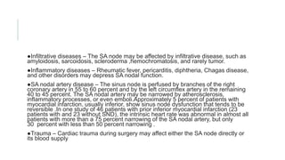
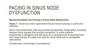
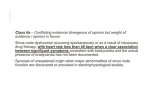
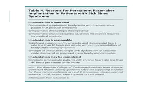
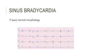
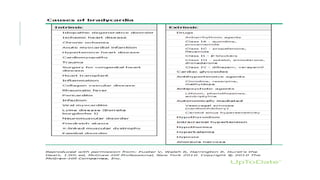
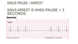
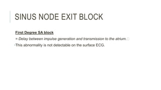
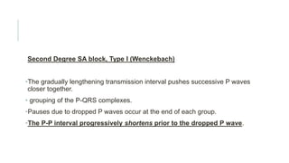
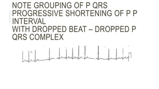
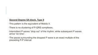
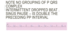
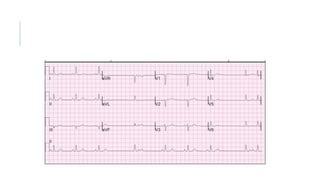
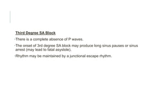
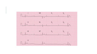
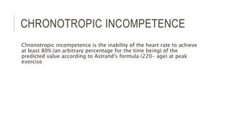
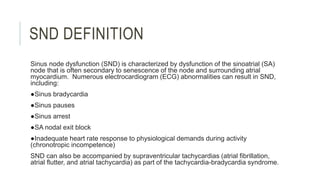
CONDUCTION
ABNORMALITIES PART 3
SINUS NODAL DYSFUNCTION
Sruthi Meenaxshi
SND DEFINITION
Sinus node dysfunction (SND) is characterized by dysfunction of the sinoatrial (SA)
node that is often secondary to senescence of the node and surrounding atrial
myocardium. Numerous electrocardiogram (ECG) abnormalities can result in SND,
including:
●Sinus bradycardia
●Sinus pauses
●Sinus arrest
●SA nodal exit block
●Inadequate heart rate response to physiological demands during activity
(chronotropic incompetence)
SND can also be accompanied by supraventricular tachycardias (atrial fibrillation,
atrial flutter, and atrial tachycardia) as part of the tachycardia-bradycardia syndrome.
CAUSES
Sinus node fibrosis — The most common cause of sinus node dysfunction is the replacement of sinus node
tissue by fibrous tissue, Medications and toxins — A number of medications and toxins can depress sinus node
function
●Beta blockers
●Non-dihydropyridine calcium channel blockers
●Digoxin
●Antiarrhythmic medications
●Acetylcholinesterase inhibitors such as donepezil (Aricept) and rivastigmine used in the treatment of Alzheimer's
disease
Other medications associated with depression of sinus node function include parasympathomimetic agents,
sympatholytic drugs (eg, methyldopa, clonidine), cimetidine, lithium, and ivabradine [35,36]. In addition,
poisoning by grayanotoxin, produced by some plants (eg, Rhododendron sp.) and found in certain varieties of
honey, has been associated with depressed sinus node function [37].
Childhood and familial disease — SND is rare in children, but when present it is most often seen in those with
congenital and acquired heart disease, particularly after corrective cardiac surgery [38-41]. Familial sinus node
dysfunction is rare, with mutations in the cardiac sodium channel gene SCN5A and the HCN4 gene (
●Infiltrative diseases – The SA node may be affected by infiltrative disease, such as
amyloidosis, sarcoidosis, scleroderma ,hemochromatosis, and rarely tumor.
●Inflammatory diseases – Rheumatic fever, pericarditis, diphtheria, Chagas disease,
and other disorders may depress SA nodal function.
●SA nodal artery disease – The sinus node is perfused by branches of the right
coronary artery in 55 to 60 percent and by the left circumflex artery in the remaining
40 to 45 percent. The SA nodal artery may be narrowed by atherosclerosis,
inflammatory processes, or even emboli.Approximately 5 percent of patients with
myocardial infarction, usually inferior, show sinus node dysfunction that tends to be
reversible .In one study of 46 patients with prior inferior myocardial infarction (23
patients with and 23 without SND), the intrinsic heart rate was abnormal in almost all
patients with more than a 75 percent narrowing of the SA nodal artery, but only
30 percent with less than 50 percent narrowing .
●Trauma – Cardiac trauma during surgery may affect either the SA node directly or
its blood supply
PACING IN SINUS NODE
DYSFUNCTION
Recommendation for Pacing in Sinus Node Dysfunction
Class I – Evidence and/or agreement that permanent pacing is useful and
effective.
•Sinus node dysfunction with documented symptomatic bradycardia, including
frequent sinus pauses that produce symptoms. In some patients,
bradycardia is iatrogenic and will occur as a consequence of essential long-
term drug therapy of a type and dose for which there are no acceptable
alternatives.
•Symptomatic chronotropic incompetence.
Class IIa – Conflicting evidence/ divergence of opinion but weight of
evidence / opinion in favour
•Sinus node dysfunction occurring spontaneously or as a result of necessary
drug therapy, with heart rate less than 40 bpm when a clear association
between significant symptoms consistent with bradycardia and the actual
presence of bradycardia has not been documented.
•Syncope of unexplained origin when major abnormalities of sinus node
function are discovered or provoked in electrophysiological studies.
SINUS BRADYCARDIA
P wave normal morphology
SINUS PAUSE / ARREST
SINUS ARREST IS WHEN PAUSE > 3
SECONDS.
SINUS NODE EXIT BLOCK
First Degree SA block
= Delay between impulse generation and transmission to the atrium.
•This abnormality is not detectable on the surface ECG.
Second Degree SA block, Type I (Wenckebach)
•The gradually lengthening transmission interval pushes successive P waves
closer together.
• grouping of the P-QRS complexes.
•Pauses due to dropped P waves occur at the end of each group.
•The P-P interval progressively shortens prior to the dropped P wave.
NOTE GROUPING OF P QRS
PROGRESSIVE SHORTENING OF P P
INTERVAL
WITH DROPPED BEAT – DROPPED P
QRS COMPLEX
Second Degree SA block, Type II
•This pattern is the equivalent of Mobitz II.
•There is no clustering of P-QRS complexes.
•Intermittent P waves “drop out” of the rhythm, while subsequent P waves
arrive “on time”.
•The pause surrounding the dropped P wave is an exact multiple of the
preceding P-P interval.
NOTE NO GROUPING OF P QRS
COMPLEX
INTERMITTENT DROPPED BEAT .
SINUS PAUSE – IS DOUBLE THE
PRECEDING PP INTERVAL
Third Degree SA Block
•There is a complete absence of P waves.
•The onset of 3rd degree SA block may produce long sinus pauses or sinus
arrest (may lead to fatal asystole).
•Rhythm may be maintained by a junctional escape rhythm.
CHRONOTROPIC INCOMPETENCE
Chronotropic incompetence is the inability of the heart rate to achieve
at least 80% (an arbitrary percentage for the time being) of the
predicted value according to Astrand's formula (220- age) at peak
exercise
THANKYOU

Sinus node dysfunction

  • 1.
    CONDUCTION ABNORMALITIES PART 3 SINUSNODAL DYSFUNCTION Sruthi Meenaxshi
  • 2.
    SND DEFINITION Sinus nodedysfunction (SND) is characterized by dysfunction of the sinoatrial (SA) node that is often secondary to senescence of the node and surrounding atrial myocardium. Numerous electrocardiogram (ECG) abnormalities can result in SND, including: ●Sinus bradycardia ●Sinus pauses ●Sinus arrest ●SA nodal exit block ●Inadequate heart rate response to physiological demands during activity (chronotropic incompetence) SND can also be accompanied by supraventricular tachycardias (atrial fibrillation, atrial flutter, and atrial tachycardia) as part of the tachycardia-bradycardia syndrome.
  • 3.
    CAUSES Sinus node fibrosis— The most common cause of sinus node dysfunction is the replacement of sinus node tissue by fibrous tissue, Medications and toxins — A number of medications and toxins can depress sinus node function ●Beta blockers ●Non-dihydropyridine calcium channel blockers ●Digoxin ●Antiarrhythmic medications ●Acetylcholinesterase inhibitors such as donepezil (Aricept) and rivastigmine used in the treatment of Alzheimer's disease Other medications associated with depression of sinus node function include parasympathomimetic agents, sympatholytic drugs (eg, methyldopa, clonidine), cimetidine, lithium, and ivabradine [35,36]. In addition, poisoning by grayanotoxin, produced by some plants (eg, Rhododendron sp.) and found in certain varieties of honey, has been associated with depressed sinus node function [37]. Childhood and familial disease — SND is rare in children, but when present it is most often seen in those with congenital and acquired heart disease, particularly after corrective cardiac surgery [38-41]. Familial sinus node dysfunction is rare, with mutations in the cardiac sodium channel gene SCN5A and the HCN4 gene (
  • 4.
    ●Infiltrative diseases –The SA node may be affected by infiltrative disease, such as amyloidosis, sarcoidosis, scleroderma ,hemochromatosis, and rarely tumor. ●Inflammatory diseases – Rheumatic fever, pericarditis, diphtheria, Chagas disease, and other disorders may depress SA nodal function. ●SA nodal artery disease – The sinus node is perfused by branches of the right coronary artery in 55 to 60 percent and by the left circumflex artery in the remaining 40 to 45 percent. The SA nodal artery may be narrowed by atherosclerosis, inflammatory processes, or even emboli.Approximately 5 percent of patients with myocardial infarction, usually inferior, show sinus node dysfunction that tends to be reversible .In one study of 46 patients with prior inferior myocardial infarction (23 patients with and 23 without SND), the intrinsic heart rate was abnormal in almost all patients with more than a 75 percent narrowing of the SA nodal artery, but only 30 percent with less than 50 percent narrowing . ●Trauma – Cardiac trauma during surgery may affect either the SA node directly or its blood supply
  • 5.
    PACING IN SINUSNODE DYSFUNCTION Recommendation for Pacing in Sinus Node Dysfunction Class I – Evidence and/or agreement that permanent pacing is useful and effective. •Sinus node dysfunction with documented symptomatic bradycardia, including frequent sinus pauses that produce symptoms. In some patients, bradycardia is iatrogenic and will occur as a consequence of essential long- term drug therapy of a type and dose for which there are no acceptable alternatives. •Symptomatic chronotropic incompetence.
  • 6.
    Class IIa –Conflicting evidence/ divergence of opinion but weight of evidence / opinion in favour •Sinus node dysfunction occurring spontaneously or as a result of necessary drug therapy, with heart rate less than 40 bpm when a clear association between significant symptoms consistent with bradycardia and the actual presence of bradycardia has not been documented. •Syncope of unexplained origin when major abnormalities of sinus node function are discovered or provoked in electrophysiological studies.
  • 8.
    SINUS BRADYCARDIA P wavenormal morphology
  • 11.
    SINUS PAUSE /ARREST SINUS ARREST IS WHEN PAUSE > 3 SECONDS.
  • 12.
    SINUS NODE EXITBLOCK First Degree SA block = Delay between impulse generation and transmission to the atrium. •This abnormality is not detectable on the surface ECG.
  • 13.
    Second Degree SAblock, Type I (Wenckebach) •The gradually lengthening transmission interval pushes successive P waves closer together. • grouping of the P-QRS complexes. •Pauses due to dropped P waves occur at the end of each group. •The P-P interval progressively shortens prior to the dropped P wave.
  • 14.
    NOTE GROUPING OFP QRS PROGRESSIVE SHORTENING OF P P INTERVAL WITH DROPPED BEAT – DROPPED P QRS COMPLEX
  • 15.
    Second Degree SAblock, Type II •This pattern is the equivalent of Mobitz II. •There is no clustering of P-QRS complexes. •Intermittent P waves “drop out” of the rhythm, while subsequent P waves arrive “on time”. •The pause surrounding the dropped P wave is an exact multiple of the preceding P-P interval.
  • 16.
    NOTE NO GROUPINGOF P QRS COMPLEX INTERMITTENT DROPPED BEAT . SINUS PAUSE – IS DOUBLE THE PRECEDING PP INTERVAL
  • 18.
    Third Degree SABlock •There is a complete absence of P waves. •The onset of 3rd degree SA block may produce long sinus pauses or sinus arrest (may lead to fatal asystole). •Rhythm may be maintained by a junctional escape rhythm.
  • 20.
    CHRONOTROPIC INCOMPETENCE Chronotropic incompetenceis the inability of the heart rate to achieve at least 80% (an arbitrary percentage for the time being) of the predicted value according to Astrand's formula (220- age) at peak exercise
  • 21.