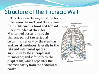
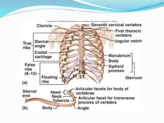
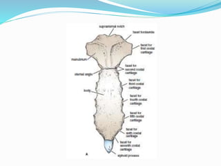
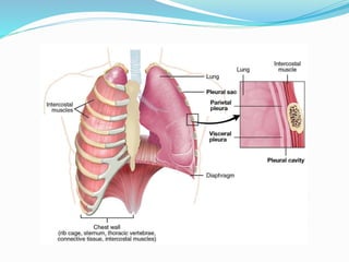
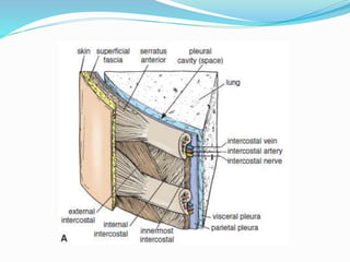
SlideShare a Scribd company logo
Thoracic wall
Dr. Abid Ullah (PT)
Lecturer PIHS
Islamabad
Thoracic Cavity
 It is the region of the body between the neck and the
abdomen
 It is bounded by 12 pairs of ribs, vertebrae and sternum.
 It contains trachea, two lungs, heart and great vessels,
esophagus and lymph nodes.
 There are two openings of the thorax, the superior thoracic
opening which is also called thoracic inlet and inferior
thoracic opening.
 Head and neck and upper limbs are attached with the
thoracic cavity through thoracic inlet.
 Inferior thoracic opening opens into the abdominal cavity.
Structure of the Thoracic Wall
is formed posteriorly by the
thoracic part of the vertebral
column; anteriorly by the sternum
and costal cartilages; laterally by the
ribs and intercostal spaces;
superiorly by the suprapleural
membrane; and inferiorly by the
diaphragm, which separates the
thoracic cavity from the abdominal
cavity
The thorax is the region of the body
between the neck and the abdomen.
It is flattened in front and behind
but rounded at the sides.
Cont…
 Superiorly, the thorax communicates with the neck,
and inferiorly it is separated from the abdomen by the
diaphragm.
 The thoracic cage protects the lungs and heart and
affords attachment for the muscles of the thorax,
upper extremity, abdomen, and back.
Cont…
 The cavity of the thorax can be divided into a median
partition, called the mediastinum, and the laterally
placed pleurae and lungs.
 The lungs are covered by a thin membrane called the
visceral pleura, which passes from each lung at its
root (i.e., where the main air passages and blood
vessels enter) to the inner surface of the chest wall,
where it is called the parietal pleura.
 In this manner, two membranous sacs called the
pleural cavities are formed, one on each side of the
thorax, between the lungs and the thoracic walls.
Sternum
 lies in the midline of the anterior chest wall
 It is a flat bone divided into three parts: manubrium
sterni, body of the sternum, and xiphoid process.
 The manubrium:
 is the upper part of the sternum.
 lies opposite the 3rd and 4th thoracic vertebrae
 articulates with the body of the sternum at the
manubriosternal joint, with the clavicles at and with the 1st
costal cartilage and the upper part of the 2nd costal cartilages
on each side.
Cont…
 The body of the sternum:
 articulates above with the manubrium at the
manubriosternal joint and below with the xiphoid process at
the xiphisternal joint.
 On each side, it articulates with the 2nd to the 7th costal
cartilages
 The xiphoid process:
 is a thin plate of cartilage that becomes ossified at its proximal
end during adult life.
 No ribs or costal cartilages are attached to it.
Ribs
 There are 12 pairs of ribs, all of which are attached
posteriorly to the thoracic vertebrae
 divided into three categories:
 True ribs:
 The upper seven pairs are attached anteriorly to the sternum by their
costal cartilages
 False ribs:
 The 8th, 9th, and 10th pairs of ribs are attached anteriorly to each
other and to the 7th rib by means of their costal cartilages and small
synovial joints.
 Floating ribs:
 The 11th and 12th pairs have no anterior attachment.
Typical Rib
 A typical rib is a long, twisted, flat bone having a
rounded, smooth superior border and a sharp, thin
inferior border
 The inferior border overhangs and forms the costal
groove, which accommodates the intercostal vessels
and nerve. The anterior end of each rib is attached to
the corresponding costal cartilage
 A rib has a head, neck, tubercle, shaft, and angle
Cont…
 The head:
 has two facets for articulation with the numerically corresponding
vertebral body and that of the vertebra immediately above
 The neck:
 is a constricted portion situated between the head and the tubercle.
 The tubercle:
 is a prominence on the outer surface of the rib at the junction of the
neck with the shaft.
 It has a facet for articulation with the transverse process of the
corresponding vertebra
 The shaft:
 is thin and flattened and twisted on its long axis. Its inferior border has
the costal groove.
 The angle:
 is where the shaft of the rib bends sharply forward.
Typical Rib
Thoracic vertebrae
Atypical Rib
 The 1st rib is important clinically because of its close
relationship to the lower nerves of the brachial plexus and
the main vessels to the arm, namely, the subclavian artery
and vein
 This rib is small and flattened from above downward.
 The scalenus anterior muscle is attached to its upper
surface and inner border.
 Anterior to the scalenus anterior, the subclavian vein
crosses the rib; posterior to the muscle attachment, the
subclavian artery and the lower trunk of the brachial plexus
cross the rib and lie in contact with the bone.
Costal Cartilages
 Costal cartilages are bars of cartilage connecting the
upper seven ribs to the lateral edge of the sternum and
the 8th, 9th, & 10th ribs to the cartilage immediately
above.
 The cartilages of the 11th and 12th ribs end in the
abdominal musculature.
 The costal cartilages contribute significantly to the
elasticity and mobility of the thoracic walls.
Joints of the Chest Wall
 Joints of the Sternum
 The manubriosternal joint is a cartilaginous joint
between the manubrium and the body of the sternum.
 A small amount of angular movement is possible during
respiration.
 The xiphisternal joint is a cartilaginous joint between
the xiphoid process and the body of the sternum.
 The xiphoid process usually fuses with the body of the
sternum during middle age
Cont…
 Joints of the Ribs
 Joints of the Heads of the Ribs (costovertebral joints)
 The 1st rib and the three lowest ribs have a single synovial joint with
their corresponding vertebral body.
 For the 2nd to 9th ribs, the head articulates by means of a synovial
joint with the corresponding vertebral body and that of the vertebra
above it.
 There is a strong intraarticular ligament that connects the head to
the intervertebral disc.
 Joints of the Tubercles of the Ribs (costotransverse joint)
 The tubercle of a rib articulates by means of a synovial joint with the
transverse process of the corresponding vertebra
 (This joint is absent on the 11th and 12th ribs.)
Cont…
 Joints of the Ribs and Costal Cartilages
 These joints are cartilaginous joints. No movement is
possible.
 Joints of the Costal Cartilages with the Sternum
 The 1st costal cartilages articulate with the manubrium, by
cartilaginous joints that permit no movement
 The 2nd to 7th costal cartilages articulate with the lateral
border of the sternum by synovial joints.
 The 6th, 7th, 8th, 9th, and 10th costal cartilages articulate
with one another along their borders by small synovial joints.
 The cartilages of the 11th and 12th ribs are embedded in the
abdominal musculature.
Openings of the Thorax
 The chest cavity communicates with the root of the
neck through an opening called the thoracic outlet.
 The important vessels and nerves emerge from the
thorax here to enter the neck and upper limbs.
 The opening is bounded posteriorly by the 1st thoracic
vertebra, laterally by the medial borders of the 1st ribs
and their costal cartilages, and anteriorly by the
superior border of the manubrium
 Through this small opening pass the esophagus and
trachea and many vessels and nerves.
Cont…
 The thoracic cavity communicates with the abdomen
through a large opening.
 The opening is bounded posteriorly by the 12th
thoracic vertebra, laterally by the curving costal
margin, and anteriorly by the xiphisternal joint
 Through this large opening, which is closed by the
diaphragm, pass the esophagus and many large vessels
and nerves, all of which pierce the diaphragm
Intercostal Spaces
 The space between two adjacent ribs is called intercostal space
 There are 9 anterior and 11 posterior intercostal spaces.
 Each intercostal space contains:
1- Intercostal muscles: External intercostal, Internal and innermost
2- An Intercostal nerve.
3- Intercostal vessels:
a. Intercostal arteries
Anterior & Posterior.
b. Intercostal veins
Anterior & Posterior
 The intercostal nerves and blood vessels run between the intermediate and
deepest layers of muscles
 They are arranged in the following order from above downward: intercostal
vein, intercostal artery, and intercostal nerve (i.e., VAN).
Suprapleural Membrane
 Superiorly, the thorax opens into the root of the neck by a narrow
aperture, the thoracic outlet.
 The outlet transmits structures that pass between the thorax and the
neck (esophagus, trachea, blood vessels, etc.) and for the most part lie
close to the midline.
 On either side of these structures, the outlet is closed by a dense fascial
layer called the suprapleural membrane.
 This tent-shaped fibrous sheet is attached laterally to the medial
border of the 1st rib and costal cartilage.
 It is attached at its apex to the tip of the transverse process of the
seventh cervical vertebra and medially to the fascia investing the
structures passing from the thorax into the neck.
 It protects the underlying cervical pleura and resists the changes in
intrathoracic pressure occurring during respiratory movements.
 Endothoracic Fascia
 is a thin layer of loose connective tissue that separates the
parietal pleura from the thoracic wall.
 The suprapleural membrane is a thickening of this fascia.
 Parietal pleura
 is the outer membrane which is attached to the inner surface
of the thoracic cavity
Intercostal Muscles
 The external intercostal muscle forms the most
superficial layer.
 Originate on ribs 1-11 and have their insertion on ribs 2-12
 Its fibers are directed downward and forward from the
inferior border of the rib above to the superior border of
the rib below
 The muscle extends forward to the costal cartilage where it
is replaced by an aponeurosis, the anterior (external)
intercostal membrane
 responsible for the elevation of the ribs thus expanding the
transverse dimensions of the thoracic cavity
Cont…
 The internal intercostal muscle forms the
intermediate layer.
 Its fibers are directed downward and backward from
the subcostal groove of the rib above to the upper
border of the rib below
 The muscle extends backward from the sternum in
front to the angles of the ribs behind, where the
muscle is replaced by an aponeurosis, the posterior
(internal) intercostal membrane
Cont…
 The innermost intercostal muscle forms the
deepest layer and corresponds to the transversus
abdominis muscle in the anterior abdominal wall.
 It is an incomplete muscle layer and crosses more than
one intercostal space within the ribs.
 It is related internally to fascia (endothoracic fascia)
and parietal pleura and externally to the intercostal
nerves and vessels.
Diaphragm
 The diaphragm is a thin muscular and tendinous
septum that separates the chest cavity above from the
abdominal cavity below
 It is pierced by the structures that pass between the
chest and the abdomen.
 The diaphragm is the most important muscle of
respiration.
 It is dome shaped and consists of a peripheral
muscular part, which arises from the margins of the
thoracic opening, and a centrally placed tendon
Cont…
 The origin of the diaphragm can be divided into three
parts:
 A sternal part arising from the posterior surface of the
xiphoid process
 A costal part arising from the deep surfaces of the lower
six ribs and their costal cartilages
 A vertebral part arising by vertical columns
Cont…
 The right crus arises from the sides of the bodies of the first three
lumbar vertebrae and the intervertebral discs
 The left crus arises from the sides of the bodies of the first two lumbar
vertebrae and the intervertebral disc
 Lateral to the crura the diaphragm arises from the medial and lateral
arcuate ligaments
 The medial arcuate ligament extends from the side of the body of
the second lumbar vertebra to the tip of the transverse process of the
first lumbar vertebra.
 The lateral arcuate ligament extends from the tip of the transverse
process of the first lumbar vertebra to the lower border of the 12th rib.
 The medial borders of the two crura are connected by a median
arcuate ligament, which crosses over the anterior surface of the aorta
Cont…
 The diaphragm is inserted into a central tendon,
which is shaped like three leaves.
 The superior surface of the tendon is partially fused
with the inferior surface of the fibrous pericardium.
 Some of the muscle fibers of the right crus pass up to
the left and surround the esophageal orifice in a sling
like loop.
Shape of the Diaphragm
 As seen from in front, the diaphragm curves up into right and left
domes, or cupulae.
 The right dome reaches as high as the upper border of the 5th rib,
and the left dome may reach the lower border of the 5th rib. (The right
dome lies at a higher level, because of the large size of the right lobe of
the liver.)
 The central tendon lies at the level of the xiphisternal joint.
 The domes support the right and left lungs, whereas the central tendon
supports the heart.
 The levels of the diaphragm vary with the phase of respiration, the
posture, and the degree of distention of the abdominal viscera.
 The diaphragm is lower when a person is sitting or standing; it is higher
in the supine position and after a large meal.
 When seen from the side, the diaphragm has the appearance of an
inverted J, the long limb extending up from the vertebral column and
the short limb extending forward to the xiphoid process
Functions of the Diaphragm
 Muscle of inspiration:
 the most important muscle used in inspiration
 On contraction, the diaphragm pulls its central tendon
down and increases the vertical diameter of the thorax
 Muscle of abdominal straining:
 The contraction of the diaphragm assists the contraction of
the muscles of the anterior abdominal wall in raising the
intra-abdominal pressure for micturition, defecation, and
parturition.
 This mechanism is further aided by the person taking a
deep breath and closing the glottis of the larynx. The
diaphragm is unable to rise because of the air trapped in
the respiratory tract.
Cont…
 Weight-lifting muscle:
 In a person taking a deep breath and holding it, the diaphragm
assists the muscles of the anterior abdominal wall in raising the
intra-abdominal pressure to such an extent that it helps support
the vertebral column and prevent flexion.
 This greatly assists the postvertebral muscles in the lifting of
heavy weights.
 Thoracoabdominal pump:
 The descent of the diaphragm decreases the intrathoracic
pressure and at the same time increases the intra-abdominal
pressure.
 This pressure change compresses the blood in the inferior vena
cava and forces it upward into the right atrium of the heart
Muscles of thorax
Intercostal Arteries
 Each intercostal space contains a large single posterior
intercostal artery and two small anterior intercostal arteries
 The posterior intercostal arteries of the first two spaces
are branches from the superior intercostal artery, which is a
branch of the costocervical trunk of the subclavian artery
 The posterior intercostal arteries of the lower nine spaces
are branches of the descending thoracic aorta.
 The anterior intercostal arteries of the first six spaces
are branches of the internal thoracic artery, which arises
from the first part of the subclavian artery.
 The anterior intercostal arteries of the lower spaces are
branches of the musculophrenic artery, one of the terminal
branches of the internal thoracic artery.
Intercostal Veins
 2 in each space
 Posterior intercostal veins
 It drain backward into the azygos or hemiazygos veins
 Anterior intercostal veins
 It drain forward into the internal thoracic and the
musculophrenic veins
Intercostal nerves
 They are the anterior branches of spinal thoracic
nerves fromT1 to T11.
 Each nerve runs in the Intercostal space inferior to
the Intercostal vessels.
 These nerves arises from the vertebrae and run
between the inner and innermost muscles of the
intercostal space.
Cont…
 The first six nerves are distributed within their
intercostal spaces (Called Typical Intercostal Nerve)
 The 7th to 9th intercostal nerves leave the anterior
ends of their intercostal spaces by passing deep to the
costal cartilages, to enter the anterior abdominal wall.
 The 10th and 11th nerves, since the corresponding ribs
are floating, pass directly into the abdominal wall
 (called Atypical)
 The 12th thoracic nerve lies in the abdomen and
runs forward in the abdominal wall as the
subcostal nerve
Branches of intercostal nerves
 Rami communicantes
 It connect the intercostal nerve to a ganglion of the sympathetic
trunk
 Gray ramus
 It joins the nerve medial at the point at which the white ramus
leaves it
 Collateral branch
 It runs forward inferiorly to the main nerve on the upper border of
the rib below
 lateral cutaneous branch
 It reaches the skin on the side of the chest. It divides into an anterior
and a posterior branch
Cont…
 Anterior cutaneous branch
 It is the terminal portion of the main trunk. It reaches the skin
near the midline.
 Muscular branches
 These runs to the intercostal muscles
 Pleural sensory branches
 These branches goes to the parietal pleura.
Peritoneal sensory branches (7th to 11th intercostal nerves
only) they run to the parietal peritoneum.
Thank
You

More Related Content

What's hot

The ribs
The ribsThe ribs
The ribs
Idris Siddiqui
 
Muscles and nerves in the thoracic wall
Muscles and nerves in the thoracic wallMuscles and nerves in the thoracic wall
Muscles and nerves in the thoracic wall
Dr. Mohammad Mahmoud
 
The pleurae
The pleuraeThe pleurae
The pleurae
Idris Siddiqui
 
Blood supply of upper limb
Blood supply of upper limbBlood supply of upper limb
Blood supply of upper limb
Idris Siddiqui
 
anatomy-lecture-3-thoracic-wall-1-slides
anatomy-lecture-3-thoracic-wall-1-slidesanatomy-lecture-3-thoracic-wall-1-slides
anatomy-lecture-3-thoracic-wall-1-slidesForensic Pathology
 
Intercostal muscles
Intercostal musclesIntercostal muscles
Intercostal muscles
Dr Asma Lashari
 
Vertebrae thoracic
Vertebrae thoracicVertebrae thoracic
Vertebrae thoracic
Idris Siddiqui
 
Anatomy of The Diaphragm
 Anatomy of The Diaphragm Anatomy of The Diaphragm
Anatomy of The Diaphragm
Trishna Kisiju
 
Front of forearm
Front of forearmFront of forearm
Bronchopulmonary segments
Bronchopulmonary segmentsBronchopulmonary segments
Bronchopulmonary segments
Manjari Mishra
 
Thorax
ThoraxThorax
Anatomy of Diaphragm
Anatomy of DiaphragmAnatomy of Diaphragm
Anatomy of Diaphragm
Dr. Mohammad Mahmoud
 
Bones and joints of the thorax
Bones and joints of the thoraxBones and joints of the thorax
Bones and joints of the thorax
Dr. Mohammad Mahmoud
 
Intercostal space
Intercostal spaceIntercostal space
Intercostal space
babar ali
 
1. pericardium
1. pericardium1. pericardium
1. pericardium
Dr. Mohammad Mahmoud
 
The anatomy of pectoral region
The  anatomy of pectoral regionThe  anatomy of pectoral region
The anatomy of pectoral region
Shaifaly madan rustagi
 
Intercostal space
Intercostal spaceIntercostal space
Intercostal space
Idris Siddiqui
 
Anatomy of the axilla
Anatomy of the axillaAnatomy of the axilla
Anatomy of the axilla
Dr. Mohammad Mahmoud
 
Pectoral region
Pectoral regionPectoral region
Pectoral region
HETA PATEL
 

What's hot (20)

The ribs
The ribsThe ribs
The ribs
 
Muscles and nerves in the thoracic wall
Muscles and nerves in the thoracic wallMuscles and nerves in the thoracic wall
Muscles and nerves in the thoracic wall
 
The pleurae
The pleuraeThe pleurae
The pleurae
 
Blood supply of upper limb
Blood supply of upper limbBlood supply of upper limb
Blood supply of upper limb
 
anatomy-lecture-3-thoracic-wall-1-slides
anatomy-lecture-3-thoracic-wall-1-slidesanatomy-lecture-3-thoracic-wall-1-slides
anatomy-lecture-3-thoracic-wall-1-slides
 
Mediastinum
MediastinumMediastinum
Mediastinum
 
Intercostal muscles
Intercostal musclesIntercostal muscles
Intercostal muscles
 
Vertebrae thoracic
Vertebrae thoracicVertebrae thoracic
Vertebrae thoracic
 
Anatomy of The Diaphragm
 Anatomy of The Diaphragm Anatomy of The Diaphragm
Anatomy of The Diaphragm
 
Front of forearm
Front of forearmFront of forearm
Front of forearm
 
Bronchopulmonary segments
Bronchopulmonary segmentsBronchopulmonary segments
Bronchopulmonary segments
 
Thorax
ThoraxThorax
Thorax
 
Anatomy of Diaphragm
Anatomy of DiaphragmAnatomy of Diaphragm
Anatomy of Diaphragm
 
Bones and joints of the thorax
Bones and joints of the thoraxBones and joints of the thorax
Bones and joints of the thorax
 
Intercostal space
Intercostal spaceIntercostal space
Intercostal space
 
1. pericardium
1. pericardium1. pericardium
1. pericardium
 
The anatomy of pectoral region
The  anatomy of pectoral regionThe  anatomy of pectoral region
The anatomy of pectoral region
 
Intercostal space
Intercostal spaceIntercostal space
Intercostal space
 
Anatomy of the axilla
Anatomy of the axillaAnatomy of the axilla
Anatomy of the axilla
 
Pectoral region
Pectoral regionPectoral region
Pectoral region
 

Similar to Thoracic wall 1

Lecture 1 thoracic wall
Lecture 1 thoracic wall  Lecture 1 thoracic wall
Lecture 1 thoracic wall
Dr. Noura El Tahawy
 
Thoracic cage Thoracic. inlet.pdf
Thoracic cage  Thoracic.       inlet.pdfThoracic cage  Thoracic.       inlet.pdf
Thoracic cage Thoracic. inlet.pdf
edwardkisembo46
 
THORAX SCS.pptx1111111111111111111111111111
THORAX SCS.pptx1111111111111111111111111111THORAX SCS.pptx1111111111111111111111111111
THORAX SCS.pptx1111111111111111111111111111
marrahmohamed33
 
Biomechanics of Thorax.pptx
Biomechanics of Thorax.pptxBiomechanics of Thorax.pptx
Biomechanics of Thorax.pptx
Dr. Meenu Verma
 
Anatomy thorax
Anatomy   thoraxAnatomy   thorax
Anatomy thorax
Abdulmalik Abdulateef
 
Introduction to thorax
Introduction to thoraxIntroduction to thorax
Introduction to thorax
Ritika Gautam
 
8th thoracic Bone.pptx
8th thoracic Bone.pptx8th thoracic Bone.pptx
8th thoracic Bone.pptx
ChangezKhan33
 
Flail chest
Flail chestFlail chest
Flail chest
DENNIS MIRITI
 
splanchnology
 splanchnology splanchnology
splanchnology
xayouluma
 
Thorax bones
Thorax bonesThorax bones
Thorax bones
mattygamer
 
Anatomy of thorax (2)
Anatomy of thorax (2)Anatomy of thorax (2)
Anatomy of thorax (2)
Dr Rajinder Dhaliwal
 
Anatomy of thorax
Anatomy of thoraxAnatomy of thorax
Anatomy of thorax
DrPinkyMaheshwari
 
Thorax (1) 2
Thorax (1) 2Thorax (1) 2
Thorax (1) 2
EducationalWorldafta
 
Thorax prt. 1.pdf
Thorax prt. 1.pdfThorax prt. 1.pdf
Thorax prt. 1.pdf
NicoleJmieMontezo
 
Respiratory system.pptx
Respiratory system.pptxRespiratory system.pptx
Respiratory system.pptx
Dr Musadiq
 
Thorax second part anatomy
Thorax second part anatomyThorax second part anatomy
Thorax second part anatomy
EducationalWorldafta
 
Vertebral Column And Contents Of The Vertebral Canal
Vertebral Column And Contents Of The Vertebral CanalVertebral Column And Contents Of The Vertebral Canal
Vertebral Column And Contents Of The Vertebral Canal
MD Specialclass
 
Anatomy of larynx
Anatomy of larynxAnatomy of larynx
Anatomy of larynx
praveen4uly
 
ANATOMY OF LARYNX.pptx
ANATOMY OF LARYNX.pptxANATOMY OF LARYNX.pptx
ANATOMY OF LARYNX.pptx
praveen4uly
 
Intercostal Mz & Nv.pdfKENNEDYLEONARYMUNISI
Intercostal Mz & Nv.pdfKENNEDYLEONARYMUNISIIntercostal Mz & Nv.pdfKENNEDYLEONARYMUNISI
Intercostal Mz & Nv.pdfKENNEDYLEONARYMUNISI
KennyjrLMunisi
 

Similar to Thoracic wall 1 (20)

Lecture 1 thoracic wall
Lecture 1 thoracic wall  Lecture 1 thoracic wall
Lecture 1 thoracic wall
 
Thoracic cage Thoracic. inlet.pdf
Thoracic cage  Thoracic.       inlet.pdfThoracic cage  Thoracic.       inlet.pdf
Thoracic cage Thoracic. inlet.pdf
 
THORAX SCS.pptx1111111111111111111111111111
THORAX SCS.pptx1111111111111111111111111111THORAX SCS.pptx1111111111111111111111111111
THORAX SCS.pptx1111111111111111111111111111
 
Biomechanics of Thorax.pptx
Biomechanics of Thorax.pptxBiomechanics of Thorax.pptx
Biomechanics of Thorax.pptx
 
Anatomy thorax
Anatomy   thoraxAnatomy   thorax
Anatomy thorax
 
Introduction to thorax
Introduction to thoraxIntroduction to thorax
Introduction to thorax
 
8th thoracic Bone.pptx
8th thoracic Bone.pptx8th thoracic Bone.pptx
8th thoracic Bone.pptx
 
Flail chest
Flail chestFlail chest
Flail chest
 
splanchnology
 splanchnology splanchnology
splanchnology
 
Thorax bones
Thorax bonesThorax bones
Thorax bones
 
Anatomy of thorax (2)
Anatomy of thorax (2)Anatomy of thorax (2)
Anatomy of thorax (2)
 
Anatomy of thorax
Anatomy of thoraxAnatomy of thorax
Anatomy of thorax
 
Thorax (1) 2
Thorax (1) 2Thorax (1) 2
Thorax (1) 2
 
Thorax prt. 1.pdf
Thorax prt. 1.pdfThorax prt. 1.pdf
Thorax prt. 1.pdf
 
Respiratory system.pptx
Respiratory system.pptxRespiratory system.pptx
Respiratory system.pptx
 
Thorax second part anatomy
Thorax second part anatomyThorax second part anatomy
Thorax second part anatomy
 
Vertebral Column And Contents Of The Vertebral Canal
Vertebral Column And Contents Of The Vertebral CanalVertebral Column And Contents Of The Vertebral Canal
Vertebral Column And Contents Of The Vertebral Canal
 
Anatomy of larynx
Anatomy of larynxAnatomy of larynx
Anatomy of larynx
 
ANATOMY OF LARYNX.pptx
ANATOMY OF LARYNX.pptxANATOMY OF LARYNX.pptx
ANATOMY OF LARYNX.pptx
 
Intercostal Mz & Nv.pdfKENNEDYLEONARYMUNISI
Intercostal Mz & Nv.pdfKENNEDYLEONARYMUNISIIntercostal Mz & Nv.pdfKENNEDYLEONARYMUNISI
Intercostal Mz & Nv.pdfKENNEDYLEONARYMUNISI
 

More from Lady Reading Hospital-Medical Teaching Institution, Peshawar

Arterial Blood Gases
Arterial Blood Gases  Arterial Blood Gases
Arterial Blood Gases
Arterial Blood Gases Arterial Blood Gases
Neurodynamics & Mobilization of Lower Limbs
Neurodynamics & Mobilization of Lower Limbs Neurodynamics & Mobilization of Lower Limbs
Neurodynamics & Mobilization of Lower Limbs
Lady Reading Hospital-Medical Teaching Institution, Peshawar
 
Neurodynamics Mobility and Mobilizations
Neurodynamics Mobility and MobilizationsNeurodynamics Mobility and Mobilizations
Neurodynamics Mobility and Mobilizations
Lady Reading Hospital-Medical Teaching Institution, Peshawar
 
Mediastinum & pleurae
Mediastinum & pleurae Mediastinum & pleurae
Muscle anatomy
Muscle anatomy Muscle anatomy
Bio-mechanics of the ankle joint
Bio-mechanics of the ankle jointBio-mechanics of the ankle joint
Biomechanics of the knee joint
Biomechanics of the knee joint Biomechanics of the knee joint
Bio-mechanics of the hip joint
Bio-mechanics of the hip jointBio-mechanics of the hip joint
Bio-mechanics of the wrist joint
Bio-mechanics of the wrist jointBio-mechanics of the wrist joint
Bio-mechanics of the Elbow Joint
Bio-mechanics of the Elbow Joint Bio-mechanics of the Elbow Joint
Biomechanics of the shoulder
Biomechanics of the shoulder Biomechanics of the shoulder
Resistance Exercises Lect 3
Resistance Exercises Lect  3Resistance Exercises Lect  3
Resistance lect... 1
Resistance lect... 1Resistance lect... 1
Mobilization
Mobilization Mobilization
Hip asseement
Hip asseementHip asseement
The spine
The spineThe spine
Wrist and hand examination
Wrist and hand examinationWrist and hand examination
Examination, evaluation & Assessment of Elbow
Examination, evaluation & Assessment of Elbow Examination, evaluation & Assessment of Elbow
Examination, evaluation & Assessment of Elbow
Lady Reading Hospital-Medical Teaching Institution, Peshawar
 
Cervical spine
Cervical spineCervical spine

More from Lady Reading Hospital-Medical Teaching Institution, Peshawar (20)

Arterial Blood Gases
Arterial Blood Gases  Arterial Blood Gases
Arterial Blood Gases
 
Arterial Blood Gases
Arterial Blood Gases Arterial Blood Gases
Arterial Blood Gases
 
Neurodynamics & Mobilization of Lower Limbs
Neurodynamics & Mobilization of Lower Limbs Neurodynamics & Mobilization of Lower Limbs
Neurodynamics & Mobilization of Lower Limbs
 
Neurodynamics Mobility and Mobilizations
Neurodynamics Mobility and MobilizationsNeurodynamics Mobility and Mobilizations
Neurodynamics Mobility and Mobilizations
 
Mediastinum & pleurae
Mediastinum & pleurae Mediastinum & pleurae
Mediastinum & pleurae
 
Muscle anatomy
Muscle anatomy Muscle anatomy
Muscle anatomy
 
Bio-mechanics of the ankle joint
Bio-mechanics of the ankle jointBio-mechanics of the ankle joint
Bio-mechanics of the ankle joint
 
Biomechanics of the knee joint
Biomechanics of the knee joint Biomechanics of the knee joint
Biomechanics of the knee joint
 
Bio-mechanics of the hip joint
Bio-mechanics of the hip jointBio-mechanics of the hip joint
Bio-mechanics of the hip joint
 
Bio-mechanics of the wrist joint
Bio-mechanics of the wrist jointBio-mechanics of the wrist joint
Bio-mechanics of the wrist joint
 
Bio-mechanics of the Elbow Joint
Bio-mechanics of the Elbow Joint Bio-mechanics of the Elbow Joint
Bio-mechanics of the Elbow Joint
 
Biomechanics of the shoulder
Biomechanics of the shoulder Biomechanics of the shoulder
Biomechanics of the shoulder
 
Resistance Exercises Lect 3
Resistance Exercises Lect  3Resistance Exercises Lect  3
Resistance Exercises Lect 3
 
Resistance lect... 1
Resistance lect... 1Resistance lect... 1
Resistance lect... 1
 
Mobilization
Mobilization Mobilization
Mobilization
 
Hip asseement
Hip asseementHip asseement
Hip asseement
 
The spine
The spineThe spine
The spine
 
Wrist and hand examination
Wrist and hand examinationWrist and hand examination
Wrist and hand examination
 
Examination, evaluation & Assessment of Elbow
Examination, evaluation & Assessment of Elbow Examination, evaluation & Assessment of Elbow
Examination, evaluation & Assessment of Elbow
 
Cervical spine
Cervical spineCervical spine
Cervical spine
 

Recently uploaded

Hemodialysis: Chapter 3, Dialysis Water Unit - Dr.Gawad
Hemodialysis: Chapter 3, Dialysis Water Unit - Dr.GawadHemodialysis: Chapter 3, Dialysis Water Unit - Dr.Gawad
Hemodialysis: Chapter 3, Dialysis Water Unit - Dr.Gawad
NephroTube - Dr.Gawad
 
Phone Us ❤85270-49040❤ #ℂall #gIRLS In Surat By Surat @ℂall @Girls Hotel With...
Phone Us ❤85270-49040❤ #ℂall #gIRLS In Surat By Surat @ℂall @Girls Hotel With...Phone Us ❤85270-49040❤ #ℂall #gIRLS In Surat By Surat @ℂall @Girls Hotel With...
Phone Us ❤85270-49040❤ #ℂall #gIRLS In Surat By Surat @ℂall @Girls Hotel With...
Savita Shen $i11
 
micro teaching on communication m.sc nursing.pdf
micro teaching on communication m.sc nursing.pdfmicro teaching on communication m.sc nursing.pdf
micro teaching on communication m.sc nursing.pdf
Anurag Sharma
 
basicmodesofventilation2022-220313203758.pdf
basicmodesofventilation2022-220313203758.pdfbasicmodesofventilation2022-220313203758.pdf
basicmodesofventilation2022-220313203758.pdf
aljamhori teaching hospital
 
heat stroke and heat exhaustion in children
heat stroke and heat exhaustion in childrenheat stroke and heat exhaustion in children
heat stroke and heat exhaustion in children
SumeraAhmad5
 
The POPPY STUDY (Preconception to post-partum cardiovascular function in prim...
The POPPY STUDY (Preconception to post-partum cardiovascular function in prim...The POPPY STUDY (Preconception to post-partum cardiovascular function in prim...
The POPPY STUDY (Preconception to post-partum cardiovascular function in prim...
Catherine Liao
 
Physiology of Special Chemical Sensation of Taste
Physiology of Special Chemical Sensation of TastePhysiology of Special Chemical Sensation of Taste
Physiology of Special Chemical Sensation of Taste
MedicoseAcademics
 
Factory Supply Best Quality Pmk Oil CAS 28578–16–7 PMK Powder in Stock
Factory Supply Best Quality Pmk Oil CAS 28578–16–7 PMK Powder in StockFactory Supply Best Quality Pmk Oil CAS 28578–16–7 PMK Powder in Stock
Factory Supply Best Quality Pmk Oil CAS 28578–16–7 PMK Powder in Stock
rebeccabio
 
Physiology of Chemical Sensation of smell.pdf
Physiology of Chemical Sensation of smell.pdfPhysiology of Chemical Sensation of smell.pdf
Physiology of Chemical Sensation of smell.pdf
MedicoseAcademics
 
BENIGN PROSTATIC HYPERPLASIA.BPH. BPHpdf
BENIGN PROSTATIC HYPERPLASIA.BPH. BPHpdfBENIGN PROSTATIC HYPERPLASIA.BPH. BPHpdf
BENIGN PROSTATIC HYPERPLASIA.BPH. BPHpdf
DR SETH JOTHAM
 
Cervical & Brachial Plexus By Dr. RIG.pptx
Cervical & Brachial Plexus By Dr. RIG.pptxCervical & Brachial Plexus By Dr. RIG.pptx
Cervical & Brachial Plexus By Dr. RIG.pptx
Dr. Rabia Inam Gandapore
 
Ocular injury ppt Upendra pal optometrist upums saifai etawah
Ocular injury  ppt  Upendra pal  optometrist upums saifai etawahOcular injury  ppt  Upendra pal  optometrist upums saifai etawah
Ocular injury ppt Upendra pal optometrist upums saifai etawah
pal078100
 
For Better Surat #ℂall #Girl Service ❤85270-49040❤ Surat #ℂall #Girls
For Better Surat #ℂall #Girl Service ❤85270-49040❤ Surat #ℂall #GirlsFor Better Surat #ℂall #Girl Service ❤85270-49040❤ Surat #ℂall #Girls
For Better Surat #ℂall #Girl Service ❤85270-49040❤ Surat #ℂall #Girls
Savita Shen $i11
 
New Directions in Targeted Therapeutic Approaches for Older Adults With Mantl...
New Directions in Targeted Therapeutic Approaches for Older Adults With Mantl...New Directions in Targeted Therapeutic Approaches for Older Adults With Mantl...
New Directions in Targeted Therapeutic Approaches for Older Adults With Mantl...
i3 Health
 
Tom Selleck Health: A Comprehensive Look at the Iconic Actor’s Wellness Journey
Tom Selleck Health: A Comprehensive Look at the Iconic Actor’s Wellness JourneyTom Selleck Health: A Comprehensive Look at the Iconic Actor’s Wellness Journey
Tom Selleck Health: A Comprehensive Look at the Iconic Actor’s Wellness Journey
greendigital
 
Superficial & Deep Fascia of the NECK.pptx
Superficial & Deep Fascia of the NECK.pptxSuperficial & Deep Fascia of the NECK.pptx
Superficial & Deep Fascia of the NECK.pptx
Dr. Rabia Inam Gandapore
 
Triangles of Neck and Clinical Correlation by Dr. RIG.pptx
Triangles of Neck and Clinical Correlation by Dr. RIG.pptxTriangles of Neck and Clinical Correlation by Dr. RIG.pptx
Triangles of Neck and Clinical Correlation by Dr. RIG.pptx
Dr. Rabia Inam Gandapore
 
Are There Any Natural Remedies To Treat Syphilis.pdf
Are There Any Natural Remedies To Treat Syphilis.pdfAre There Any Natural Remedies To Treat Syphilis.pdf
Are There Any Natural Remedies To Treat Syphilis.pdf
Little Cross Family Clinic
 
Surat @ℂall @Girls ꧁❤8527049040❤꧂@ℂall @Girls Service Vip Top Model Safe
Surat @ℂall @Girls ꧁❤8527049040❤꧂@ℂall @Girls Service Vip Top Model SafeSurat @ℂall @Girls ꧁❤8527049040❤꧂@ℂall @Girls Service Vip Top Model Safe
Surat @ℂall @Girls ꧁❤8527049040❤꧂@ℂall @Girls Service Vip Top Model Safe
Savita Shen $i11
 
Ozempic: Preoperative Management of Patients on GLP-1 Receptor Agonists
Ozempic: Preoperative Management of Patients on GLP-1 Receptor Agonists  Ozempic: Preoperative Management of Patients on GLP-1 Receptor Agonists
Ozempic: Preoperative Management of Patients on GLP-1 Receptor Agonists
Saeid Safari
 

Recently uploaded (20)

Hemodialysis: Chapter 3, Dialysis Water Unit - Dr.Gawad
Hemodialysis: Chapter 3, Dialysis Water Unit - Dr.GawadHemodialysis: Chapter 3, Dialysis Water Unit - Dr.Gawad
Hemodialysis: Chapter 3, Dialysis Water Unit - Dr.Gawad
 
Phone Us ❤85270-49040❤ #ℂall #gIRLS In Surat By Surat @ℂall @Girls Hotel With...
Phone Us ❤85270-49040❤ #ℂall #gIRLS In Surat By Surat @ℂall @Girls Hotel With...Phone Us ❤85270-49040❤ #ℂall #gIRLS In Surat By Surat @ℂall @Girls Hotel With...
Phone Us ❤85270-49040❤ #ℂall #gIRLS In Surat By Surat @ℂall @Girls Hotel With...
 
micro teaching on communication m.sc nursing.pdf
micro teaching on communication m.sc nursing.pdfmicro teaching on communication m.sc nursing.pdf
micro teaching on communication m.sc nursing.pdf
 
basicmodesofventilation2022-220313203758.pdf
basicmodesofventilation2022-220313203758.pdfbasicmodesofventilation2022-220313203758.pdf
basicmodesofventilation2022-220313203758.pdf
 
heat stroke and heat exhaustion in children
heat stroke and heat exhaustion in childrenheat stroke and heat exhaustion in children
heat stroke and heat exhaustion in children
 
The POPPY STUDY (Preconception to post-partum cardiovascular function in prim...
The POPPY STUDY (Preconception to post-partum cardiovascular function in prim...The POPPY STUDY (Preconception to post-partum cardiovascular function in prim...
The POPPY STUDY (Preconception to post-partum cardiovascular function in prim...
 
Physiology of Special Chemical Sensation of Taste
Physiology of Special Chemical Sensation of TastePhysiology of Special Chemical Sensation of Taste
Physiology of Special Chemical Sensation of Taste
 
Factory Supply Best Quality Pmk Oil CAS 28578–16–7 PMK Powder in Stock
Factory Supply Best Quality Pmk Oil CAS 28578–16–7 PMK Powder in StockFactory Supply Best Quality Pmk Oil CAS 28578–16–7 PMK Powder in Stock
Factory Supply Best Quality Pmk Oil CAS 28578–16–7 PMK Powder in Stock
 
Physiology of Chemical Sensation of smell.pdf
Physiology of Chemical Sensation of smell.pdfPhysiology of Chemical Sensation of smell.pdf
Physiology of Chemical Sensation of smell.pdf
 
BENIGN PROSTATIC HYPERPLASIA.BPH. BPHpdf
BENIGN PROSTATIC HYPERPLASIA.BPH. BPHpdfBENIGN PROSTATIC HYPERPLASIA.BPH. BPHpdf
BENIGN PROSTATIC HYPERPLASIA.BPH. BPHpdf
 
Cervical & Brachial Plexus By Dr. RIG.pptx
Cervical & Brachial Plexus By Dr. RIG.pptxCervical & Brachial Plexus By Dr. RIG.pptx
Cervical & Brachial Plexus By Dr. RIG.pptx
 
Ocular injury ppt Upendra pal optometrist upums saifai etawah
Ocular injury  ppt  Upendra pal  optometrist upums saifai etawahOcular injury  ppt  Upendra pal  optometrist upums saifai etawah
Ocular injury ppt Upendra pal optometrist upums saifai etawah
 
For Better Surat #ℂall #Girl Service ❤85270-49040❤ Surat #ℂall #Girls
For Better Surat #ℂall #Girl Service ❤85270-49040❤ Surat #ℂall #GirlsFor Better Surat #ℂall #Girl Service ❤85270-49040❤ Surat #ℂall #Girls
For Better Surat #ℂall #Girl Service ❤85270-49040❤ Surat #ℂall #Girls
 
New Directions in Targeted Therapeutic Approaches for Older Adults With Mantl...
New Directions in Targeted Therapeutic Approaches for Older Adults With Mantl...New Directions in Targeted Therapeutic Approaches for Older Adults With Mantl...
New Directions in Targeted Therapeutic Approaches for Older Adults With Mantl...
 
Tom Selleck Health: A Comprehensive Look at the Iconic Actor’s Wellness Journey
Tom Selleck Health: A Comprehensive Look at the Iconic Actor’s Wellness JourneyTom Selleck Health: A Comprehensive Look at the Iconic Actor’s Wellness Journey
Tom Selleck Health: A Comprehensive Look at the Iconic Actor’s Wellness Journey
 
Superficial & Deep Fascia of the NECK.pptx
Superficial & Deep Fascia of the NECK.pptxSuperficial & Deep Fascia of the NECK.pptx
Superficial & Deep Fascia of the NECK.pptx
 
Triangles of Neck and Clinical Correlation by Dr. RIG.pptx
Triangles of Neck and Clinical Correlation by Dr. RIG.pptxTriangles of Neck and Clinical Correlation by Dr. RIG.pptx
Triangles of Neck and Clinical Correlation by Dr. RIG.pptx
 
Are There Any Natural Remedies To Treat Syphilis.pdf
Are There Any Natural Remedies To Treat Syphilis.pdfAre There Any Natural Remedies To Treat Syphilis.pdf
Are There Any Natural Remedies To Treat Syphilis.pdf
 
Surat @ℂall @Girls ꧁❤8527049040❤꧂@ℂall @Girls Service Vip Top Model Safe
Surat @ℂall @Girls ꧁❤8527049040❤꧂@ℂall @Girls Service Vip Top Model SafeSurat @ℂall @Girls ꧁❤8527049040❤꧂@ℂall @Girls Service Vip Top Model Safe
Surat @ℂall @Girls ꧁❤8527049040❤꧂@ℂall @Girls Service Vip Top Model Safe
 
Ozempic: Preoperative Management of Patients on GLP-1 Receptor Agonists
Ozempic: Preoperative Management of Patients on GLP-1 Receptor Agonists  Ozempic: Preoperative Management of Patients on GLP-1 Receptor Agonists
Ozempic: Preoperative Management of Patients on GLP-1 Receptor Agonists
 

Thoracic wall 1

  • 1. Thoracic wall Dr. Abid Ullah (PT) Lecturer PIHS Islamabad
  • 2. Thoracic Cavity  It is the region of the body between the neck and the abdomen  It is bounded by 12 pairs of ribs, vertebrae and sternum.  It contains trachea, two lungs, heart and great vessels, esophagus and lymph nodes.  There are two openings of the thorax, the superior thoracic opening which is also called thoracic inlet and inferior thoracic opening.  Head and neck and upper limbs are attached with the thoracic cavity through thoracic inlet.  Inferior thoracic opening opens into the abdominal cavity.
  • 3. Structure of the Thoracic Wall is formed posteriorly by the thoracic part of the vertebral column; anteriorly by the sternum and costal cartilages; laterally by the ribs and intercostal spaces; superiorly by the suprapleural membrane; and inferiorly by the diaphragm, which separates the thoracic cavity from the abdominal cavity The thorax is the region of the body between the neck and the abdomen. It is flattened in front and behind but rounded at the sides.
  • 4. Cont…  Superiorly, the thorax communicates with the neck, and inferiorly it is separated from the abdomen by the diaphragm.  The thoracic cage protects the lungs and heart and affords attachment for the muscles of the thorax, upper extremity, abdomen, and back.
  • 5.
  • 6. Cont…  The cavity of the thorax can be divided into a median partition, called the mediastinum, and the laterally placed pleurae and lungs.  The lungs are covered by a thin membrane called the visceral pleura, which passes from each lung at its root (i.e., where the main air passages and blood vessels enter) to the inner surface of the chest wall, where it is called the parietal pleura.  In this manner, two membranous sacs called the pleural cavities are formed, one on each side of the thorax, between the lungs and the thoracic walls.
  • 7. Sternum  lies in the midline of the anterior chest wall  It is a flat bone divided into three parts: manubrium sterni, body of the sternum, and xiphoid process.  The manubrium:  is the upper part of the sternum.  lies opposite the 3rd and 4th thoracic vertebrae  articulates with the body of the sternum at the manubriosternal joint, with the clavicles at and with the 1st costal cartilage and the upper part of the 2nd costal cartilages on each side.
  • 8.
  • 9. Cont…  The body of the sternum:  articulates above with the manubrium at the manubriosternal joint and below with the xiphoid process at the xiphisternal joint.  On each side, it articulates with the 2nd to the 7th costal cartilages  The xiphoid process:  is a thin plate of cartilage that becomes ossified at its proximal end during adult life.  No ribs or costal cartilages are attached to it.
  • 10. Ribs  There are 12 pairs of ribs, all of which are attached posteriorly to the thoracic vertebrae  divided into three categories:  True ribs:  The upper seven pairs are attached anteriorly to the sternum by their costal cartilages  False ribs:  The 8th, 9th, and 10th pairs of ribs are attached anteriorly to each other and to the 7th rib by means of their costal cartilages and small synovial joints.  Floating ribs:  The 11th and 12th pairs have no anterior attachment.
  • 11. Typical Rib  A typical rib is a long, twisted, flat bone having a rounded, smooth superior border and a sharp, thin inferior border  The inferior border overhangs and forms the costal groove, which accommodates the intercostal vessels and nerve. The anterior end of each rib is attached to the corresponding costal cartilage  A rib has a head, neck, tubercle, shaft, and angle
  • 12. Cont…  The head:  has two facets for articulation with the numerically corresponding vertebral body and that of the vertebra immediately above  The neck:  is a constricted portion situated between the head and the tubercle.  The tubercle:  is a prominence on the outer surface of the rib at the junction of the neck with the shaft.  It has a facet for articulation with the transverse process of the corresponding vertebra  The shaft:  is thin and flattened and twisted on its long axis. Its inferior border has the costal groove.  The angle:  is where the shaft of the rib bends sharply forward.
  • 15. Atypical Rib  The 1st rib is important clinically because of its close relationship to the lower nerves of the brachial plexus and the main vessels to the arm, namely, the subclavian artery and vein  This rib is small and flattened from above downward.  The scalenus anterior muscle is attached to its upper surface and inner border.  Anterior to the scalenus anterior, the subclavian vein crosses the rib; posterior to the muscle attachment, the subclavian artery and the lower trunk of the brachial plexus cross the rib and lie in contact with the bone.
  • 16.
  • 17.
  • 18. Costal Cartilages  Costal cartilages are bars of cartilage connecting the upper seven ribs to the lateral edge of the sternum and the 8th, 9th, & 10th ribs to the cartilage immediately above.  The cartilages of the 11th and 12th ribs end in the abdominal musculature.  The costal cartilages contribute significantly to the elasticity and mobility of the thoracic walls.
  • 19. Joints of the Chest Wall  Joints of the Sternum  The manubriosternal joint is a cartilaginous joint between the manubrium and the body of the sternum.  A small amount of angular movement is possible during respiration.  The xiphisternal joint is a cartilaginous joint between the xiphoid process and the body of the sternum.  The xiphoid process usually fuses with the body of the sternum during middle age
  • 20. Cont…  Joints of the Ribs  Joints of the Heads of the Ribs (costovertebral joints)  The 1st rib and the three lowest ribs have a single synovial joint with their corresponding vertebral body.  For the 2nd to 9th ribs, the head articulates by means of a synovial joint with the corresponding vertebral body and that of the vertebra above it.  There is a strong intraarticular ligament that connects the head to the intervertebral disc.  Joints of the Tubercles of the Ribs (costotransverse joint)  The tubercle of a rib articulates by means of a synovial joint with the transverse process of the corresponding vertebra  (This joint is absent on the 11th and 12th ribs.)
  • 21.
  • 22. Cont…  Joints of the Ribs and Costal Cartilages  These joints are cartilaginous joints. No movement is possible.  Joints of the Costal Cartilages with the Sternum  The 1st costal cartilages articulate with the manubrium, by cartilaginous joints that permit no movement  The 2nd to 7th costal cartilages articulate with the lateral border of the sternum by synovial joints.  The 6th, 7th, 8th, 9th, and 10th costal cartilages articulate with one another along their borders by small synovial joints.  The cartilages of the 11th and 12th ribs are embedded in the abdominal musculature.
  • 23. Openings of the Thorax  The chest cavity communicates with the root of the neck through an opening called the thoracic outlet.  The important vessels and nerves emerge from the thorax here to enter the neck and upper limbs.  The opening is bounded posteriorly by the 1st thoracic vertebra, laterally by the medial borders of the 1st ribs and their costal cartilages, and anteriorly by the superior border of the manubrium  Through this small opening pass the esophagus and trachea and many vessels and nerves.
  • 24. Cont…  The thoracic cavity communicates with the abdomen through a large opening.  The opening is bounded posteriorly by the 12th thoracic vertebra, laterally by the curving costal margin, and anteriorly by the xiphisternal joint  Through this large opening, which is closed by the diaphragm, pass the esophagus and many large vessels and nerves, all of which pierce the diaphragm
  • 25. Intercostal Spaces  The space between two adjacent ribs is called intercostal space  There are 9 anterior and 11 posterior intercostal spaces.  Each intercostal space contains: 1- Intercostal muscles: External intercostal, Internal and innermost 2- An Intercostal nerve. 3- Intercostal vessels: a. Intercostal arteries Anterior & Posterior. b. Intercostal veins Anterior & Posterior  The intercostal nerves and blood vessels run between the intermediate and deepest layers of muscles  They are arranged in the following order from above downward: intercostal vein, intercostal artery, and intercostal nerve (i.e., VAN).
  • 26.
  • 27.
  • 28. Suprapleural Membrane  Superiorly, the thorax opens into the root of the neck by a narrow aperture, the thoracic outlet.  The outlet transmits structures that pass between the thorax and the neck (esophagus, trachea, blood vessels, etc.) and for the most part lie close to the midline.  On either side of these structures, the outlet is closed by a dense fascial layer called the suprapleural membrane.  This tent-shaped fibrous sheet is attached laterally to the medial border of the 1st rib and costal cartilage.  It is attached at its apex to the tip of the transverse process of the seventh cervical vertebra and medially to the fascia investing the structures passing from the thorax into the neck.  It protects the underlying cervical pleura and resists the changes in intrathoracic pressure occurring during respiratory movements.
  • 29.
  • 30.  Endothoracic Fascia  is a thin layer of loose connective tissue that separates the parietal pleura from the thoracic wall.  The suprapleural membrane is a thickening of this fascia.  Parietal pleura  is the outer membrane which is attached to the inner surface of the thoracic cavity
  • 31. Intercostal Muscles  The external intercostal muscle forms the most superficial layer.  Originate on ribs 1-11 and have their insertion on ribs 2-12  Its fibers are directed downward and forward from the inferior border of the rib above to the superior border of the rib below  The muscle extends forward to the costal cartilage where it is replaced by an aponeurosis, the anterior (external) intercostal membrane  responsible for the elevation of the ribs thus expanding the transverse dimensions of the thoracic cavity
  • 32.
  • 33.
  • 34. Cont…  The internal intercostal muscle forms the intermediate layer.  Its fibers are directed downward and backward from the subcostal groove of the rib above to the upper border of the rib below  The muscle extends backward from the sternum in front to the angles of the ribs behind, where the muscle is replaced by an aponeurosis, the posterior (internal) intercostal membrane
  • 35.
  • 36. Cont…  The innermost intercostal muscle forms the deepest layer and corresponds to the transversus abdominis muscle in the anterior abdominal wall.  It is an incomplete muscle layer and crosses more than one intercostal space within the ribs.  It is related internally to fascia (endothoracic fascia) and parietal pleura and externally to the intercostal nerves and vessels.
  • 37.
  • 38. Diaphragm  The diaphragm is a thin muscular and tendinous septum that separates the chest cavity above from the abdominal cavity below  It is pierced by the structures that pass between the chest and the abdomen.  The diaphragm is the most important muscle of respiration.  It is dome shaped and consists of a peripheral muscular part, which arises from the margins of the thoracic opening, and a centrally placed tendon
  • 39.
  • 40.
  • 41. Cont…  The origin of the diaphragm can be divided into three parts:  A sternal part arising from the posterior surface of the xiphoid process  A costal part arising from the deep surfaces of the lower six ribs and their costal cartilages  A vertebral part arising by vertical columns
  • 42. Cont…  The right crus arises from the sides of the bodies of the first three lumbar vertebrae and the intervertebral discs  The left crus arises from the sides of the bodies of the first two lumbar vertebrae and the intervertebral disc  Lateral to the crura the diaphragm arises from the medial and lateral arcuate ligaments  The medial arcuate ligament extends from the side of the body of the second lumbar vertebra to the tip of the transverse process of the first lumbar vertebra.  The lateral arcuate ligament extends from the tip of the transverse process of the first lumbar vertebra to the lower border of the 12th rib.  The medial borders of the two crura are connected by a median arcuate ligament, which crosses over the anterior surface of the aorta
  • 43.
  • 44. Cont…  The diaphragm is inserted into a central tendon, which is shaped like three leaves.  The superior surface of the tendon is partially fused with the inferior surface of the fibrous pericardium.  Some of the muscle fibers of the right crus pass up to the left and surround the esophageal orifice in a sling like loop.
  • 45. Shape of the Diaphragm  As seen from in front, the diaphragm curves up into right and left domes, or cupulae.  The right dome reaches as high as the upper border of the 5th rib, and the left dome may reach the lower border of the 5th rib. (The right dome lies at a higher level, because of the large size of the right lobe of the liver.)  The central tendon lies at the level of the xiphisternal joint.  The domes support the right and left lungs, whereas the central tendon supports the heart.  The levels of the diaphragm vary with the phase of respiration, the posture, and the degree of distention of the abdominal viscera.  The diaphragm is lower when a person is sitting or standing; it is higher in the supine position and after a large meal.  When seen from the side, the diaphragm has the appearance of an inverted J, the long limb extending up from the vertebral column and the short limb extending forward to the xiphoid process
  • 46. Functions of the Diaphragm  Muscle of inspiration:  the most important muscle used in inspiration  On contraction, the diaphragm pulls its central tendon down and increases the vertical diameter of the thorax  Muscle of abdominal straining:  The contraction of the diaphragm assists the contraction of the muscles of the anterior abdominal wall in raising the intra-abdominal pressure for micturition, defecation, and parturition.  This mechanism is further aided by the person taking a deep breath and closing the glottis of the larynx. The diaphragm is unable to rise because of the air trapped in the respiratory tract.
  • 47.
  • 48. Cont…  Weight-lifting muscle:  In a person taking a deep breath and holding it, the diaphragm assists the muscles of the anterior abdominal wall in raising the intra-abdominal pressure to such an extent that it helps support the vertebral column and prevent flexion.  This greatly assists the postvertebral muscles in the lifting of heavy weights.  Thoracoabdominal pump:  The descent of the diaphragm decreases the intrathoracic pressure and at the same time increases the intra-abdominal pressure.  This pressure change compresses the blood in the inferior vena cava and forces it upward into the right atrium of the heart
  • 50. Intercostal Arteries  Each intercostal space contains a large single posterior intercostal artery and two small anterior intercostal arteries  The posterior intercostal arteries of the first two spaces are branches from the superior intercostal artery, which is a branch of the costocervical trunk of the subclavian artery  The posterior intercostal arteries of the lower nine spaces are branches of the descending thoracic aorta.  The anterior intercostal arteries of the first six spaces are branches of the internal thoracic artery, which arises from the first part of the subclavian artery.  The anterior intercostal arteries of the lower spaces are branches of the musculophrenic artery, one of the terminal branches of the internal thoracic artery.
  • 51.
  • 52.
  • 53.
  • 54.
  • 55.
  • 56. Intercostal Veins  2 in each space  Posterior intercostal veins  It drain backward into the azygos or hemiazygos veins  Anterior intercostal veins  It drain forward into the internal thoracic and the musculophrenic veins
  • 57.
  • 58.
  • 59. Intercostal nerves  They are the anterior branches of spinal thoracic nerves fromT1 to T11.  Each nerve runs in the Intercostal space inferior to the Intercostal vessels.  These nerves arises from the vertebrae and run between the inner and innermost muscles of the intercostal space.
  • 60.
  • 61. Cont…  The first six nerves are distributed within their intercostal spaces (Called Typical Intercostal Nerve)  The 7th to 9th intercostal nerves leave the anterior ends of their intercostal spaces by passing deep to the costal cartilages, to enter the anterior abdominal wall.  The 10th and 11th nerves, since the corresponding ribs are floating, pass directly into the abdominal wall  (called Atypical)  The 12th thoracic nerve lies in the abdomen and runs forward in the abdominal wall as the subcostal nerve
  • 62.
  • 63. Branches of intercostal nerves  Rami communicantes  It connect the intercostal nerve to a ganglion of the sympathetic trunk  Gray ramus  It joins the nerve medial at the point at which the white ramus leaves it  Collateral branch  It runs forward inferiorly to the main nerve on the upper border of the rib below  lateral cutaneous branch  It reaches the skin on the side of the chest. It divides into an anterior and a posterior branch
  • 64. Cont…  Anterior cutaneous branch  It is the terminal portion of the main trunk. It reaches the skin near the midline.  Muscular branches  These runs to the intercostal muscles  Pleural sensory branches  These branches goes to the parietal pleura. Peritoneal sensory branches (7th to 11th intercostal nerves only) they run to the parietal peritoneum.
  • 65.
  • 66.
  • 67.