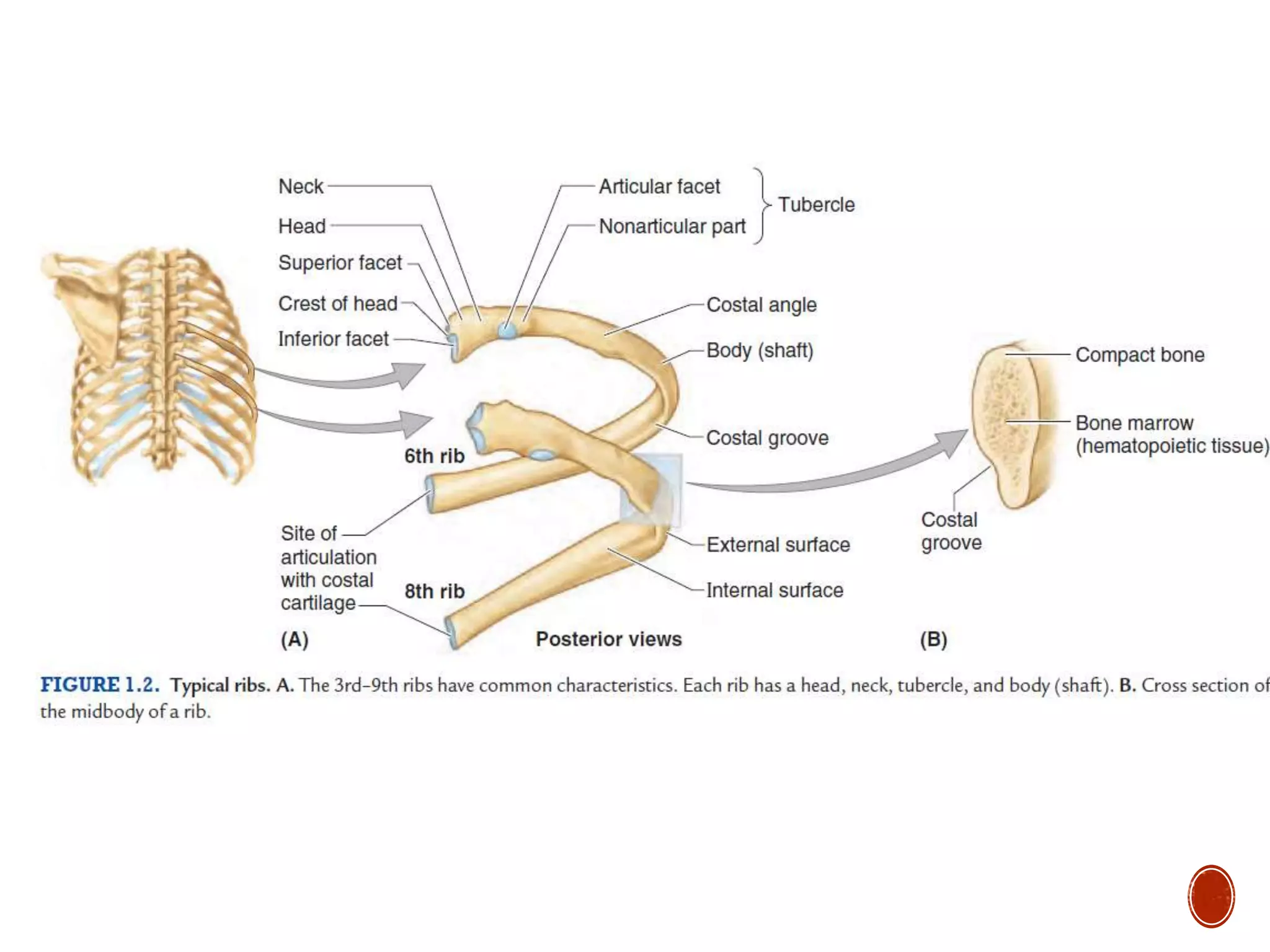
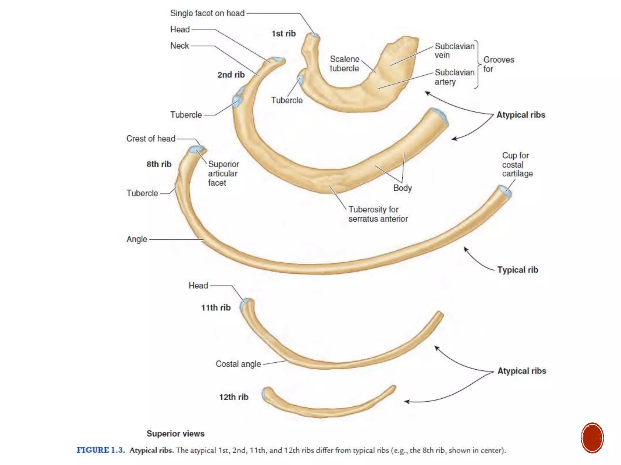
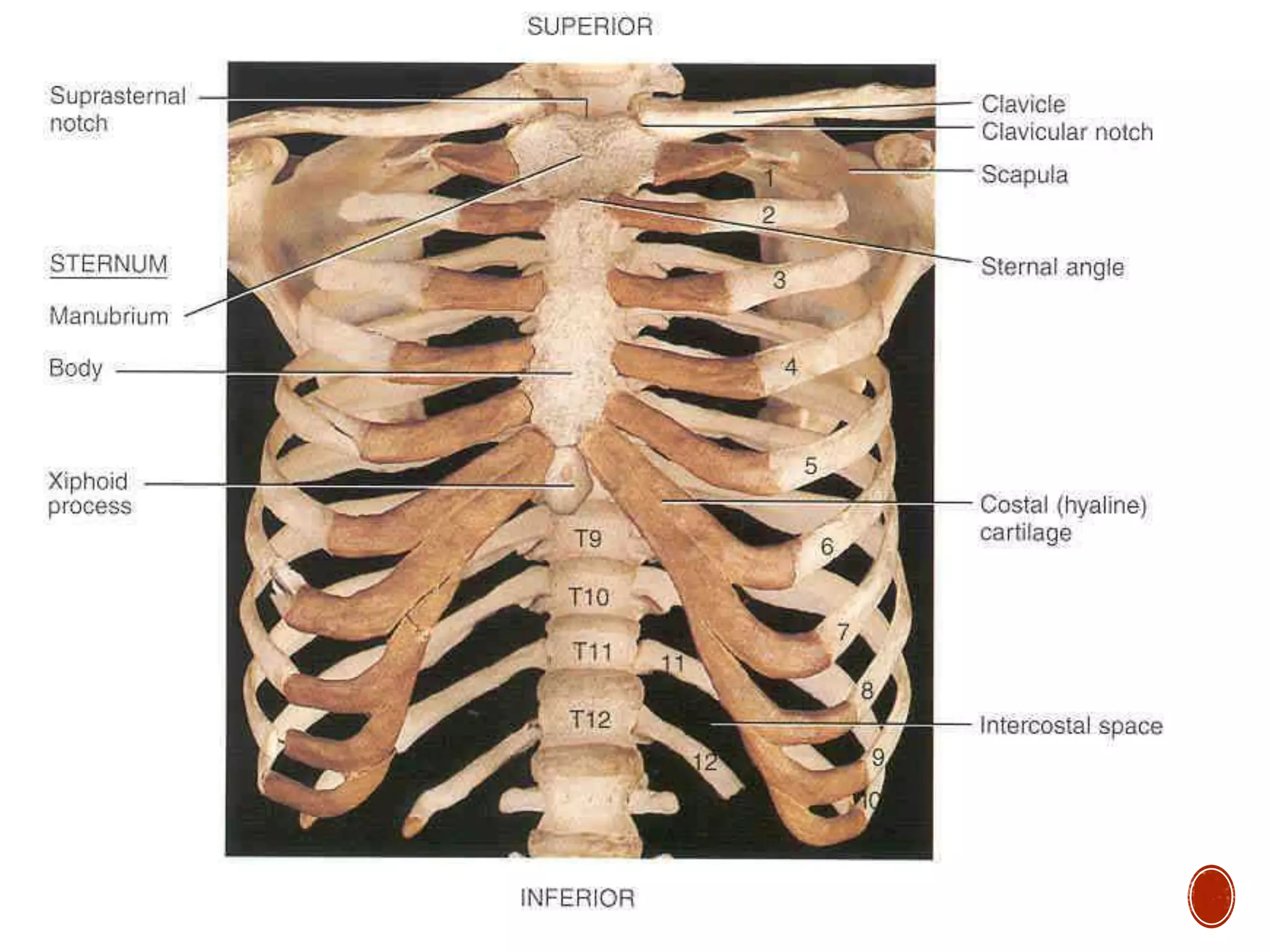
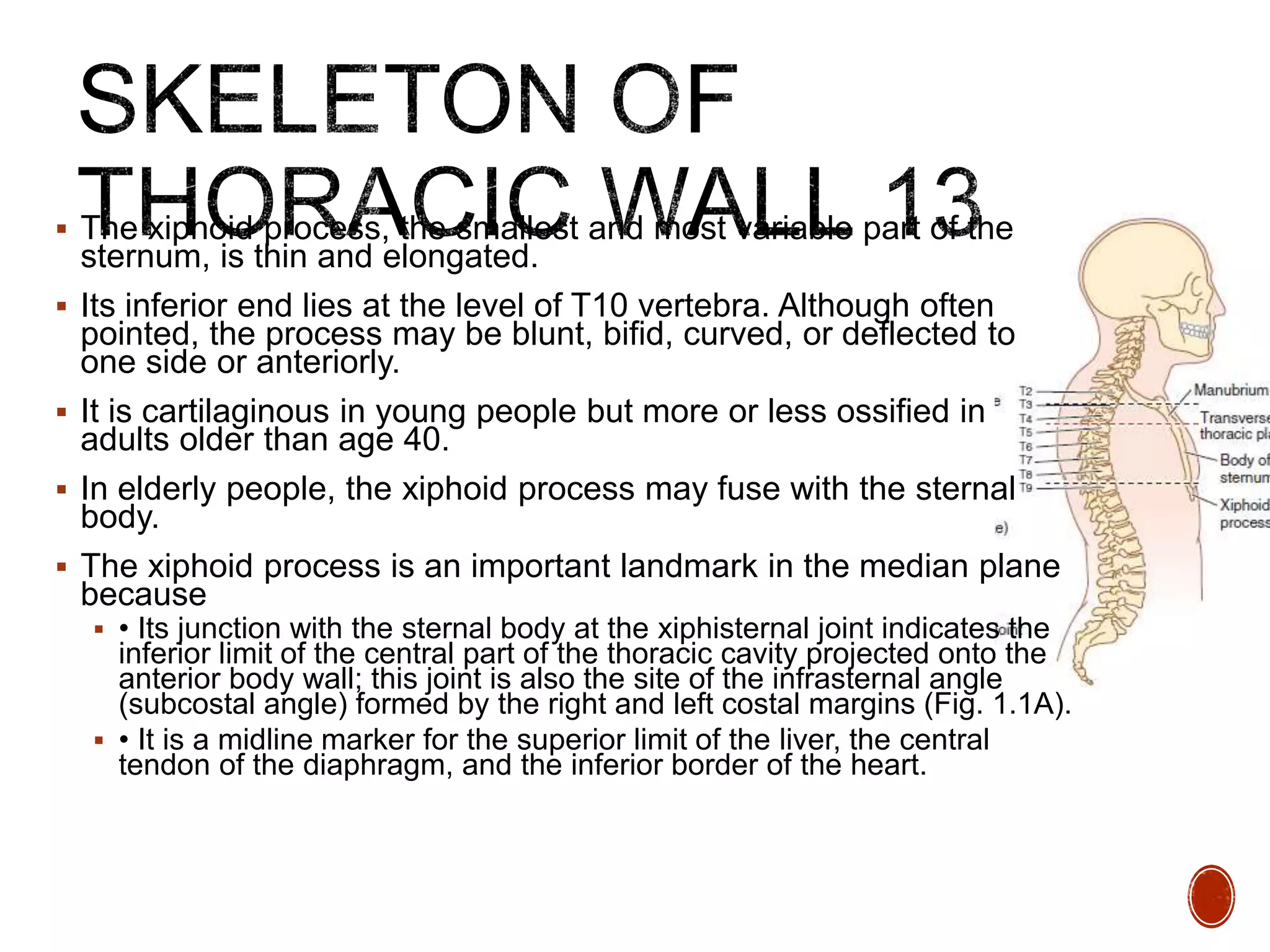
3. The thoracic cavity and its wall have the shape of a truncated cone
The wall of the thoracic cavity is relatively thin, almost as thick as its
skeleton.
The thoracic skeleton takes the form of a domed birdcage. The
thoracic cage, formed by ribs and costal cartilages, is also supported
by the vertical sternum and thoracic vertebrae
The floor of the thoracic cavity (thoracic diaphragm) is deeply
invaginated inferiorly (pushed upward) by viscera of the abdominal
cavity.
So nearly the lower half of the thoracic wall surrounds and protects
abdominal rather than thoracic viscera
The thorax and its cavity are much smaller compared to external
appearances of the chest.
4.
5.
6.
7. The thorax includes the primary organs of the respiratory and
cardiovascular systems.
The thoracic cavity is divided into three major spaces:
the central compartment or mediastinum that houses the thoracic
viscera except for the lungs and
on each side, the right and left pulmonary cavities housing the
lungs.
The majority of the thoracic cavity is occupied by the lungs,
which provide for the exchange of oxygen and carbon dioxide
between the air and blood.
Rest of the thoracic cavity is occupied by the heart and
structures involved in conducting the air and blood to and from
the lungs.
Nutrients (food) traverse the thoracic cavity via the esophagus,
passing from the site of entry in the head to the site of digestion
and absorption in the abdomen.
8.
9. The true thoracic wall includes the thoracic cage and the
muscles that extend between the ribs as well as the skin,
subcutaneous tissue, muscles, and fascia covering its
anterolateral aspect.
The same structures covering its posterior aspect are
considered to belong to the back.
The anterolateral axio-appendicular muscles that overlie the
thoracic cage and form the bed of the breast are distinctly upper
limb muscles based on function and innervation.
10. The domed shape of the thoracic cage provides remarkable
rigidity, given the light weight of its components, enabling it to:
• Protect vital thoracic and abdominal organs (most air or fluid filled)
from external forces.
• Resist the negative (sub-atmospheric) internal pressures
generated by the elastic recoil of the lungs
• Provide attachment for and support the weight of the upper limbs.
• Provide the anchoring attachment (origin) of many of the muscles
that move and maintain the position of the upper limbs relative to
the trunk,
Provide the attachments for muscles of the abdomen, neck, back,
and respiration.
11. Although the shape of the thoracic cage provides rigidity, its joints
and the thinness and flexibility of the ribs allow it to absorb many
external blows and compressions and to change its shape for
respiration.
Because the most important structures within the thorax (heart,
great vessels, lungs, and trachea), as well as its floor and walls,
are constantly in motion, the thorax is one of the most dynamic
regions of the body.
With each breath, the muscles of the thoracic wall—working in
concert with the diaphragm and muscles of the abdominal wall—
vary the volume of the thoracic cavity, first by expanding the
capacity of the cavity, thereby causing the lungs to expand and
draw air in and then, due to lung elasticity and muscle relaxation,
decreasing the volume of the cavity and causing them to expel air.
12.
13. The thoracic skeleton forms the osteocartilaginous thoracic cage which
protects the thoracic viscera and some abdominal organs.
The thoracic skeleton includes 12 pairs of ribs and associated costal
cartilages, 12 thoracic vertebrae and the intervertebral (IV) discs
interposed between them, and the sternum.
The ribs and costal cartilages form the largest part of the thoracic cage;
both are identified numerically, from the most superior (1st rib or costal
cartilage) to the most inferior (12th).
14. RIBS, COSTAL CARTILAGES, AND INTERCOSTAL SPACES
Ribs (L. costae) are curved, flat bones that form most of the
thoracic cage
They are remarkably light in weight yet highly resilient.
Each rib has a spongy interior containing bone marrow
(hematopoietic tissue), which forms blood cells.
15. There are three types of ribs that can be classified as typical or
atypical:
1. True (vertebrosternal) ribs (1st–7th ribs): They attach directly to
the sternum through their own costal cartilages.
2. False (vertebrochondral) ribs (8th, 9th, and usually 10th ribs):
Their cartilages are connected to the cartilage of the rib above
them; thus their connection with the sternum is indirect.
3. Floating (vertebral, free) ribs (11th, 12th, and sometimes 10th
ribs): The rudimentary cartilages of these ribs do not connect even
indirectly with the sternum; instead they end in the posterior
abdominal musculature.
16. Typical ribs (3rd–9th) have the following components:
• Head: wedge-shaped and has two facets, separated by the crest of
the head one facet for articulation with the numerically corresponding
vertebra and one facet for the vertebra superior to it.
• Neck: connects the head of the rib with the body at the level of the
tubercle.
• Tubercle: located at the junction of the neck and body; a smooth
articular part articulates with the corresponding transverse process of
the vertebra, and a rough nonarticular part provides attachment for the
costotransverse ligament (see Fig. 1.8B).
• Body (shaft): thin, flat, and curved, most markedly at the costal
angle where the rib turns anterolaterally. The angle also demarcates
the lateral limit of attachment of the deep back muscles to the ribs. The
concave internal surface of the body has a costal groove paralleling
the inferior border of the rib, which provides some protection for the
intercostal nerve and vessels.
17.
18. Atypical ribs (1st, 2nd, and 10th–12th) are dissimilar (Fig. 1.3):
• The 1st rib is the broadest (i.e., its body is widest and nearly
horizontal), shortest, and most sharply curved of the seven true ribs. It
has a single facet on its head for articulation with the T1 vertebra only
and two transversely directed grooves crossing its superior surface for
the subclavian vessels; the grooves are separated by a scalene
tubercle and ridge, to which the anterior scalene muscle is attached.
• The 2nd rib has a thinner, less curved body and is substantially
longer than the 1st rib. Its head has two facets for articulation with the
bodies of the T1 and T2 vertebrae; its main atypical feature is a rough
area on its upper surface, the tuberosity for serratus anterior, from
which part of that muscle originates.
• The 10th–12th ribs, like the 1st rib, have only one facet on their
heads and articulate with a single vertebra.
• The 11th and 12th ribs are short and have no neck or tubercle.
19.
20. Costal cartilages prolong the ribs anteriorly and contribute to the
elasticity of the thoracic wall, providing a flexible attachment for their
anterior ends (tips).
The cartilages increase in length through the first 7 and then gradually
decrease.
The first 7 costal cartilages attach directly and independently to the
sternum; the 8th, 9th, and 10th articulate with the costal cartilages just
superior to them, forming a continuous, articulated, cartilaginous costal
margin.
The 11th and 12th costal cartilages form caps on the anterior ends of
the corresponding ribs and do not reach or attach to any other bone or
cartilage.
The costal cartilages of ribs 1–10 clearly anchor the anterior end of the
rib to the sternum, limiting its overall movement as the posterior end
rotates around the transverse axis of the rib.
21.
22. Intercostal spaces separate the ribs and their costal cartilages from one
another (Fig. 1.1A).
The spaces are named according to the rib forming the superior border of
the space—for example, the 4th intercostal space lies between ribs 4 and
5. There are 11 intercostal spaces and 11 intercostal nerves.
Intercostal spaces are occupied by intercostal muscles and membranes,
and two sets (main and collateral) of intercostal blood vessels and nerves,
identified by the same number assigned to the space.
The space below the 12th rib does not lie between ribs and thus is referred
to as the subcostal space, and the anterior ramus (branch) of spinal nerve
T12 is the subcostal nerve.
The intercostal spaces are widest anterolaterally, and they widen further
with inspiration.
They can also be further widened by extension and/or lateral flexion of the
thoracic vertebral column to the contra lateral side.
23.
24. Most thoracic vertebrae are typical in that they are independent,
have bodies, vertebral arches, and seven processes for
muscular and articular connections
Characteristic features of thoracic vertebrae include:
• Bilateral costal facets (demifacets) on the vertebral bodies, usually
occurring in inferior and superior pairs, for articulation with the
heads of ribs.
• Costal facets on the transverse processes for articulation with the
tubercles of ribs, except for the inferior two or three thoracic
vertebrae.
• Long, inferiorly slanting spinous processes
25. Superior and inferior costal facets, most of which are small
demifacets, occur as bilaterally paired, planar surfaces on the
superior and inferior posterolateral margins of the bodies of
typical thoracic vertebrae (T2–T9).
Functionally, the facets are arranged in pairs on adjacent vertebrae,
flanking an interposed IV disc: an inferior (demi)facet of the superior
vertebra and a superior (demi)facet of the inferior vertebra.
Typically, two demifacets paired in this manner and the
posterolateral margin of the IV disc between them form a single
socket to receive the head of the rib of the same identifying number
as the inferior vertebra (e.g., head of rib 6 with the superior costal
facet of vertebra T6).
26. Atypical thoracic vertebrae bear whole costal facets in place
of demifacets:
• The superior costal facets of vertebra T1 are not demifacets
because there are no demifacets on the C7 vertebra above, and rib
1 articulates only with vertebra T1. T1 has a typical inferior costal
(demi)facet.
• T10 has only one bilateral pair of (whole) costal facets, located
partly on its body and partly on its pedicle.
• T11 and T12 also have only a single pair of (whole) costal facets,
located on their pedicles.
27. The spinous processes projecting from the vertebral arches of
typical thoracic vertebrae (e.g., vertebrae T6 or T7) are long and
slope inferiorly, usually overlapping the vertebra below (Figs. 1.4D
and 1.5).
They cover the intervals between the laminae of adjacent
vertebrae, thereby preventing sharp objects such as a knife from
entering the vertebral canal and injuring the spinal cord.
The convex superior articular facets of the superior articular
processes face mainly posteriorly and slightly laterally, whereas
the concave inferior articular facets of the inferior articular
processes face mainly anteriorly and slightly medially.
The bilateral joint planes between the respective articular facets of
adjacent thoracic vertebrae define an arc, centering on an axis of
rotation within the vertebral body (Fig. 1.4A–C). Thus small
rotatory movements are permitted between adjacent vertebrae,
limited by the attached rib cage.
28. The sternum (G. sternon, chest) is the flat, elongated bone that
forms the middle of the anterior part of the thoracic cage (Fig.
1.6).
It directly overlies and affords protection for mediastinal viscera
in general and much of the heart in particular.
The sternum consists of three parts: manubrium, body, and
xiphoid process.
In adolescents and young adults, the three parts are connected
together by cartilaginous joints (synchondroses) that ossify
during middle to late adulthood.
29. The manubrium (L. handle, as in the handle of a sword, with the sternal
body forming the blade) is a roughly trapezoidal bone.
The manubrium is the widest and thickest of the three parts of the
sternum.
The easily palpated concave center of the superior border of the
manubrium is the jugular notch (suprasternal notch).
The notch is deepened by the medial (sternal) ends of the clavicles, which
are much larger than the relatively small clavicular notches in the
manubrium that receive them, forming the sternoclavicular (SC) joints (Fig.
1.1A).
Inferolateral to the clavicular notch, the costal cartilage of the 1st rib is
tightly attached to the lateral border of the manubrium—the synchondrosis
of the first rib (Figs. 1.1A and 1.6A). The manubrium and body of the
sternum lie in slightly different planes superior and inferior to their junction
The manubriosternal joint (Fig. 1.6A & B); hence, their junction forms a
projecting sternal angle (of Louis).
30. The body of the sternum, is longer, narrower, and thinner than
the manubrium, and is located at the level of the T5–T9
vertebrae (Fig. 1.6A–C).
Its width varies because of the scalloping of its lateral borders
by the costal notches.
In young people, four sternebrae (primordial segments of the
sternum) are obvious.
The sternebrae articulate with each other at primary
cartilaginous joints (sternal synchondroses).
These joints begin to fuse from the inferior end between
puberty (sexual maturity) and age 25.
The nearly flat anterior surface of the body of the sternum is
marked in adults by three variable transverse ridges (Fig. 1.6A),
which represent the lines of fusion (synostosis) of its four
originally separate sternebrae.
31. The xiphoid process, the smallest and most variable part of the
sternum, is thin and elongated.
Its inferior end lies at the level of T10 vertebra. Although often
pointed, the process may be blunt, bifid, curved, or deflected to
one side or anteriorly.
It is cartilaginous in young people but more or less ossified in
adults older than age 40.
In elderly people, the xiphoid process may fuse with the sternal
body.
The xiphoid process is an important landmark in the median plane
because
• Its junction with the sternal body at the xiphisternal joint indicates the
inferior limit of the central part of the thoracic cavity projected onto the
anterior body wall; this joint is also the site of the infrasternal angle
(subcostal angle) formed by the right and left costal margins (Fig. 1.1A).
• It is a midline marker for the superior limit of the liver, the central
tendon of the diaphragm, and the inferior border of the heart.