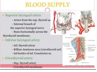
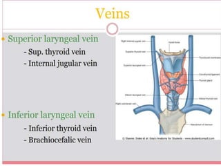
Download to read offline
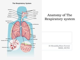

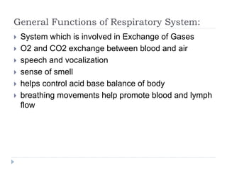
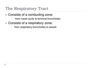
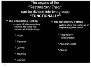
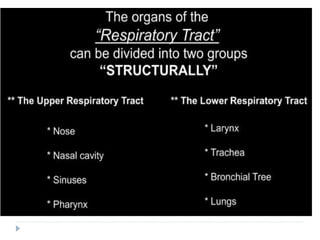
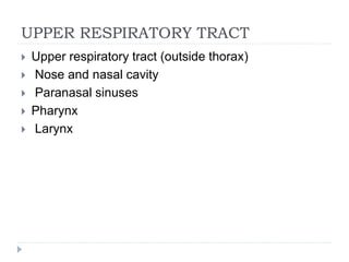
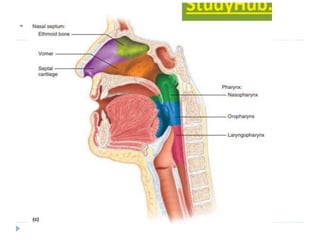
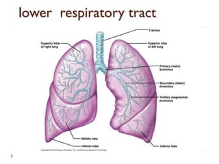
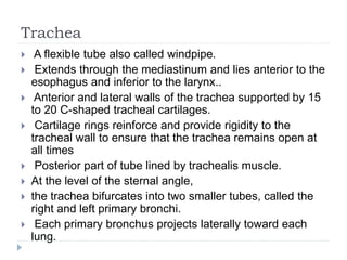
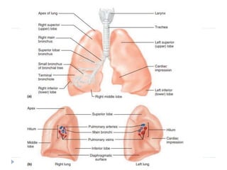


The document summarizes the anatomy and functions of the respiratory system. It describes the major components including the thorax, sternum, ribs, lungs, trachea, bronchi, nose, pharynx, and larynx. It explains that the respiratory system is involved in gas exchange, breathing, speech, smell, and acid-base balance. The conducting zone extends from the nose to terminal bronchioles, while the respiratory zone contains the alveoli in the lungs.


![L12__Respiratory_system_anatomy[1].pptx](https://cdn.slidesharecdn.com/ss_thumbnails/l12respiratorysystemanatomy1-230531143920-02738076-thumbnail.jpg?width=640&height=640&fit=bounds)