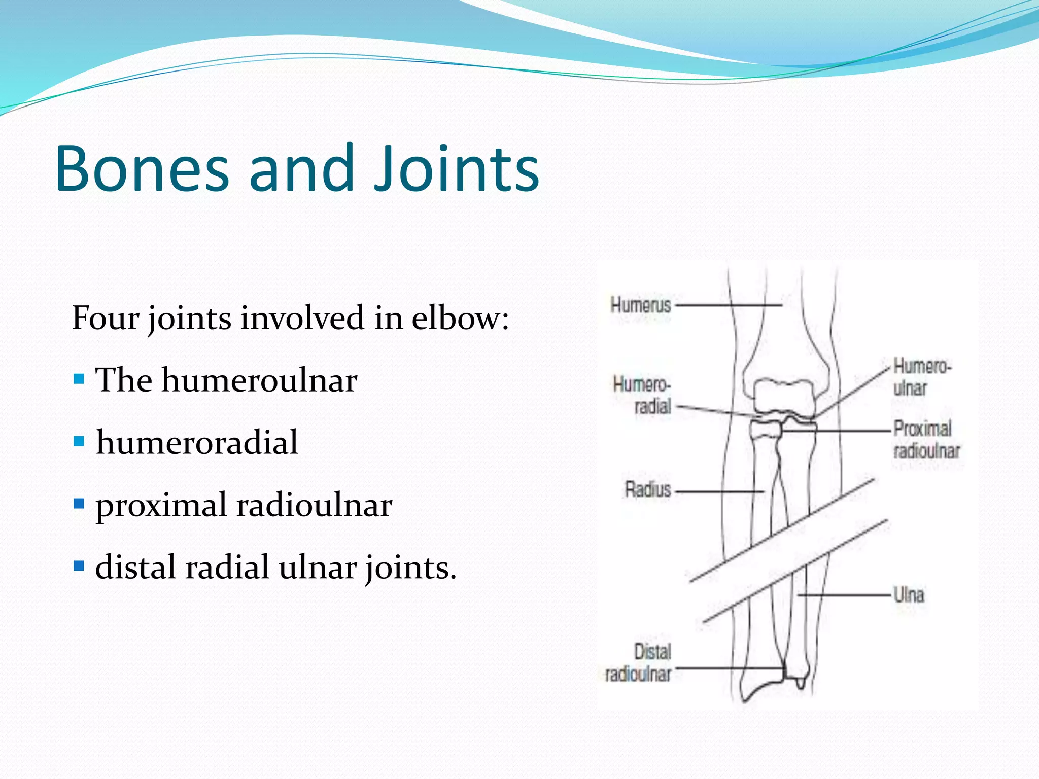
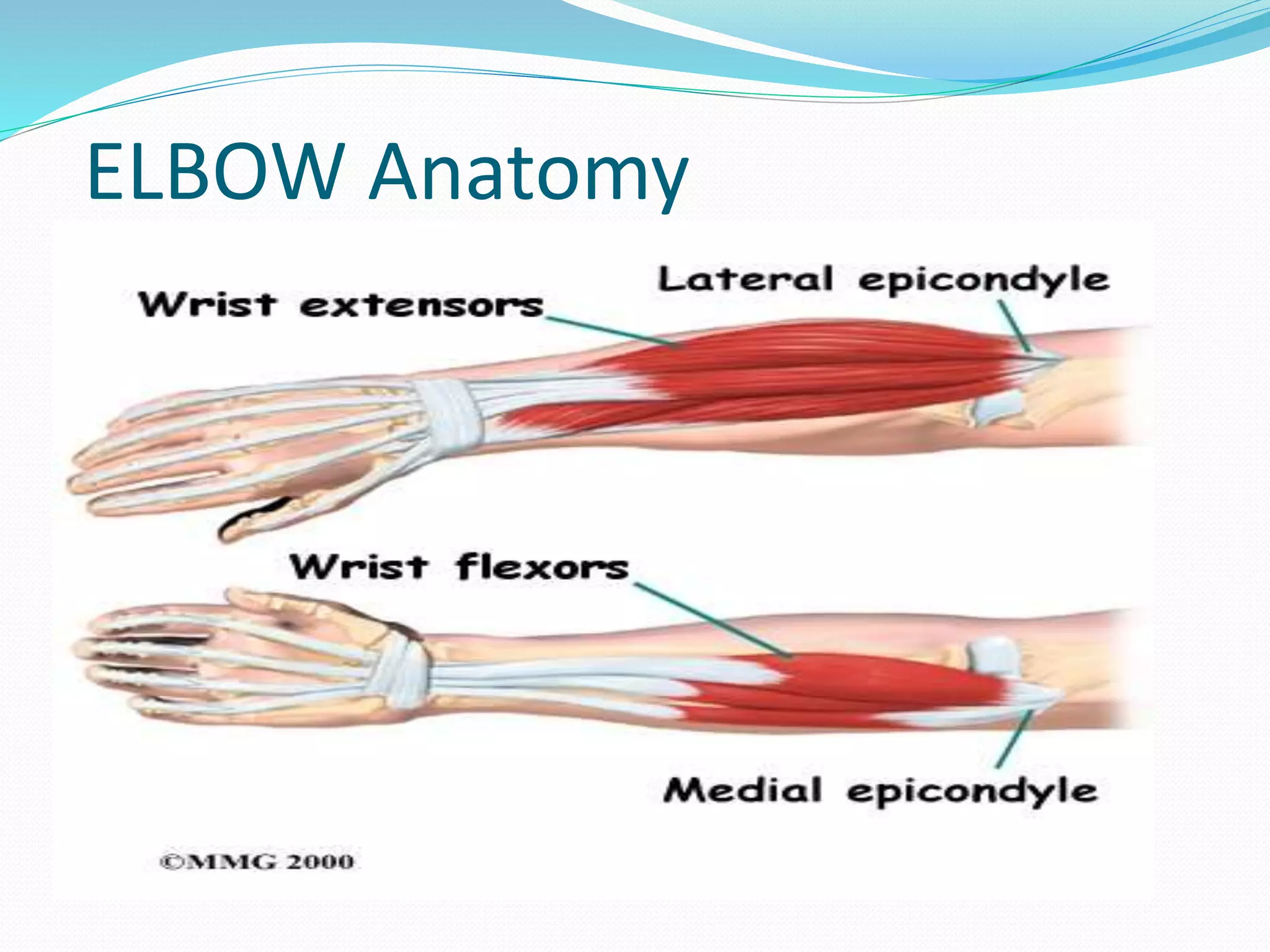
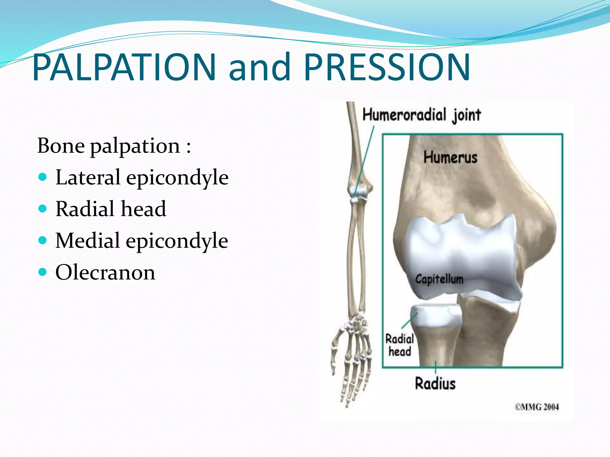
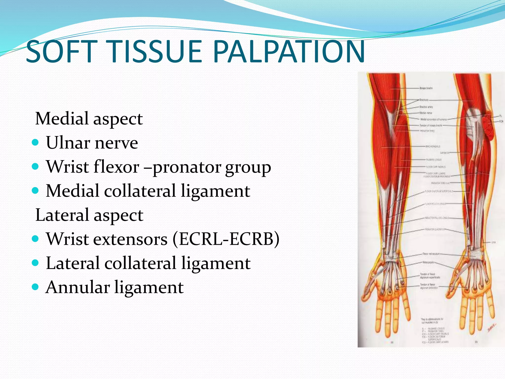
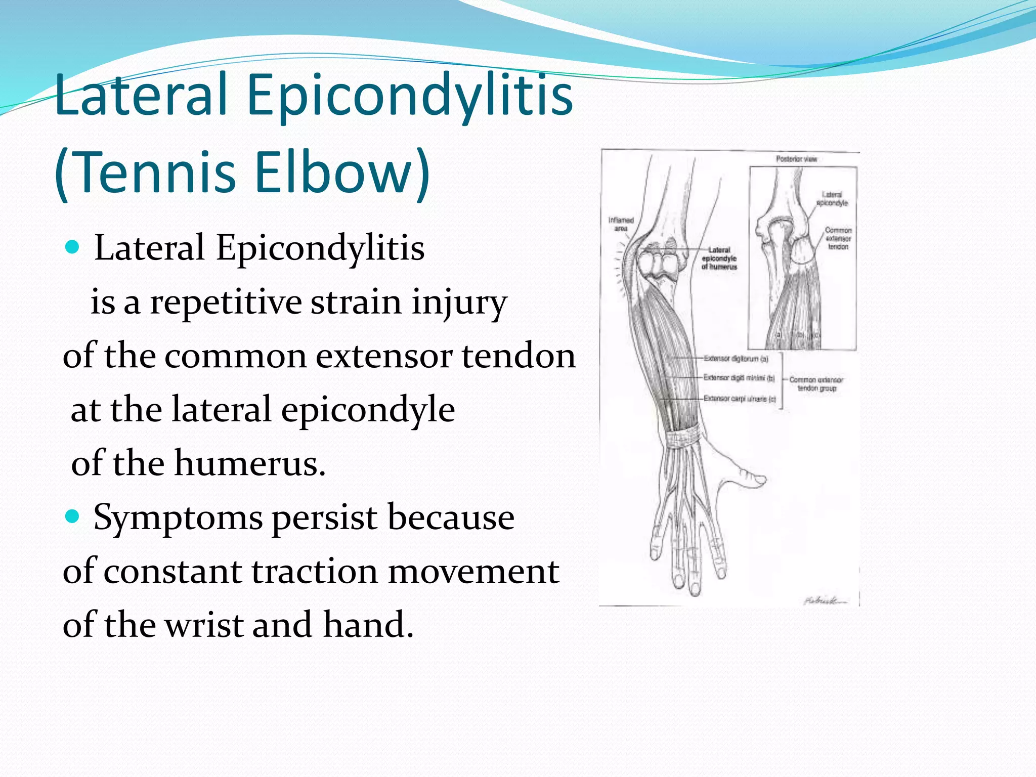
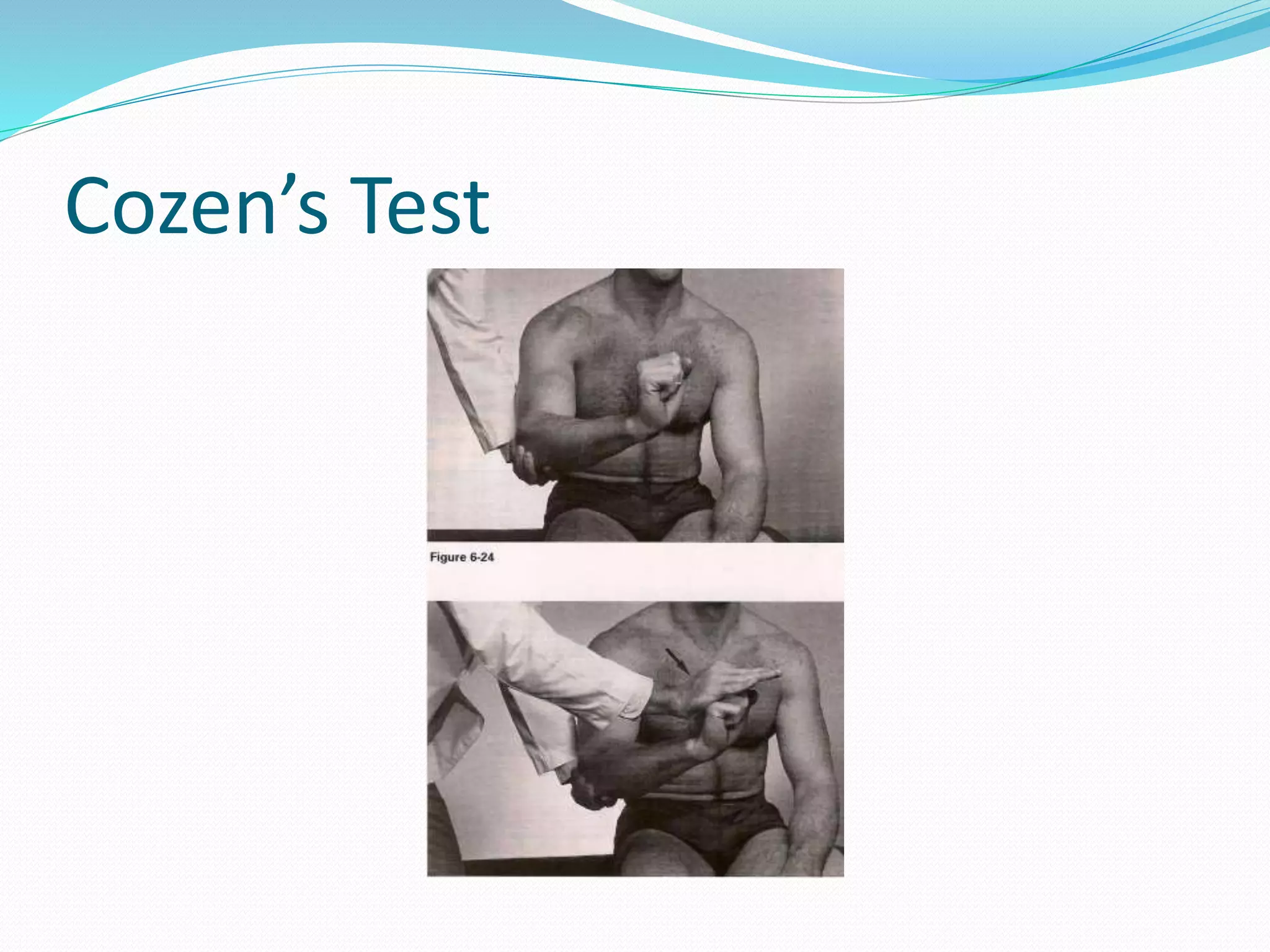
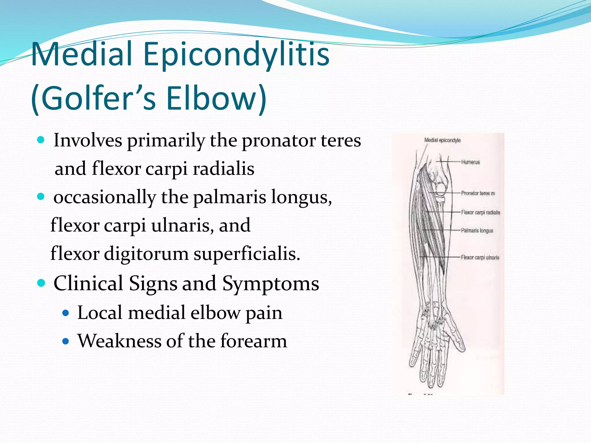
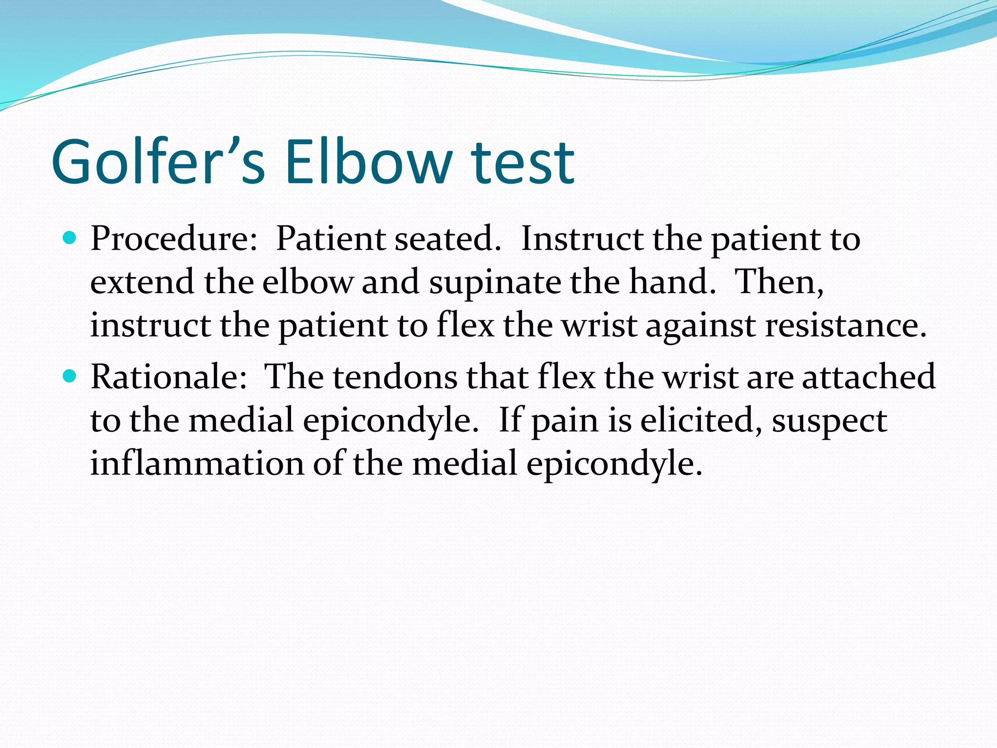
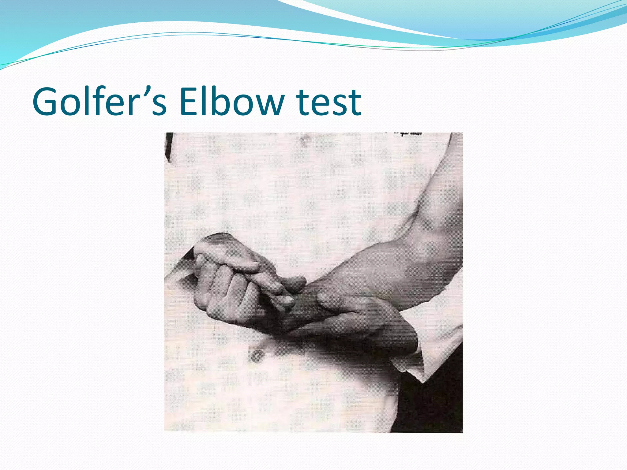
Examination evaluation & Assessment
of Elbow
Dr. Abid Ullah PT
Lecturer FIMS
Abbottabad
Email:
dr.abidullahpt@yahoo.com
ELBOW EXAMINATION
 Anatomy
 Evaluation
 Inspection-Observation
 Palpation-Pression
 Range of motion
 Common clinical conditions
 Neurological examination
 Special tests
ELBOW ANATOMY
 Bones
 Joints
 Ligaments
 Muscles
Bones and Joints
Four joints involved in elbow:
 The humeroulnar
 humeroradial
 proximal radioulnar
 distal radial ulnar joints.
Elbow Joint Characteristics and Arthrokinematics
 Humeroulnar Articulation: (HU) is hinge joint.
 Formed b/w the trochlear notch of the proximal ulna &
trochlea of the humerus.
 The angulation of this joint form the “ carrying angle”
 the distal end of the humerus is convex and faces anteriorly
and downward 45 from the shaft of the humerus.
 The concave trochlear fossa, on the proximal ulna, faces
upward and anteriorly 45 from the ulna.
 The primary motion at this articulation is flexion and
extension
Arthrokinematics
 During flexion/extension the concave fossa slides in
the same direction in which the ulna moves,
 with elbow flexion ___ an anterior and distal direction.
 With elbow extension___ in a posterior and proximal
direction.
 There is slight medial and lateral sliding of the ulna,
for full elbow range of motion (ROM);
 valgus angulation of the joint with elbow extension.
 varus angulation with elbow flexion.
Humeroradial Articulation
 The (HR) is a hinge-pivot joint.
 The laterally placed, spherical capitulum at the distal
end of the humerus is convex. The concave bony
partner, (head of radius) is at the proximal end of the
radius.
 Flexion/extension and pronation/supination occur at
this articulation.
Arthrokinematics
 As the elbow flexes and extends, the concave radial
head slides in the same direction as the bone motion.
 with elbow flexion the concave head slides anteriorly.
 with elbow extension it slides posteriorly.
 With pronation and supination of the forearm, the
radial head spins on the capitulum
Elbow Anatomy
Medial Elbow
Medial (ulnar) collateral ligament
 The MCL ___ 3 components: anterior (taut in
extension), posterior (taut in flexion) and transverse
portions ( provide valgus stability & help with joint
approximation)
 providing medial support to the elbow against valgus
stresses and limiting end-range elbow extension.
 Keeps the joint surfaces in approximation.
 Throwing and golfing impose significant stresses to
the medial collateral ligament complex.
 Valgus stress test is used to assess.
Elbow Anatomy
Lateral Elbow
Lateral (radial) collateral ligament
 The LCL is a fan-shaped on the lateral surface of the
elbow.
 Lateral ( radial) collateral ligament
 Lateral ulnar collateral ligament
 Annular ligament.
 provides stability to the lateral aspect of the elbow
against varus forces
 prevents posterior translation of the radial head (Fig.
18.2B).
 Elbow varus instability stress test.
MUSCLES
 Elbow Flexion
 Bicep
 Brachialis
 Brachioradialis
 Pronator teres
 Elbow Extension
 Triceps
 Anconeus
MUSCLES (cont…)
 Wrist extensor ( lateral epicondyle)
 Extensor carpi radialis longus
 Extensor carpi radialis brevis
 Extensor carpi ulnaris
 Wrist flexors ( medial epicondyle)
 Flexor carpi radialis longus
 Flexor carpi ulnaris
 Palmaris longus
ELBOW Anatomy
MUSCLES (cont…)
 Pronatars
 Pronator teres
 Pronator quadratus
 Supinators
 Bicep brachii
 Supinator
EVALUATION
INSPECTION
 Anterior –posterior side
 Medial-lateral side
 Carrying angle
male 7- 10 deg
female 10-15 deg
 Swelling
PALPATION and PRESSION
Bone palpation :
 Lateral epicondyle
 Radial head
 Medial epicondyle
 Olecranon
SOFT TISSUE PALPATION
Medial aspect
 Ulnar nerve
 Wrist flexor –pronator group
 Medial collateral ligament
Lateral aspect
 Wrist extensors (ECRL-ECRB)
 Lateral collateral ligament
 Annular ligament
SOFT TISSUE PALPATION
Anterior aspect
 Cubital fossa
 Brachial artery
 Median nerve
 Musculo-cutaneus nerve
Posterior aspect
 Olecranon bursa
 Triceps tendon
ELBOW ROM
 Flexion -135 degree
 Extension -0 to 10 degree
 Pronation -90 degree
 Supination -90 degree
 Functional motion
 30-100 (ext-flx)
 50-50 (pronation-supination)
Elbow contractures
 Loss of motion in capsular pattern (flexion –
extension)
 Loss of motion in noncapsular pattern
 Examination by comparing of AROM, PROM, Resistive
test and Plpation
Lateral Epicondylitis
(Tennis Elbow)
 Lateral Epicondylitis
is a repetitive strain injury
of the common extensor tendon
at the lateral epicondyle
of the humerus.
 Symptoms persist because
of constant traction movement
of the wrist and hand.
Lateral Epicondylitis
(Tennis Elbow)
 involves primarily the extensor carpi radialis brevis
 occasionally the extensor digitorum, extensor carpi
radialis longus
 More rarely, the extensor carpi ulnaris.
 Manual laborers
 Plumbers, painters, gardeners and carpenters
 Sports participants
Clinical Signs and Symptoms
• Local lateral elbow pain
 Weakness of the forearm
 Worsened by grasping objects
 Pain when lifting objects
 Pain radiating down the forearm
 Pain with resisted wrist extension
 Pain with resisted middle finger extension
 Pain with passive wrist flexion
Cozen’s Test
 Procedure: Patient seated. Stabilize forearm. Patient
should make a fist and extend it against resistance.
 Rationale: The tendons that extend the wrist attach to
the lateral epicondyle. Forcing the extended wrist into
flexion will exacerbate the pain if the tendons are
inflamed.
Cozen’s Test
Medial Epicondylitis
(Golfer’s Elbow)
 Medial epicondylitis is a repetitive injury of the
common flexor tendon at the medial epicondyle of the
humerus.
 Symptoms persist due to constant traction and
movement of the wrist and hand.
Medial Epicondylitis
(Golfer’s Elbow)
 Involves primarily the pronator teres
and flexor carpi radialis
 occasionally the palmaris longus,
flexor carpi ulnaris, and
flexor digitorum superficialis.
 Clinical Signs and Symptoms
 Local medial elbow pain
 Weakness of the forearm
Golfer’s Elbow test
 Procedure: Patient seated. Instruct the patient to
extend the elbow and supinate the hand. Then,
instruct the patient to flex the wrist against resistance.
 Rationale: The tendons that flex the wrist are attached
to the medial epicondyle. If pain is elicited, suspect
inflammation of the medial epicondyle.
Golfer’s Elbow test
Ligamentous Instability
 Ligamentous instability of the elbow is relatively
uncommon.
 The injury may be caused by forced elbow
hyperextension, forced abduction of the extended
arm, or forced adduction of the extended arm.
Ligamentous Instability
 Forced adduction will damage the radial collateral
ligament.
 Forced abduction will damage the Ulnar collateral
ligament.
 Clinical Signs and Symptoms
 Medial or Lateral elbow pain
 Local swelling
Adduction Stress Test
 Procedure: Patient seated. Stabilize the medial arm
and place adduction pressure on the patient’s lateral
forearm.
 Rationale: Adduction pressure will stress the radial
collateral ligament. Gapping and pain indicate radial
collateral ligament instability.
Adduction Stress Test
Abduction Stress Test
 Procedure: Patient seated. Stabilize the lateral arm
and place abduction pressure on the medial forearm.
 Rationale: Abduction pressure on the medial forearm
applies stress to the Ulnar collateral ligament.
Gapping and pain indicate Ulnar collateral ligament
instability.
Abduction Stress Test
Elbow contracture
Neuropathy / Compression
Syndromes
 Neuropathy and compression syndromes of the elbow
are peripheral neurological disorders.
 They are caused by trauma, overuse, arthritis, and
postural considerations.
 Paresthesia and weakness of the forearm and/or hand.
 The ulnar nerve is most often affected.
 Compression occurs in the groove between the
olecranon process and the medial epicondyle or the
cubital tunnel syndrome.
Neuropathy / Compression
Syndromes
 Clinical Signs and Symptoms
 Forearm and/or hand paresthesia
 Forearm and/or hand weakness
 Numbness & tingling of the ring & small fingers
Tinel’s Sign
 Procedure: Patient seated. Tap the ulnar nerve in the
groove between the olecranon process and the medial
epicondyle with a neurological reflex hammer.
 Rationale: If pain is elicited, it suggests a neuritis or
neuroma of the ulnar nerve.
 Excessive use or repetitive motion injuries.
 Arthritis of the elbow joint.
 Cubital tunnel compression, between the heads of the
flexor carpi ulnaris muscle.
 Postural habits that compress the nerve, such as
sleeping with elbows flexed and hands under head.
 Recurrent nerve subluxations or dislocations
Causes of Ulnar Nerve Damage

Examination, evaluation & Assessment of Elbow

  • 1.
    Examination evaluation &Assessment of Elbow Dr. Abid Ullah PT Lecturer FIMS Abbottabad Email: dr.abidullahpt@yahoo.com
  • 2.
    ELBOW EXAMINATION  Anatomy Evaluation  Inspection-Observation  Palpation-Pression  Range of motion  Common clinical conditions  Neurological examination  Special tests
  • 3.
    ELBOW ANATOMY  Bones Joints  Ligaments  Muscles
  • 4.
    Bones and Joints Fourjoints involved in elbow:  The humeroulnar  humeroradial  proximal radioulnar  distal radial ulnar joints.
  • 5.
    Elbow Joint Characteristicsand Arthrokinematics  Humeroulnar Articulation: (HU) is hinge joint.  Formed b/w the trochlear notch of the proximal ulna & trochlea of the humerus.  The angulation of this joint form the “ carrying angle”  the distal end of the humerus is convex and faces anteriorly and downward 45 from the shaft of the humerus.  The concave trochlear fossa, on the proximal ulna, faces upward and anteriorly 45 from the ulna.  The primary motion at this articulation is flexion and extension
  • 6.
    Arthrokinematics  During flexion/extensionthe concave fossa slides in the same direction in which the ulna moves,  with elbow flexion ___ an anterior and distal direction.  With elbow extension___ in a posterior and proximal direction.  There is slight medial and lateral sliding of the ulna, for full elbow range of motion (ROM);  valgus angulation of the joint with elbow extension.  varus angulation with elbow flexion.
  • 7.
    Humeroradial Articulation  The(HR) is a hinge-pivot joint.  The laterally placed, spherical capitulum at the distal end of the humerus is convex. The concave bony partner, (head of radius) is at the proximal end of the radius.  Flexion/extension and pronation/supination occur at this articulation.
  • 8.
    Arthrokinematics  As theelbow flexes and extends, the concave radial head slides in the same direction as the bone motion.  with elbow flexion the concave head slides anteriorly.  with elbow extension it slides posteriorly.  With pronation and supination of the forearm, the radial head spins on the capitulum
  • 9.
  • 10.
    Medial (ulnar) collateralligament  The MCL ___ 3 components: anterior (taut in extension), posterior (taut in flexion) and transverse portions ( provide valgus stability & help with joint approximation)  providing medial support to the elbow against valgus stresses and limiting end-range elbow extension.  Keeps the joint surfaces in approximation.  Throwing and golfing impose significant stresses to the medial collateral ligament complex.  Valgus stress test is used to assess.
  • 11.
  • 12.
    Lateral (radial) collateralligament  The LCL is a fan-shaped on the lateral surface of the elbow.  Lateral ( radial) collateral ligament  Lateral ulnar collateral ligament  Annular ligament.  provides stability to the lateral aspect of the elbow against varus forces  prevents posterior translation of the radial head (Fig. 18.2B).  Elbow varus instability stress test.
  • 13.
    MUSCLES  Elbow Flexion Bicep  Brachialis  Brachioradialis  Pronator teres  Elbow Extension  Triceps  Anconeus
  • 14.
    MUSCLES (cont…)  Wristextensor ( lateral epicondyle)  Extensor carpi radialis longus  Extensor carpi radialis brevis  Extensor carpi ulnaris  Wrist flexors ( medial epicondyle)  Flexor carpi radialis longus  Flexor carpi ulnaris  Palmaris longus
  • 15.
  • 16.
    MUSCLES (cont…)  Pronatars Pronator teres  Pronator quadratus  Supinators  Bicep brachii  Supinator
  • 17.
    EVALUATION INSPECTION  Anterior –posteriorside  Medial-lateral side  Carrying angle male 7- 10 deg female 10-15 deg  Swelling
  • 18.
    PALPATION and PRESSION Bonepalpation :  Lateral epicondyle  Radial head  Medial epicondyle  Olecranon
  • 19.
    SOFT TISSUE PALPATION Medialaspect  Ulnar nerve  Wrist flexor –pronator group  Medial collateral ligament Lateral aspect  Wrist extensors (ECRL-ECRB)  Lateral collateral ligament  Annular ligament
  • 20.
    SOFT TISSUE PALPATION Anterioraspect  Cubital fossa  Brachial artery  Median nerve  Musculo-cutaneus nerve Posterior aspect  Olecranon bursa  Triceps tendon
  • 21.
    ELBOW ROM  Flexion-135 degree  Extension -0 to 10 degree  Pronation -90 degree  Supination -90 degree  Functional motion  30-100 (ext-flx)  50-50 (pronation-supination)
  • 22.
    Elbow contractures  Lossof motion in capsular pattern (flexion – extension)  Loss of motion in noncapsular pattern  Examination by comparing of AROM, PROM, Resistive test and Plpation
  • 23.
    Lateral Epicondylitis (Tennis Elbow) Lateral Epicondylitis is a repetitive strain injury of the common extensor tendon at the lateral epicondyle of the humerus.  Symptoms persist because of constant traction movement of the wrist and hand.
  • 24.
    Lateral Epicondylitis (Tennis Elbow) involves primarily the extensor carpi radialis brevis  occasionally the extensor digitorum, extensor carpi radialis longus  More rarely, the extensor carpi ulnaris.  Manual laborers  Plumbers, painters, gardeners and carpenters  Sports participants
  • 25.
    Clinical Signs andSymptoms • Local lateral elbow pain  Weakness of the forearm  Worsened by grasping objects  Pain when lifting objects  Pain radiating down the forearm  Pain with resisted wrist extension  Pain with resisted middle finger extension  Pain with passive wrist flexion
  • 26.
    Cozen’s Test  Procedure:Patient seated. Stabilize forearm. Patient should make a fist and extend it against resistance.  Rationale: The tendons that extend the wrist attach to the lateral epicondyle. Forcing the extended wrist into flexion will exacerbate the pain if the tendons are inflamed.
  • 27.
  • 28.
    Medial Epicondylitis (Golfer’s Elbow) Medial epicondylitis is a repetitive injury of the common flexor tendon at the medial epicondyle of the humerus.  Symptoms persist due to constant traction and movement of the wrist and hand.
  • 29.
    Medial Epicondylitis (Golfer’s Elbow) Involves primarily the pronator teres and flexor carpi radialis  occasionally the palmaris longus, flexor carpi ulnaris, and flexor digitorum superficialis.  Clinical Signs and Symptoms  Local medial elbow pain  Weakness of the forearm
  • 30.
    Golfer’s Elbow test Procedure: Patient seated. Instruct the patient to extend the elbow and supinate the hand. Then, instruct the patient to flex the wrist against resistance.  Rationale: The tendons that flex the wrist are attached to the medial epicondyle. If pain is elicited, suspect inflammation of the medial epicondyle.
  • 31.
  • 32.
    Ligamentous Instability  Ligamentousinstability of the elbow is relatively uncommon.  The injury may be caused by forced elbow hyperextension, forced abduction of the extended arm, or forced adduction of the extended arm.
  • 33.
    Ligamentous Instability  Forcedadduction will damage the radial collateral ligament.  Forced abduction will damage the Ulnar collateral ligament.  Clinical Signs and Symptoms  Medial or Lateral elbow pain  Local swelling
  • 34.
    Adduction Stress Test Procedure: Patient seated. Stabilize the medial arm and place adduction pressure on the patient’s lateral forearm.  Rationale: Adduction pressure will stress the radial collateral ligament. Gapping and pain indicate radial collateral ligament instability.
  • 35.
  • 36.
    Abduction Stress Test Procedure: Patient seated. Stabilize the lateral arm and place abduction pressure on the medial forearm.  Rationale: Abduction pressure on the medial forearm applies stress to the Ulnar collateral ligament. Gapping and pain indicate Ulnar collateral ligament instability.
  • 37.
  • 38.
  • 39.
    Neuropathy / Compression Syndromes Neuropathy and compression syndromes of the elbow are peripheral neurological disorders.  They are caused by trauma, overuse, arthritis, and postural considerations.  Paresthesia and weakness of the forearm and/or hand.  The ulnar nerve is most often affected.  Compression occurs in the groove between the olecranon process and the medial epicondyle or the cubital tunnel syndrome.
  • 40.
    Neuropathy / Compression Syndromes Clinical Signs and Symptoms  Forearm and/or hand paresthesia  Forearm and/or hand weakness  Numbness & tingling of the ring & small fingers
  • 41.
    Tinel’s Sign  Procedure:Patient seated. Tap the ulnar nerve in the groove between the olecranon process and the medial epicondyle with a neurological reflex hammer.  Rationale: If pain is elicited, it suggests a neuritis or neuroma of the ulnar nerve.
  • 43.
     Excessive useor repetitive motion injuries.  Arthritis of the elbow joint.  Cubital tunnel compression, between the heads of the flexor carpi ulnaris muscle.  Postural habits that compress the nerve, such as sleeping with elbows flexed and hands under head.  Recurrent nerve subluxations or dislocations Causes of Ulnar Nerve Damage