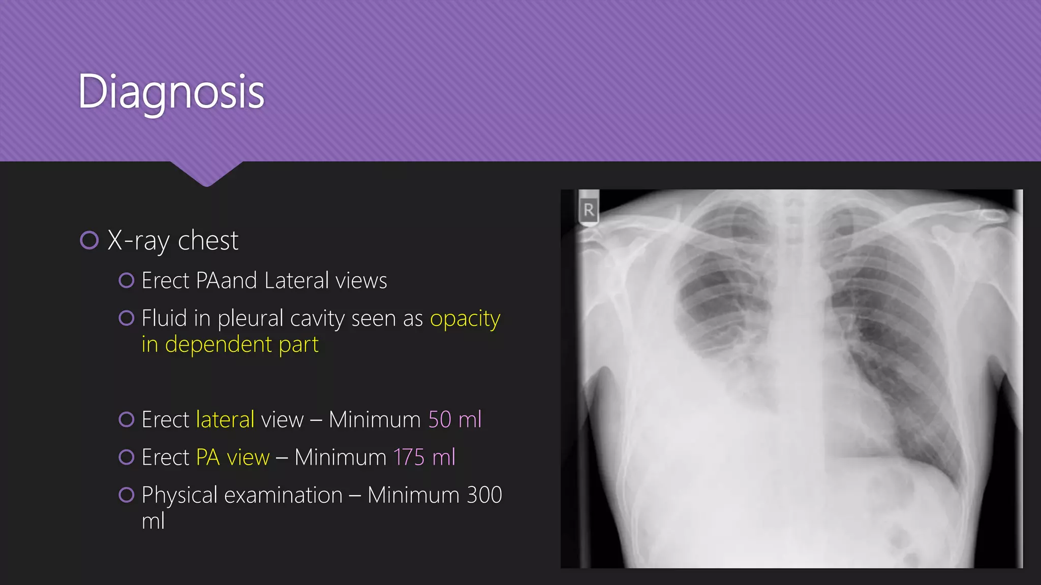
Empyema Thoracis
Dr. Sunil K S Gaur
Senior Resident
Introduction
 “Pus in the plural space”
 End stage of pleural infection from any cause
 Hippocrates credited with first description of natural history and
treatment
 Streptococcal or Pneumococcal – m/c
 Gram negative and anaerobic
 Tubercular
Aetiology
 Unresolved pneumonia – m/c
 Other pulmonary infections – bronchiectasis, tuberculosis, fungal infections, lung
abscess
 Trauma – haemothorax becomes infected, oesophageal injury
 Complication of any thoracic operation
 Pus under the diaphragm
 Osteomyelitis of ribs or vertebrae
Pathology
Pathology
 Presence of thick pus and
 Thick cortex of fibrin and coagulum over the lung
 When presents de novo it usually follows pneumonia, and three phases
are described:
1. Exudative phase (1-3 days)
2. Fibrino- purulent phase (4-14 days)
3. Organizing/Chronic phase (after 14 days)
1. Exudative phase
 Protein-rich (>30 g/L) effusion
 If this becomes infected with the organisms from the lung the scene is set for empyema
 Increased neutrophils in fluid
 However normal glucose, LDH level and pH
 Antibiotics may be all that is required
 Aspiration or drainage to dryness in addition is preferred
2. Fibrino-purulent phase
 Pleural fluid becomes thick & fibrin deposits over the pleural surfaces
 Bacterial stains present, frank pus
 pH and glucose levels become low
 LDH, neutrophils and protein levels increase
 Drainage at this stage is prudent as antibiotics alone are ineffective
3. Organizing/Chronic Phase
 Fibrin converted in to fibroblasts, new capillary ingrowth begins
 Causes the lung to be trapped by a thick peel or ‘cortex’
 Effusion grossly purulent, thick like curd
 Contraction of thorax - scoliosis
 Surgical management may be required
Clinical features
Clinical Features
 History
 Malaise
 Chest pain
 Fever
 Cough
 Dyspnea
 Loss of appetite
 Weight loss
Clinical Features (contd.)
Examination:
Inspection:
Asymmetric chest expansion
May be discharging wound
Tracheal deviation may be seen
Bulging ICS
Palpation:
Raised local temperature in acute
phase
Local tenderness
Decreased tactile fremitus
Clinical Features (contd.)
Examination:
Percussion:
Dull note
Auscultation:
Egophony
Pleural friction rub
Decreased breath sounds
Diagnosis
Diagnosis
 X-ray chest
 Erect PAand Lateral views
 Fluid in pleural cavity seen as opacity
in dependent part
 Erect lateral view – Minimum 50 ml
 Erect PA view – Minimum 175 ml
 Physical examination – Minimum 300
ml
Diagnosis (contd.)
 Ultrasonography:
 Amount of fluid, and loculations are
imaged
 USG guided diagnostic/therapeutic
thoracocentesis
 Can detect physiological amount of
pleural fluid, i.e. 5 ml
 100% sensitivity for effusion >100 ml
Diagnosis (contd.)
 CT scan:
 Reference standard in plural diseases
 Distinguish pleural with parenchymal
abnormalities
 Determine precise location, extent
and loculations
 Split pleura sign – Enhancing,
thickened visceral and parietal
pleural layers separated by an
intervening layer of low attenuation
fluid
Diagnosis (contd.)
 Aspiration and Pus analysis:
 Gram staining
 Culture & sensitivity
 AFB/ZN staining
 CBNAAT for mycobacterium
 KOH mount for fungal
 Blood tests:
 CBC – TLC, DLC
 Blood sugar – to rule out DM
 Culture & sensitivity
 CRP
 Arterial blood gas
Treatment
Treatment Options
Non-surgical
 Moist O2 inhalation
 Chest physiotherapy
 Antimicrobials
 NSAIDs
 Anti-pyretics
 Pleural tap
 Mechanical ventilation
Surgical
 Chest tube drainage
 Talc pleurodesis
 Fibrinolysis
 VATS + Debridement
 Thoracotomy + Decortication
Treatment Phase 1
 USG guided pleural tap – Diagnostic + Therapeutic
 Chest tube insertion
 Antibiotics/ATT
 Chest physiotherapy
 Thoracocentesis without pleural drain placement is not
recommended in empyema
Treatment Phase 2
 Chest tube insertion + Fibrinolysis (or)
 VATS + debridement + Chest tube in
situ
 Antibiotics/ATT
 Chest physiotherapy
 Fibrinolytic agent – Streptokinase,
urokinase
Treatment Phase 3
 Thoracotomy + Decortication + Chest
tube in situ
 Antibiotics/ATT
 Chest physiotherapy
 If feasible, VATS is a good option
Surgical management of pleural
effusions and infections
Thoracoscopy
 Direct-vision thoracoscope was used for many years
 Use was limited mainly to performing biopsies
 had a limited view
 uncomfortable to use for any length of time
 one hand used up for holding the thoracoscope
VATS (Video-assisted
thoracoscopic surgery)
 Camera is attached to the thoracoscope,
which can be operated by an assistant with
the image displayed on a screen
 Surgeon’s hands are freed
 Can manipulate instruments with both hands
to perform a variety of procedures
 lung is isolated using a double lumen tube
 the patient is positioned disease side up, and
the pleural cavity is entered
VATS
(contd.)
VATS - Benefits
 Less blood loss
 Small scar
 Less postoperative morbidity
 Decreased need of post-op mechanical ventilation
 Earlier chest physiotherapy
 Reduction in 30 days mortality
Drainage and Talc Pleurodesis
 Early in its evolution, requires drainage
 Direct visualization of the pleural cavity
for complete drainage
 Excellent talc insufflation to achieve
pleurodesis in malignant pleural effusion
 Multiple pleural biopsies can be taken
for undiagnosed cases
Debridement of empyema
 Once the fluid component becomes
fibrinopurulent and loculated it requires
surgical debridement
 Can often be achieved through a VATS
approach
 Fluid and debris are vigorously
debrided, freeing the lung and allowing
for re-expansion
Decortication
 fibrous cortex or peel from the
entrapped underlying lung is removed
 usually performed through a
posterolateral thoracotomy
 though in selected cases it can be
performed as a VATS procedure
 requires careful dissection to remove the
parietal and visceral cortex
 taking care not to damage the visceral
pleura
Complications
Complications
 Septicemia and MODS
 Respiratory failure
 Secondary scoliosis
 Empyema necessitans
 Bronchopleural fistula
 Pericarditis
 Peritonitis
Empyema
necessitans
 Extension of a pus out of the pleura and
into the neighboring chest wall and soft
tissues
 Fluctuant swelling over chest wall
 Now rare; only in immuno-compromised
 Tx – Closed drainage; rest same
Bronchopleural fistula
 Abnormal connection between airway
and pleural cavity
 Leads to cough with copious amounts of
purulent expectoration
 Tx – Adequate drainage of empyema
+/- repair
Thank
you!

Empyema Thoracis

  • 1.
    Empyema Thoracis Dr. SunilK S Gaur Senior Resident
  • 2.
    Introduction  “Pus inthe plural space”  End stage of pleural infection from any cause  Hippocrates credited with first description of natural history and treatment  Streptococcal or Pneumococcal – m/c  Gram negative and anaerobic  Tubercular
  • 3.
    Aetiology  Unresolved pneumonia– m/c  Other pulmonary infections – bronchiectasis, tuberculosis, fungal infections, lung abscess  Trauma – haemothorax becomes infected, oesophageal injury  Complication of any thoracic operation  Pus under the diaphragm  Osteomyelitis of ribs or vertebrae
  • 4.
  • 5.
    Pathology  Presence ofthick pus and  Thick cortex of fibrin and coagulum over the lung  When presents de novo it usually follows pneumonia, and three phases are described: 1. Exudative phase (1-3 days) 2. Fibrino- purulent phase (4-14 days) 3. Organizing/Chronic phase (after 14 days)
  • 6.
    1. Exudative phase Protein-rich (>30 g/L) effusion  If this becomes infected with the organisms from the lung the scene is set for empyema  Increased neutrophils in fluid  However normal glucose, LDH level and pH  Antibiotics may be all that is required  Aspiration or drainage to dryness in addition is preferred
  • 7.
    2. Fibrino-purulent phase Pleural fluid becomes thick & fibrin deposits over the pleural surfaces  Bacterial stains present, frank pus  pH and glucose levels become low  LDH, neutrophils and protein levels increase  Drainage at this stage is prudent as antibiotics alone are ineffective
  • 8.
    3. Organizing/Chronic Phase Fibrin converted in to fibroblasts, new capillary ingrowth begins  Causes the lung to be trapped by a thick peel or ‘cortex’  Effusion grossly purulent, thick like curd  Contraction of thorax - scoliosis  Surgical management may be required
  • 9.
  • 10.
    Clinical Features  History Malaise  Chest pain  Fever  Cough  Dyspnea  Loss of appetite  Weight loss
  • 11.
    Clinical Features (contd.) Examination: Inspection: Asymmetricchest expansion May be discharging wound Tracheal deviation may be seen Bulging ICS Palpation: Raised local temperature in acute phase Local tenderness Decreased tactile fremitus
  • 12.
    Clinical Features (contd.) Examination: Percussion: Dullnote Auscultation: Egophony Pleural friction rub Decreased breath sounds
  • 13.
  • 14.
    Diagnosis  X-ray chest Erect PAand Lateral views  Fluid in pleural cavity seen as opacity in dependent part  Erect lateral view – Minimum 50 ml  Erect PA view – Minimum 175 ml  Physical examination – Minimum 300 ml
  • 15.
    Diagnosis (contd.)  Ultrasonography: Amount of fluid, and loculations are imaged  USG guided diagnostic/therapeutic thoracocentesis  Can detect physiological amount of pleural fluid, i.e. 5 ml  100% sensitivity for effusion >100 ml
  • 16.
    Diagnosis (contd.)  CTscan:  Reference standard in plural diseases  Distinguish pleural with parenchymal abnormalities  Determine precise location, extent and loculations  Split pleura sign – Enhancing, thickened visceral and parietal pleural layers separated by an intervening layer of low attenuation fluid
  • 17.
    Diagnosis (contd.)  Aspirationand Pus analysis:  Gram staining  Culture & sensitivity  AFB/ZN staining  CBNAAT for mycobacterium  KOH mount for fungal  Blood tests:  CBC – TLC, DLC  Blood sugar – to rule out DM  Culture & sensitivity  CRP  Arterial blood gas
  • 18.
  • 19.
    Treatment Options Non-surgical  MoistO2 inhalation  Chest physiotherapy  Antimicrobials  NSAIDs  Anti-pyretics  Pleural tap  Mechanical ventilation Surgical  Chest tube drainage  Talc pleurodesis  Fibrinolysis  VATS + Debridement  Thoracotomy + Decortication
  • 20.
    Treatment Phase 1 USG guided pleural tap – Diagnostic + Therapeutic  Chest tube insertion  Antibiotics/ATT  Chest physiotherapy  Thoracocentesis without pleural drain placement is not recommended in empyema
  • 21.
    Treatment Phase 2 Chest tube insertion + Fibrinolysis (or)  VATS + debridement + Chest tube in situ  Antibiotics/ATT  Chest physiotherapy  Fibrinolytic agent – Streptokinase, urokinase
  • 22.
    Treatment Phase 3 Thoracotomy + Decortication + Chest tube in situ  Antibiotics/ATT  Chest physiotherapy  If feasible, VATS is a good option
  • 23.
    Surgical management ofpleural effusions and infections
  • 24.
    Thoracoscopy  Direct-vision thoracoscopewas used for many years  Use was limited mainly to performing biopsies  had a limited view  uncomfortable to use for any length of time  one hand used up for holding the thoracoscope
  • 25.
    VATS (Video-assisted thoracoscopic surgery) Camera is attached to the thoracoscope, which can be operated by an assistant with the image displayed on a screen  Surgeon’s hands are freed  Can manipulate instruments with both hands to perform a variety of procedures  lung is isolated using a double lumen tube  the patient is positioned disease side up, and the pleural cavity is entered
  • 26.
  • 27.
    VATS - Benefits Less blood loss  Small scar  Less postoperative morbidity  Decreased need of post-op mechanical ventilation  Earlier chest physiotherapy  Reduction in 30 days mortality
  • 28.
    Drainage and TalcPleurodesis  Early in its evolution, requires drainage  Direct visualization of the pleural cavity for complete drainage  Excellent talc insufflation to achieve pleurodesis in malignant pleural effusion  Multiple pleural biopsies can be taken for undiagnosed cases
  • 29.
    Debridement of empyema Once the fluid component becomes fibrinopurulent and loculated it requires surgical debridement  Can often be achieved through a VATS approach  Fluid and debris are vigorously debrided, freeing the lung and allowing for re-expansion
  • 30.
    Decortication  fibrous cortexor peel from the entrapped underlying lung is removed  usually performed through a posterolateral thoracotomy  though in selected cases it can be performed as a VATS procedure  requires careful dissection to remove the parietal and visceral cortex  taking care not to damage the visceral pleura
  • 31.
  • 32.
    Complications  Septicemia andMODS  Respiratory failure  Secondary scoliosis  Empyema necessitans  Bronchopleural fistula  Pericarditis  Peritonitis
  • 33.
    Empyema necessitans  Extension ofa pus out of the pleura and into the neighboring chest wall and soft tissues  Fluctuant swelling over chest wall  Now rare; only in immuno-compromised  Tx – Closed drainage; rest same
  • 34.
    Bronchopleural fistula  Abnormalconnection between airway and pleural cavity  Leads to cough with copious amounts of purulent expectoration  Tx – Adequate drainage of empyema +/- repair
  • 35.