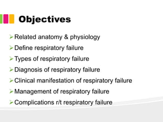
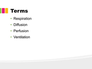
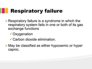
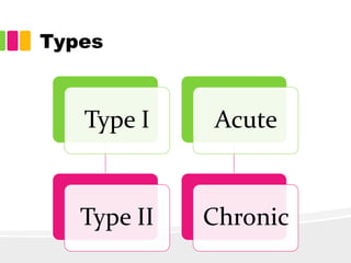
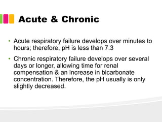
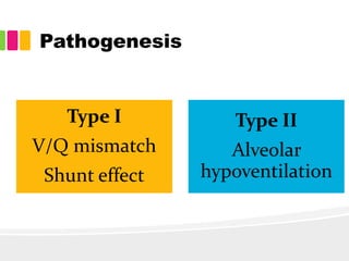
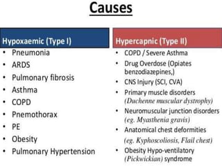
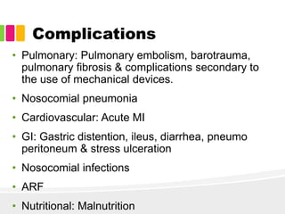
Respiratory failure
Presentor
Anjani Walia
Msc N 1st year
Objectives
Related anatomy & physiology
Define respiratory failure
Types of respiratory failure
Diagnosis of respiratory failure
Clinical manifestation of respiratory failure
Management of respiratory failure
Complications r/t respiratory failure
Respiratory tract
Terms
• Respiration
• Diffusion
• Perfusion
• Ventilation
Control
• Resting respiration is the result of cyclical excitation
of the respiratory muscles by the phrenic nerve.
• Apneustic center
• Pneumotaxic center
• Central chemoreceptors
• Peripheral chemoreceptors
• Baroreceptors
Respiratory failure
Respiratory failure is a syndrome in which the
respiratory system fails in one or both of its gas
exchange functions
Oxygenation
Carbon dioxide elimination.
May be classified as either hypoxemic or hyper
capnic.
Types
Type I
Type II
Acute
Chronic
Types
Pathogenic
mechanism
Ventilatory
disorders
Obstructive Restrictive
Gas
exchange
disorders
Diffusion
disorder
V/Q
mismatchi
ng
Acute & Chronic
• Acute respiratory failure develops over minutes to
hours; therefore, pH is less than 7.3
• Chronic respiratory failure develops over several
days or longer, allowing time for renal
compensation & an increase in bicarbonate
concentration. Therefore, the pH usually is only
slightly decreased.
Pathogenesis
Type I
V/Q mismatch
Shunt effect
Type II
Alveolar
hypoventilation
Clinical manifestations
• Neurologic manifestations
• Tachycardia & arrhythmias
• Cyanosis
• Dyspnea
• Confusion & somnolence
• Cor pulmonale.
Diagnostic Evaluation
Laboratory Studies
• ABG
• CBC
• RFT & LFT
• Electrolytes such as potassium, magnesium &
phosphate
• S. creatine kinase, troponin I
Radiography
• Frequently reveals the cause
Echocardiography
• Not be performed routinely, useful test when a
cardiac cause of acute respiratory failure is
suspected.
Management
• Cardiac monitoring, blood pressure, pulse oximetry
& capnometry are recommended.
• Reverse/ prevent tissue hypoxia.
• Appropriate management of the underlying
disease
• Treated with O2 supplementation & ventilatory
assist devices
Diuretics
• First-line therapy generally includes a loop
diuretic such as furosemide, which inhibits
sodium chloride reabsorption in the ascending
loop of Henle.
Nitrates
• Nitrates reduce myocardial oxygen demand by
lowering preload & afterload.
Inotropic Agents
• The principal inotropic agents are dopamine,
dobutamine & digoxin.
Opioid Analgesics
• Morphine IV is an excellent adjunct.
• Reduces preload
• Causes arterial dilatation, which reduces
systemic vascular resistance and may increase
cardiac output.
Corticosteroids
• Effective in accelerating recovery from acute
COPD exacerbations & are an important anti-
inflammatory therapy in asthma.
Beta 2 Agonists
• These agents act to decrease muscle tone in
both small and large airways in the lungs.
• Includes beta-adrenergics, methylxanthines &
anticholinergics.
Anticholinergics
• Antagonize the action of acetylcholine with
muscarinic receptor on bronchial smooth
muscle.
Complications
• Pulmonary: Pulmonary embolism, barotrauma,
pulmonary fibrosis & complications secondary to
the use of mechanical devices.
• Nosocomial pneumonia
• Cardiovascular: Acute MI
• GI: Gastric distention, ileus, diarrhea, pneumo
peritoneum & stress ulceration
• Nosocomial infections
• ARF
• Nutritional: Malnutrition
Nursing diagnosis
• Impaired gas exchange
• Ineffective breathing pattern
• Impaired verbal communication
• Activity intolerance
• Self care deficit
• Impaired physical mobility
• Risk for impaired skin integrity
• Risk of imbalanced nutrition: less than body
requirement
Summary
Conclusion
• Issues involved in timely recognition of &
response to clinical deterioration remain complex,
yet patient safety relies on nurses’ timely
assessments and actions.
References
1. Hinkle LJ, Cheever HK. Brunner & Sudharth's textbook of
medical surgical nursing. 13th Edition. I volume .New
Delhi: Wolters Kluwer Publications; 2014.Pp 545-47
2. Respiratory Failure: Background, Pathophysiology,
Etiology [Internet]. [cited 2018 Mar 6]. Available from:
https://emedicine.medscape.com/article/167981-
overview#showall
3. Pathophysiology of acute respiratory failure. - PubMed -
NCBI [Internet]. [cited 2018 Mar 6]. Available from:
https://www.ncbi.nlm.nih.gov/pubmed/8200186
4. NCP Nursing Care Plan for Acute Respiratory Failure
[Internet]. [cited 2018 Mar 6]. Available from: http://nurse-
thought.blogspot.in/2011/03/ncp-nursing-care-plan-for-
acute_18.html
Thank you

Respiratory failure

  • 1.
  • 2.
    Objectives Related anatomy &physiology Define respiratory failure Types of respiratory failure Diagnosis of respiratory failure Clinical manifestation of respiratory failure Management of respiratory failure Complications r/t respiratory failure
  • 3.
  • 4.
    Terms • Respiration • Diffusion •Perfusion • Ventilation
  • 5.
    Control • Resting respirationis the result of cyclical excitation of the respiratory muscles by the phrenic nerve. • Apneustic center • Pneumotaxic center • Central chemoreceptors • Peripheral chemoreceptors • Baroreceptors
  • 6.
    Respiratory failure Respiratory failureis a syndrome in which the respiratory system fails in one or both of its gas exchange functions Oxygenation Carbon dioxide elimination. May be classified as either hypoxemic or hyper capnic.
  • 7.
  • 8.
  • 9.
    Acute & Chronic •Acute respiratory failure develops over minutes to hours; therefore, pH is less than 7.3 • Chronic respiratory failure develops over several days or longer, allowing time for renal compensation & an increase in bicarbonate concentration. Therefore, the pH usually is only slightly decreased.
  • 10.
    Pathogenesis Type I V/Q mismatch Shunteffect Type II Alveolar hypoventilation
  • 12.
    Clinical manifestations • Neurologicmanifestations • Tachycardia & arrhythmias • Cyanosis • Dyspnea • Confusion & somnolence • Cor pulmonale.
  • 13.
    Diagnostic Evaluation Laboratory Studies •ABG • CBC • RFT & LFT • Electrolytes such as potassium, magnesium & phosphate • S. creatine kinase, troponin I
  • 14.
    Radiography • Frequently revealsthe cause Echocardiography • Not be performed routinely, useful test when a cardiac cause of acute respiratory failure is suspected.
  • 15.
    Management • Cardiac monitoring,blood pressure, pulse oximetry & capnometry are recommended. • Reverse/ prevent tissue hypoxia. • Appropriate management of the underlying disease • Treated with O2 supplementation & ventilatory assist devices
  • 16.
    Diuretics • First-line therapygenerally includes a loop diuretic such as furosemide, which inhibits sodium chloride reabsorption in the ascending loop of Henle. Nitrates • Nitrates reduce myocardial oxygen demand by lowering preload & afterload. Inotropic Agents • The principal inotropic agents are dopamine, dobutamine & digoxin.
  • 17.
    Opioid Analgesics • MorphineIV is an excellent adjunct. • Reduces preload • Causes arterial dilatation, which reduces systemic vascular resistance and may increase cardiac output. Corticosteroids • Effective in accelerating recovery from acute COPD exacerbations & are an important anti- inflammatory therapy in asthma.
  • 18.
    Beta 2 Agonists •These agents act to decrease muscle tone in both small and large airways in the lungs. • Includes beta-adrenergics, methylxanthines & anticholinergics. Anticholinergics • Antagonize the action of acetylcholine with muscarinic receptor on bronchial smooth muscle.
  • 19.
    Complications • Pulmonary: Pulmonaryembolism, barotrauma, pulmonary fibrosis & complications secondary to the use of mechanical devices. • Nosocomial pneumonia • Cardiovascular: Acute MI • GI: Gastric distention, ileus, diarrhea, pneumo peritoneum & stress ulceration • Nosocomial infections • ARF • Nutritional: Malnutrition
  • 20.
    Nursing diagnosis • Impairedgas exchange • Ineffective breathing pattern • Impaired verbal communication • Activity intolerance • Self care deficit • Impaired physical mobility • Risk for impaired skin integrity • Risk of imbalanced nutrition: less than body requirement
  • 21.
  • 22.
    Conclusion • Issues involvedin timely recognition of & response to clinical deterioration remain complex, yet patient safety relies on nurses’ timely assessments and actions.
  • 23.
    References 1. Hinkle LJ,Cheever HK. Brunner & Sudharth's textbook of medical surgical nursing. 13th Edition. I volume .New Delhi: Wolters Kluwer Publications; 2014.Pp 545-47 2. Respiratory Failure: Background, Pathophysiology, Etiology [Internet]. [cited 2018 Mar 6]. Available from: https://emedicine.medscape.com/article/167981- overview#showall 3. Pathophysiology of acute respiratory failure. - PubMed - NCBI [Internet]. [cited 2018 Mar 6]. Available from: https://www.ncbi.nlm.nih.gov/pubmed/8200186 4. NCP Nursing Care Plan for Acute Respiratory Failure [Internet]. [cited 2018 Mar 6]. Available from: http://nurse- thought.blogspot.in/2011/03/ncp-nursing-care-plan-for- acute_18.html
  • 24.

Editor's Notes

  • #4 left lung consists of an upper & lower lobe, whereas the right lung has an upper, middle, and lower lobe. Each lobe is further subdivided into two to five segments separated by fissures, which are extensions of the pleura. lobar bronchi (3R, 2L). segmental bronchi(10 R 8L) subsegmental bronchi, bronchioles, terminal bronchioles
  • #5 whole process of gas exchange between the atmospheric air and the blood and between the blood and cells of the body is called respiration. Diffusion- oxygen and carbon dioxide are exchanged at the air–blood interface. Pulmonary perfusion is the actual blood flow through the pulmonary circulation. Ventilation is the flow of gas in and out of the lungs, and perfusion is the filling of the pulmonary capillaries with blood.
  • #6 lower pons stimulates the inspiratory medullary center to promote deep, prolonged inspirations upper pons, control the pattern of respirations. medulla & respond to chemical changes in the CSF fluid, an increase or decrease in the pH & change the depth & rate of ventilation to correct the imbalance. aortic arch and the carotid arteries and respond first to changes in PaO2, then to PaCO2 and Ph aortic and carotid bodies, respond to an increase or decrease in arterial blood pressure and cause reflex hypoventilation or hyperventilation
  • #8 Hypoxemic respiratory failure is characterized by an arterial oxygen tension (PaO2) lower than 60 mm Hg with a normal or low arterial carbon dioxide tension (PaCO2).  Hypercapnic respiratory failure is characterized by a PaCO2 higher than 50 mm Hg
  • #9 Obstructive- hard to exhale. Asthma, emphysema, bronchitis restrictive- difficulty fully expanding their lungs. Fracture, deformity
  • #11 perfusion but no ventilation- shunt ventilation but no perfusion- dead space Alveolar hypoventilation- generator (CNS, Drug, lesions), pump(chest wall, NM system, Myasthenia, polio, Gbs), effector organs(lung, airways, copd,ards)
  • #13 include restlessness, anxiety, confusion, seizures, or coma. may result from hypoxemia and acidosis. Enlargement of the right ventricle from hypertrophy or dilation or as a secondary response to disorders that affect the lungs
  • #14 suspected on clinical grounds, This helps assess the severity of respiratory failure and helps guide management. indicate anemia, contribute to tissue hypoxia, polycythemia may indicate chronic hypoxemic respiratory failure. To exclude MI
  • #16 Once the airway is secured, attention is turned toward correcting the underlying hypoxemia, the most life-threatening facet of acute respiratory failure. The goal is to assure adequate oxygen delivery  Correction of Hypoxemia
  • #20 renal hypoperfusion & the use of nephrotoxic drugs