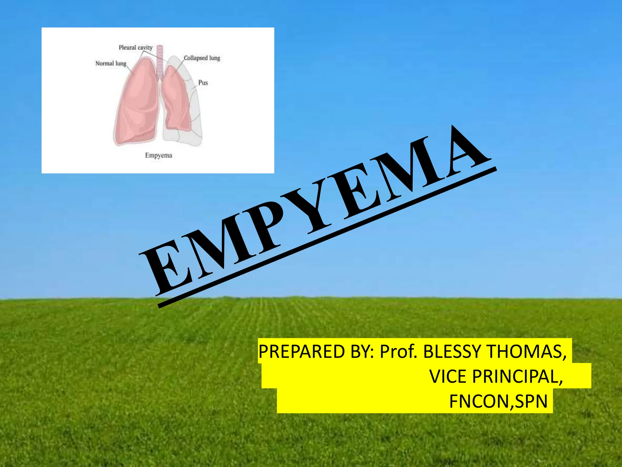
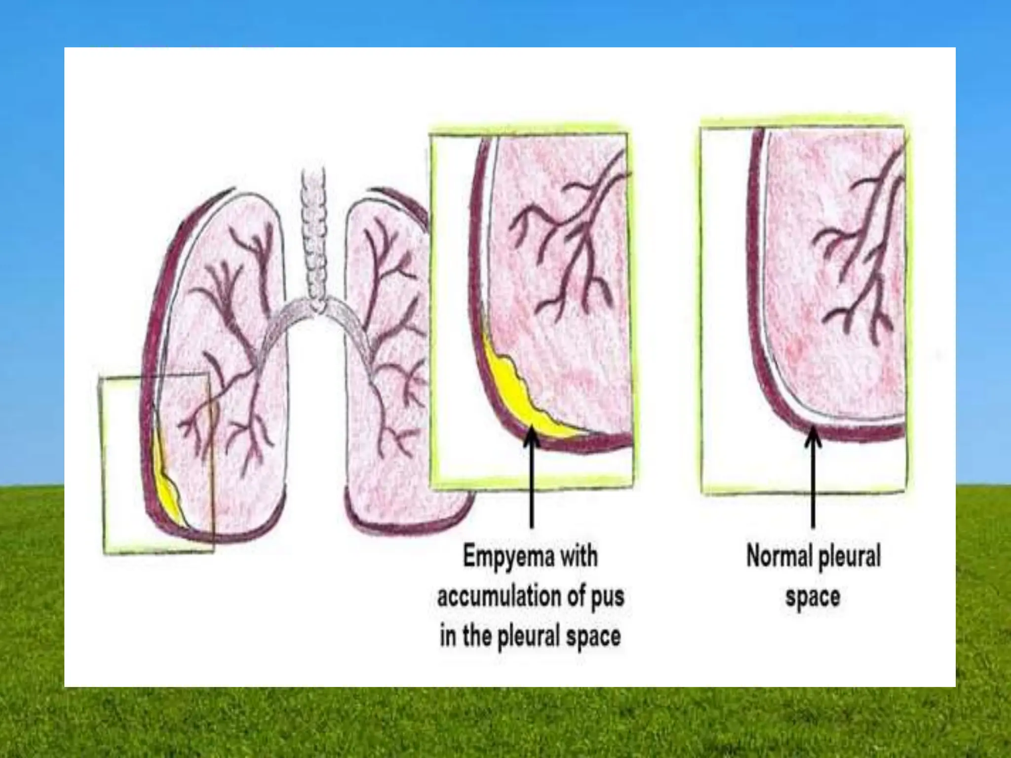
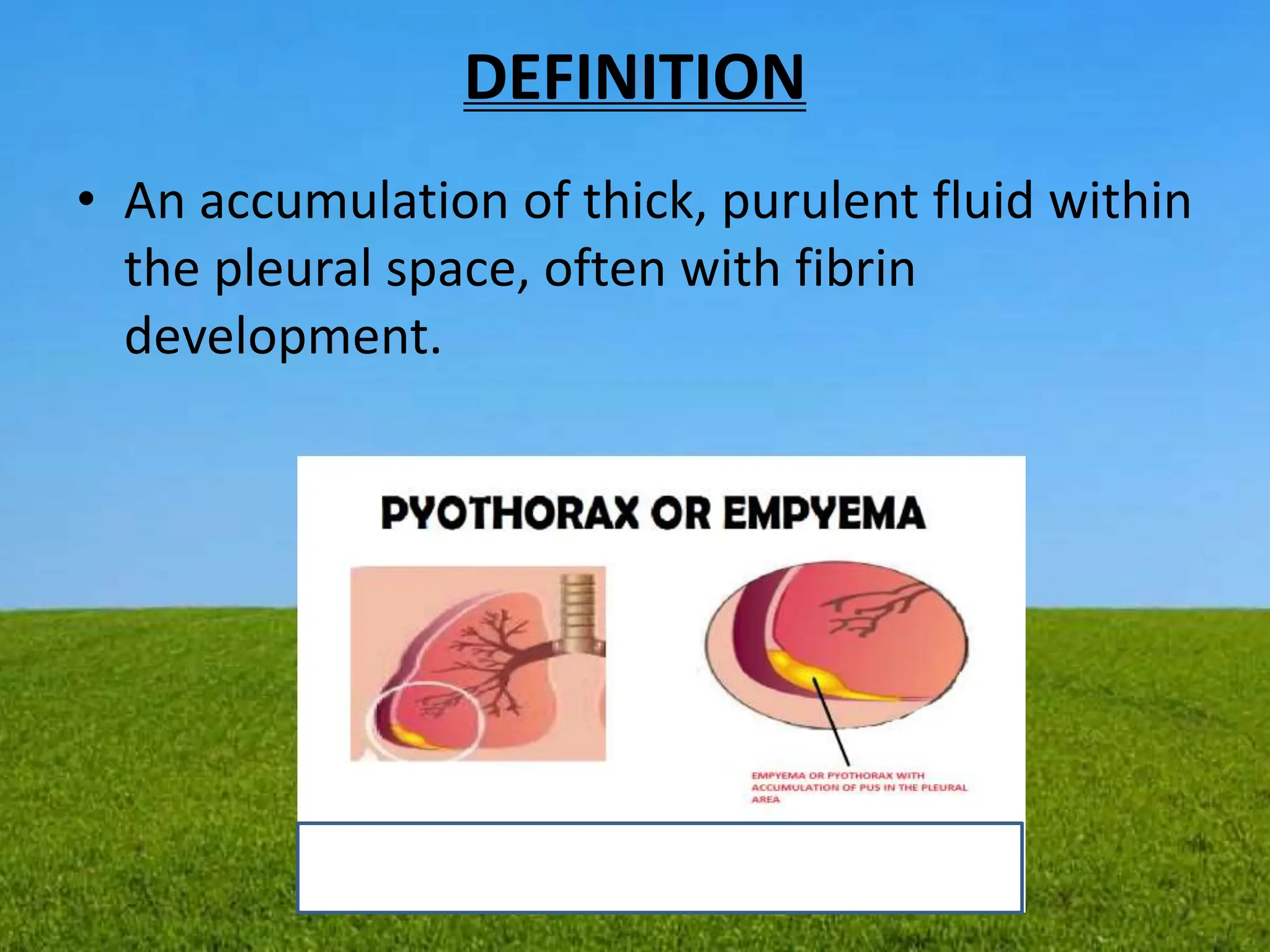
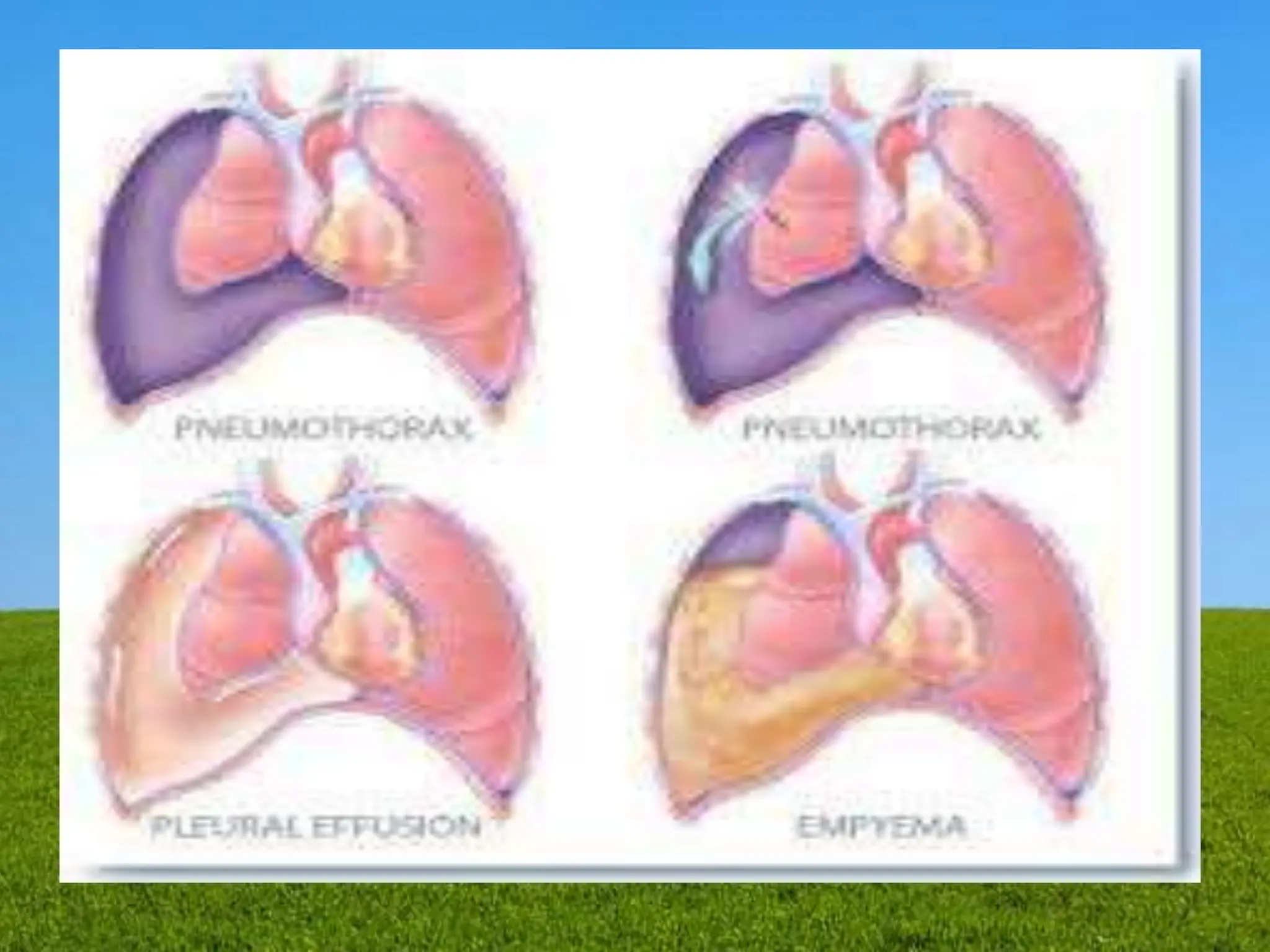
PREPARED BY: Prof. BLESSY THOMAS,
VICE PRINCIPAL,
FNCON,SPN
DEFINITION
• An accumulation of thick, purulent fluid within
the pleural space, often with fibrin
development.
• Empyema is also called pyothorax or purulent
pleuritis.
• It’s a condition in which pus gathers in the area
between the lungs and the inner surface of the
chest wall. This area is known as the pleural space.
• Pus is a fluid that’s filled with immune cells, dead
cells, and bacteria.
• Pus in the pleural space can’t be coughed out.
Instead, it needs to be drained by a needle or
surgery.
• Empyema usually develops after pneumonia,
which is an infection of the lung tissue.
CAUSES
• Causative organisms
• Staphylococcus
aureus,
• Streptococcus
pneumoniae
• Streptococcus
pyogenes
• Iatrogenic causes
• As a complication of
bacterial pneumonia,
lung abscess
• Penetrating chest
trauma.
• Non bacterial
infection.
• Bacterial infection.
• Chest trauma.
SIMPLE EMPYEMA
• Simple empyema occurs in the early stages of the
illness.
• A person has this type if the pus is free-flowing.
• shortness of breath
• dry cough
• fever
• sweating
• chest pain when breathing that may be described as
stabbing
• headache
• confusion
• loss of appetite
COMPLEX EMPYEMA
• Complex empyema occurs in the later stage of
the illness.
• In complex empyema, the inflammation is more
severe. Scar tissue may form and divide the chest
cavity into smaller cavities. This is called
loculation, and it’s more difficult to treat.
• If the infection continues to get worse, it can lead
to the formation of a thick peel over the pleura,
called a pleural peel.
• This peel prevents the lung from expanding.
• Surgery is required to fix it.
Other symptoms in complex Empyema
include:
• difficulty breathing
• decreased breath sounds
• weight loss
• chest pain
PATHOPHYSIOLOGY
Due to etiological factors.
Presence of Para pneumonic effusion.
Release of inflammatory mediators.
Increase permeability of the capillaries.
Attracts WBCs to the site.
Escape of albumin & others protein from capillaries
Increase amount of pleural fluid.
STAGE I
• Presence of free flowing protein rich pleural
fluid
• Inflammation worsens.
• Attract more WBG to the site.
• Extensive purulent exudate production.
STAGE II
• Initiation of fibroblastic activity.
STAGE III
• Adherence of the two pleural membranes
• Formation of pus
STAGES OF EMPYEMA
• Exudative stage (1-3 days )
• Fibrino purulentstage (4 to 14 days)
• Organizing stage (after 14 days)
Exudative stage (1-3 days)
• Immediate response with outpouring of the
fluid.
• It is simple parapneumonic effusion with
normal pH and glucose levels.
• Swelling in pleural space
• Fibrinous materials forms on both pleural
surface
Fibrino purulent stage (4 to 14 days)
• Large number of poly-morphonuclear
leukocytes and fibrin accumulates.
• Acumulation of neutrophills and fibrin,
effusion becomes purulent and viscous
leading to development of empyema.
Organizing stage (after 14 days)
• Fibro-blasts grow into exudates on both the
visceral and parietal pleural surfaces
• Development of an inelastic membrane "the
peel".
• Most common in S. aureus infection.
• Thickened pleural peel can restrict lung
movement and it is commonly termed as
trapped lung
Common Clinical Manifestations
• Chest pain –which worsens when taking a deep breath.
• Dry cough.
• Excessive sweating night sweats.
• Fever with chills.
• General discomfort.
• Uneasiness.
• Malaise
• Shortness of breath.
• Weight loss.
• Clubbing of nails.
• Dull percussion sounds.
• Blood in sputum.
• Coma.
DIAGNOSTIC MEASURES
• History Collection.
• Physical examination.
• X ray.
• CT Scan.
• Diagnostic thoracenthesis .
Chest X-rays and CT scans will show whether or not
there’s fluid in the pleural space.
An ultrasound of the chest will show the amount of
fluid and its exact location.
Blood tests can help check your white blood cell count,
look for the C-reactive protein, and identify the bacteria
causing the infection. White cell count can be elevated
when you have an infection.
During a thoracentesis, a needle is inserted through
the back of your ribcage into the pleural space to take a
sample of fluid. The fluid is then analyzed under a
microscope to look for bacteria, protein, and other cells.
MEDICAL MANAGEMENT
• Objectives – to the drain the pleural cavity to achieve full
expansion of lung.
• PHARMACOLOGICAL MANAGEMENT
• Antibiotics (10 -14 days)
• Cefotaxime (4-6 wks)
• Metronidazole 500 mg iv
• Benzyl pencillin 12gm iv
• Ciprofloxacin 400 mg iv
• Metronidazole 500 mg iv
• Meropenam 1 gm
• Amoxicillin + Clavulinic aid
• Metronidazole or clindamycin others drugs for symptomatic
management.
SURGICAL MANAGEMENT
• Thoracentesis -it is a procedure to remove fluid
from the space between the lining of the outside
of the lungs (pleura) and the wall of the chest
• Needle Aspiration – Thoracentesis – it the fluid
not too purulent or thick.
• Tube thoracostomy – Chest drainage using.
• A large – Diameters intercostals tube attached to
water seal drainage. In the fibrinolytic agents
installed through the chest tube in patient with
computers.
Open Chest Drainage Via
• Thoracotomy
• Rib resection
• Remove Thickened pleura pus & debris
• Removal of diseases part of lung tissue.
• In case of long standing inflammation
• Exudates will be formed over the lung and it
will interfere with normal expansion.
• Rx: - Removal of exudates surgically
• : - Drainage tube is left in place until the
pus cleared off completely.
Thoracotomy
• Thoracostomy: In this procedure, doctor will
insert a plastic tube into your chest between two
ribs. Then connect the tube to a suction device
and remove the fluid. Also inject medication to
help drain the fluid.
• Video-assisted thoracic surgery- surgeon will
remove the affected tissue around the lung and
then insert a drainage tube or use medication to
remove the fluid. They will create three small
incisions and use a tiny camera called a
thoracoscope for this process.
• Open decortication: In this surgery, your surgeon
will peel away the pleural peel.
• Stoma
• A chest drain isn't suitable for all patients. Some
will instead opt to have an opening made in their
chest, known as a stoma.
• A special bag is placed over the stoma to collect
the fluid that leaks from the empyema.
• This is worn on the body, and may be more
discreet and interfere less with your lifestyle than
a chest drain.
• But with modern treatments, getting a stoma is
uncommon.
NURSING MANAGEMENT
• Lung expanding & creating excursive
• Follow up care
• Teaching family and patient about the care of the drainage
tube.
• Drainage tube care
– Check the position of tube.
– Measurement of drainage.
– Color of drainage to check for any complications.
• NURSING DIAGNOSIS
• Risk for Activity Intolerance r/t hypoxia secondary to
Empyema.
• Acute Pain r/t infection of the pleura.
• Impaired Gas Exchange r/t compressed lung.
Complications
• In rare instances, a case of complex empyema can lead to
more severe complications. These include sepsis and a
collapsed lung, also called a pneumothorax. The symptoms
of sepsis include:
• high fever
• chills
• rapid breathing
• fast heart rate
• low blood pressure
• A collapsed lung can cause sudden, sharp chest pain and
shortness of breath that gets worse when coughing or
breathing.
nursing management of patient with Empyema ppt

nursing management of patient with Empyema ppt

  • 1.
    PREPARED BY: Prof.BLESSY THOMAS, VICE PRINCIPAL, FNCON,SPN
  • 3.
    DEFINITION • An accumulationof thick, purulent fluid within the pleural space, often with fibrin development.
  • 4.
    • Empyema isalso called pyothorax or purulent pleuritis. • It’s a condition in which pus gathers in the area between the lungs and the inner surface of the chest wall. This area is known as the pleural space. • Pus is a fluid that’s filled with immune cells, dead cells, and bacteria. • Pus in the pleural space can’t be coughed out. Instead, it needs to be drained by a needle or surgery. • Empyema usually develops after pneumonia, which is an infection of the lung tissue.
  • 6.
    CAUSES • Causative organisms •Staphylococcus aureus, • Streptococcus pneumoniae • Streptococcus pyogenes • Iatrogenic causes • As a complication of bacterial pneumonia, lung abscess • Penetrating chest trauma. • Non bacterial infection. • Bacterial infection. • Chest trauma.
  • 7.
    SIMPLE EMPYEMA • Simpleempyema occurs in the early stages of the illness. • A person has this type if the pus is free-flowing. • shortness of breath • dry cough • fever • sweating • chest pain when breathing that may be described as stabbing • headache • confusion • loss of appetite
  • 8.
    COMPLEX EMPYEMA • Complexempyema occurs in the later stage of the illness. • In complex empyema, the inflammation is more severe. Scar tissue may form and divide the chest cavity into smaller cavities. This is called loculation, and it’s more difficult to treat. • If the infection continues to get worse, it can lead to the formation of a thick peel over the pleura, called a pleural peel. • This peel prevents the lung from expanding. • Surgery is required to fix it.
  • 9.
    Other symptoms incomplex Empyema include: • difficulty breathing • decreased breath sounds • weight loss • chest pain
  • 10.
    PATHOPHYSIOLOGY Due to etiologicalfactors. Presence of Para pneumonic effusion. Release of inflammatory mediators. Increase permeability of the capillaries. Attracts WBCs to the site. Escape of albumin & others protein from capillaries Increase amount of pleural fluid.
  • 11.
    STAGE I • Presenceof free flowing protein rich pleural fluid • Inflammation worsens. • Attract more WBG to the site. • Extensive purulent exudate production.
  • 12.
    STAGE II • Initiationof fibroblastic activity. STAGE III • Adherence of the two pleural membranes • Formation of pus
  • 13.
    STAGES OF EMPYEMA •Exudative stage (1-3 days ) • Fibrino purulentstage (4 to 14 days) • Organizing stage (after 14 days)
  • 14.
    Exudative stage (1-3days) • Immediate response with outpouring of the fluid. • It is simple parapneumonic effusion with normal pH and glucose levels. • Swelling in pleural space • Fibrinous materials forms on both pleural surface
  • 15.
    Fibrino purulent stage(4 to 14 days) • Large number of poly-morphonuclear leukocytes and fibrin accumulates. • Acumulation of neutrophills and fibrin, effusion becomes purulent and viscous leading to development of empyema.
  • 16.
    Organizing stage (after14 days) • Fibro-blasts grow into exudates on both the visceral and parietal pleural surfaces • Development of an inelastic membrane "the peel". • Most common in S. aureus infection. • Thickened pleural peel can restrict lung movement and it is commonly termed as trapped lung
  • 17.
    Common Clinical Manifestations •Chest pain –which worsens when taking a deep breath. • Dry cough. • Excessive sweating night sweats. • Fever with chills. • General discomfort. • Uneasiness. • Malaise • Shortness of breath. • Weight loss. • Clubbing of nails. • Dull percussion sounds. • Blood in sputum. • Coma.
  • 18.
    DIAGNOSTIC MEASURES • HistoryCollection. • Physical examination. • X ray. • CT Scan. • Diagnostic thoracenthesis .
  • 19.
    Chest X-rays andCT scans will show whether or not there’s fluid in the pleural space. An ultrasound of the chest will show the amount of fluid and its exact location. Blood tests can help check your white blood cell count, look for the C-reactive protein, and identify the bacteria causing the infection. White cell count can be elevated when you have an infection. During a thoracentesis, a needle is inserted through the back of your ribcage into the pleural space to take a sample of fluid. The fluid is then analyzed under a microscope to look for bacteria, protein, and other cells.
  • 20.
    MEDICAL MANAGEMENT • Objectives– to the drain the pleural cavity to achieve full expansion of lung. • PHARMACOLOGICAL MANAGEMENT • Antibiotics (10 -14 days) • Cefotaxime (4-6 wks) • Metronidazole 500 mg iv • Benzyl pencillin 12gm iv • Ciprofloxacin 400 mg iv • Metronidazole 500 mg iv • Meropenam 1 gm • Amoxicillin + Clavulinic aid • Metronidazole or clindamycin others drugs for symptomatic management.
  • 21.
    SURGICAL MANAGEMENT • Thoracentesis-it is a procedure to remove fluid from the space between the lining of the outside of the lungs (pleura) and the wall of the chest • Needle Aspiration – Thoracentesis – it the fluid not too purulent or thick. • Tube thoracostomy – Chest drainage using. • A large – Diameters intercostals tube attached to water seal drainage. In the fibrinolytic agents installed through the chest tube in patient with computers.
  • 22.
    Open Chest DrainageVia • Thoracotomy • Rib resection • Remove Thickened pleura pus & debris • Removal of diseases part of lung tissue. • In case of long standing inflammation • Exudates will be formed over the lung and it will interfere with normal expansion. • Rx: - Removal of exudates surgically • : - Drainage tube is left in place until the pus cleared off completely.
  • 23.
  • 26.
    • Thoracostomy: Inthis procedure, doctor will insert a plastic tube into your chest between two ribs. Then connect the tube to a suction device and remove the fluid. Also inject medication to help drain the fluid. • Video-assisted thoracic surgery- surgeon will remove the affected tissue around the lung and then insert a drainage tube or use medication to remove the fluid. They will create three small incisions and use a tiny camera called a thoracoscope for this process. • Open decortication: In this surgery, your surgeon will peel away the pleural peel.
  • 27.
    • Stoma • Achest drain isn't suitable for all patients. Some will instead opt to have an opening made in their chest, known as a stoma. • A special bag is placed over the stoma to collect the fluid that leaks from the empyema. • This is worn on the body, and may be more discreet and interfere less with your lifestyle than a chest drain. • But with modern treatments, getting a stoma is uncommon.
  • 30.
    NURSING MANAGEMENT • Lungexpanding & creating excursive • Follow up care • Teaching family and patient about the care of the drainage tube. • Drainage tube care – Check the position of tube. – Measurement of drainage. – Color of drainage to check for any complications. • NURSING DIAGNOSIS • Risk for Activity Intolerance r/t hypoxia secondary to Empyema. • Acute Pain r/t infection of the pleura. • Impaired Gas Exchange r/t compressed lung.
  • 31.
    Complications • In rareinstances, a case of complex empyema can lead to more severe complications. These include sepsis and a collapsed lung, also called a pneumothorax. The symptoms of sepsis include: • high fever • chills • rapid breathing • fast heart rate • low blood pressure • A collapsed lung can cause sudden, sharp chest pain and shortness of breath that gets worse when coughing or breathing.