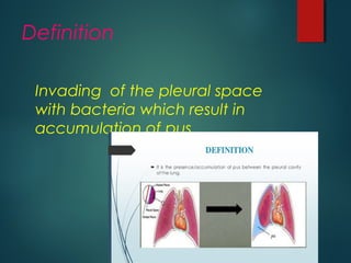
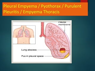
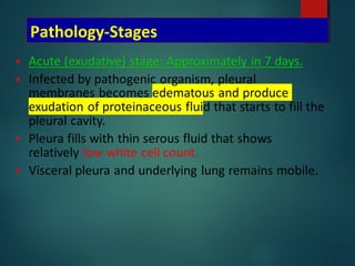
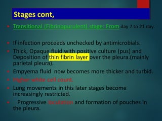
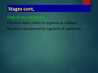
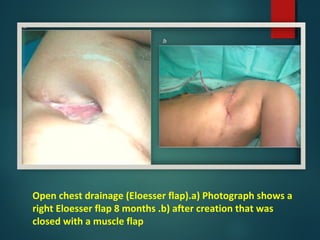
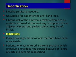
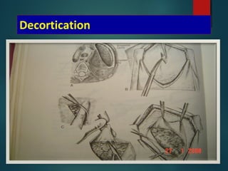
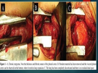
EMPYEMA
Mohammad Tailakh
Contents

Definition

Etiology

Stages

Symptoms & signs

Complications

Investigations

Management
Definition
Invading of the pleural space
with bacteria which result in
accumulation of pus .
Pleural Empyema / Pyothorax / Purulent
Pleuritis / Empyema Thoracis
Etiology (Introduction of infection)
EMPYEMA
THORACIC
SEPSIS
EXTRATHORACIC
SEPSIS IATROGENIC
NON-
IATROGENIC
SUBPHRENIC
ABSCESS,
HEPATIC
ABSCESS
MEDIASTINITIS
PULMONARY
DISEASE
OSTEOMYELI
TIS
LUNG
RESECTION,
OESOPHAGEAL
TEARS,
PARACETESIS
THORACIS,
LIVER BIOPSY
STABBINGS,G
UNSHOT
WOUNDS,ETC
PNEUMONIA, TB,
BRONCHIECTASIS
,LUNG ABCESS
STERNUM,
VERTEBRAE,
RIBS
etiology
 PARAPNEUMONIC( secondary to a
pneumonia)the most common
 Post trauma.
 Post surgery(esophageal or pulmonary(
 Subphrenic Abscess
Bacteriological data.
 Streptococcus pneumoniae: most common
Increased resistance
 Staphylococcus:15-30%
 Streptococcus spp
 Gram Negative: 20-50%
Klebsiella, Enterobacter,
Pseudomonas, Hemophilus, E.Coli
 Anaerobes:
Fusobacterium, Bacteroides fragilis
Influence of predisposing factors
 In adults – empyema arises as a complication of
CAP,often pneumococcal.
 Most common empyema in children post-pneumonia
parcent 80% ,adult 20%.
 Aerobic gram negative bacilli infection likely to affect
pleura – from below diaphragm or as a result of
oesophageal instrumentation.
 Mycobacteria and fungi more common in
immunocompromised.
Uncommon microbial causes
 Tuberculous
 Fungal – Aspergillous,Cryptococcus,Blastomyces,
Histoplasmosis.
 Actinomyces – aerobic gram negative filamentous
bacteria.
 Clostridia – anaerobic organism.
 Hydatid disease – Echinococcus.
 Lung fluke – Paragonimus westermani.
 Protozoa – Trichomonas,Entamoeba histolytica.
Pathology-Stages
Stages cont,
Stages cont,
 Stage of vascularization:
 Fibrinous layers starts to organize as collagen.
 Becomes vascularized by ingrowth of capillaries.
Stages cont,
 Organizing (chronic) Stage: after 21 days.
 Usually 4-6 weeks.
 Empyema cavity becomes surrounded by a cortex.
 Contains pus.
 Inner layers shows inflammatory cells.
 Outer layers gets fibrous – exerts restrictive effect.
 Compressing the underlying lung (trapped lung
effect).
 Draws the ribs together producing chest deformity.
 Later on gets calcified – fibrothorax.
RISK FACTORS
 alcoholism.
drug use.
 HIV infection.
 neoplasm .
pre-existent pulmonary disease
.
Symptoms & signs
 Depends on nature of infecting organism
 competence of patients immune system.
 Ranges from complete absence of symptoms to a severe
illness with all usual manifestations of systemic toxicity.
 Fever
 Cough & Expectoration.
 Pleuretic chest pain.
 Dyspnoea
 Easy fatiguability.
 Loss of weight.
 Night sweating.
Signs of pleural effusion.
Finger clubbing.
Complications
 Rupture into the lung; BronchoPleural
fistula.
 Spread to the subcutaneous tissue;
Empyema necessitans.
 Septicaemia & septic shock.
Diagnosis
 LRTI)lower respiratory tract infection) – possibility of
complicating empyema.
 History and physical findings may be suggestive.
 CXR,USG(ultrasonogrophy),CT.
 Thoracentesis- PH < 7.4
Glucose <40 mg/dl
LDH> 1000 iu/dl
Protein > 2.5 gm/dl
Sp.gravity >1.018
 Other findings (non specific):neutrophil leucocytosis
Chest x ray
 In early stages same as
uncomplicated pleural
effusion.
 As time passes, fibrosis
develops around empyema
cavity.
 Fluid contained in one
location.
 Air fluid level
 Homogenous shadow
extending upwards.
Empyema Ct scan
empyema
ultrasonography
Thoracentesis:PH < 7.4
Glucose <40 mg/dl
LDH> 1000 iu/dl
Protein > 2.5 gm/dl
Sp.gravity >1.018
Lateral cxr
 opacity convex anteriorly.
Tapering at its upper and
lower ends
Extending into the thorax.
D – shaped shadow.
Pleural Empyema
Management
Goals of the treatment
 Treat the infection.
 Drain the purulent effusion adequately and
completely.
 Re-expand the lung to fill the pleural space.
 Eliminate complications and avoid chronicity.
Antimicrobial Therapy
 Choice of antibiotic – microbiological C/S testing.
 Anaerobes- may be treated with Benzylpenicillin.
 If resistant – add metronidazole.
 Better response – Clindamycin + Penicillin ( active
against Bacteroids fragilis and other penicillin-
resistant anaerobes
 Pneumococcus
 Responds to high dose benzylpenicillin
initially,continuing with oral phenoxy methyl
penicillin(penicillin V) or amoxycillin.
 Alternatives for penicillin allergic individuals- Cefradin or
Clarithromycin.
 Staphylococcus aureus
 Dicloxacillin,oxacillin for parenteral use.
 First generation cephalosporins – cefradine.
 MRSA- vancomycin,Linezolid.
 Gram negative aerobes
 Serious aerobic infections may be treated with the
combination of a third generation cephalosporin –
Ceftazidime and an amynoglycoside such as gentamycin.
 Mixed infection,including anaerobes – piperacillin.
 Adults with empyema who are admitted from the
community, and in whom infecting organism have not
yet been identified may be treated initially with a
combination that includes co-amoxyclav,metronidazole
and flucloxacillin.
 This regimen is modified in the light of cultures and the
patients clinical response.
 Duration of therapy is likely to be several weeks.
 It can be continued for at least 3 weeks after all
drainage has ceased.
BTS guidelines for the management of empyemaOrigin of infection Intravenous antibiotic
treatment
Oral antibiotic treatment
Community acquired culture
negative pleural infection
Cefuroxime 1.5 g tds iv +
metronidazole 400 mg tds
orally or 500 mg tds iv
Amoxycillin 1 g tds +
clavulanic acid 125 mg tds
Benzyl penicillin 1.2 g qds iv
+ ciprofloxacin 400 mg bd iv
Amoxycillin 1 g tds +
metronidazole 400 mg tds
Meropenem 1 g tds iv +
metronidazole 400 mg tds
orally or 500 mg tds iv
Clindamycin 300 mg qds
Hospital acquired culture
negative pleural infection
Piperacillin + tazobactam 4.5
g qds iv
Not applicable
Ceftazidime 2 g tds iv,
Meropenem 1 g tds iv ±
metronidazole 400 mg tds
orally or 500 mg tds iv
Tuberculous Empyema
 Rare entity.
 Purulent fluid loaded with tuberculous organisms.
 Usually develops in fibrous scar tissue resulting from
pleurisy, artificial pneumothorax or thoracoplasty.
 Underlying pleura is heavily calcified.
 Sub acute or chronic illness
 Fatigue, low grade fever and weight loss.
 Radigraphically – obvious pleural effusion, pleural
thickening.
 CT scan – thick calcified pleural rind and rib thickening
surrounded by loculated pleural fluid.
Tuberculous Empyema
 Diagnosis – thoracentesis, AFB smear and
culture.
 Treatment – intensive chemotherapy coupled
with serial thoracentesis can be curative at
times.
 Multiple drug regimen at their maximal tolerated
dosages.
 Strong tendency to develop resistant organisms.
 ATT frequently do not reach there normal levels
in the pleural space owing to the thick, fibrous
and often calcified pleura.
 VATS/Decortication.
Primary treatment options
 Antibiotics alone;
 Recurrent thoracocentesis
 Insertion of chest drain alone or in combination with fibrinolytics
 VATS.
 Open decortication
Thoracocenthesis
 Big caliber needle.
 Repeated aspiration is carried out.
 Use of Abrams punch biopsy needle
is useful initially. Wide callibre
allow easy aspiration and also
permits pleural biopsy.
 Mostly diagnosis technique
 Therapeutically used if the liquid
remains fluid
 Helps in pleural lavage also.
Chest Tube
 Closed tube thoracostomy.
 As soon as the fluid is thick.
 Localization
 loculated: Chest imaging using
ultrasonography and/or computed
tomography
 Size: 20 - 28 F
 Passed under USG guidance,helps in
breaking fibrinous septa and pus
rapidly gets removed
 Bedside
Pleural Lavage
 Isotonic saline
 +/- Noxyflex (noxytioline)
 Modalités
 3 way stopcock
 Directly through the CT: 250 to 500 ml
 Cautiously if suspicion of broncho-pleural
fistula
 Timing:
 Immediately after CT placement+++
 Once a day until the liquid is clear
Fibrinolytics
Intrapleural Streptokinase;
 Indications
 Acute or fibrino purulent stage
 Presence of loculations.
 Incomplete drainage after tube insertion
 Contraindications:
 Chronic stage
 Post-operative empyema
 Empyema with BPF.
Fibrinolytics
 Was reported in 1949.
 Then was abandoned due to allergic reactions,but taken up
again due to availability of purer forms of
streptokinase,urokinase.
 (Davies RJO,Trail ZC Thorax 1997; 52:416.)
 Urokinase: 100 000 or 300 000 IU .
 Streptokinase: 250000 IU .
 250.000 IU in 10-20 ml isotonic saline.
 Don’t evacuate before 24 to 48 hours.
 Constantly associated with fever (38-39°C).
 Then evacuate.
 Local antibiotics
 Intrapleural instillation of antibiotics, especially
metronidazole,Colimycin.
 Still debated.
 Do not replace systemic treatment.
Video-assisted thoracic surgery
 VATS.
 If closed drainage does not result in
prompt re-expansion of the lung and
especially if loculi have been identified
by USG.
 Decision to intervene early is made.
 Debridement and drainage.
 Breakage of loculi,evacuating pus,debris
and freeing lung.
 Helps in re expansion of lung.
Compare Chest Tube + Streptokinase
(n=9) vs VATS (n=11)
Wait et al, Chest 1997
Bronchoscopy
 Recommended following the successful conclusion
of closed drainage.
 In order to exclude any endobronchial causes of
obstruction, such as tumour or foreign body.
Open drainage
 If empyema persists both clinically and radiologically.
 In whom closed drainage has proved unsuccessful.
 If VATS unavailable, unsuccessful or considered
inappropriate.
 Rib Resection Drainage.
 Eloesser Flap .
Open chest drainage (Eloesser flap).a) Photograph shows a
right Eloesser flap 8 months .b) after creation that was
closed with a muscle flap
Decortication
 Elective surgical procedure.
 Unsuitable for patients who are ill and toxic.
 Fibrous wall of the empyema cavity,reffered to as
cortex is exposed at thoracotomy is stripped off and
adjacent visceral and parietal pleura may be left
intact.
 Indications
 Closed drainage/thoracoscopic methods have been
unsuccessful.
 Patients who has entered a chronic phase in which
underlying lung does not expand because of failure
of cortex to become reabsorbed.
 There is no optimal time for decortication.
 Some surgeons arguing for early intervention and
others opting for a conservative approach.
 Early surgical intervention in pleural empyema.thorac cardiovascular surg
1985.
Decortication
Indications
Thoracocentesis
Clear liquid Not clear or purulent effusion
pH>7.20 pH<7.20
No intervention
Not loculated Loculated
Drainage
Pleural lavage
Fibrinolytics 24-48h
Drainage
Fibrinolytics
Pleural lavage
VATS
Drainage
Pleural lavage
Failure
VATS
Surgery
Failure
Surgery
Reccurent
thoracocentesis
Hamm et al, ERJ 1997
Empyema presentation

Empyema presentation

  • 1.
  • 2.
  • 3.
    Definition Invading of thepleural space with bacteria which result in accumulation of pus .
  • 4.
    Pleural Empyema /Pyothorax / Purulent Pleuritis / Empyema Thoracis
  • 5.
    Etiology (Introduction ofinfection) EMPYEMA THORACIC SEPSIS EXTRATHORACIC SEPSIS IATROGENIC NON- IATROGENIC SUBPHRENIC ABSCESS, HEPATIC ABSCESS MEDIASTINITIS PULMONARY DISEASE OSTEOMYELI TIS LUNG RESECTION, OESOPHAGEAL TEARS, PARACETESIS THORACIS, LIVER BIOPSY STABBINGS,G UNSHOT WOUNDS,ETC PNEUMONIA, TB, BRONCHIECTASIS ,LUNG ABCESS STERNUM, VERTEBRAE, RIBS
  • 6.
    etiology  PARAPNEUMONIC( secondaryto a pneumonia)the most common  Post trauma.  Post surgery(esophageal or pulmonary(  Subphrenic Abscess
  • 7.
    Bacteriological data.  Streptococcuspneumoniae: most common Increased resistance  Staphylococcus:15-30%  Streptococcus spp  Gram Negative: 20-50% Klebsiella, Enterobacter, Pseudomonas, Hemophilus, E.Coli  Anaerobes: Fusobacterium, Bacteroides fragilis
  • 8.
    Influence of predisposingfactors  In adults – empyema arises as a complication of CAP,often pneumococcal.  Most common empyema in children post-pneumonia parcent 80% ,adult 20%.  Aerobic gram negative bacilli infection likely to affect pleura – from below diaphragm or as a result of oesophageal instrumentation.  Mycobacteria and fungi more common in immunocompromised.
  • 9.
    Uncommon microbial causes Tuberculous  Fungal – Aspergillous,Cryptococcus,Blastomyces, Histoplasmosis.  Actinomyces – aerobic gram negative filamentous bacteria.  Clostridia – anaerobic organism.  Hydatid disease – Echinococcus.  Lung fluke – Paragonimus westermani.  Protozoa – Trichomonas,Entamoeba histolytica.
  • 10.
  • 11.
  • 12.
    Stages cont,  Stageof vascularization:  Fibrinous layers starts to organize as collagen.  Becomes vascularized by ingrowth of capillaries.
  • 13.
    Stages cont,  Organizing(chronic) Stage: after 21 days.  Usually 4-6 weeks.  Empyema cavity becomes surrounded by a cortex.  Contains pus.  Inner layers shows inflammatory cells.  Outer layers gets fibrous – exerts restrictive effect.  Compressing the underlying lung (trapped lung effect).  Draws the ribs together producing chest deformity.  Later on gets calcified – fibrothorax.
  • 14.
    RISK FACTORS  alcoholism. drug use.  HIVinfection.  neoplasm . pre-existent pulmonary disease .
  • 15.
    Symptoms & signs Depends on nature of infecting organism  competence of patients immune system.  Ranges from complete absence of symptoms to a severe illness with all usual manifestations of systemic toxicity.  Fever  Cough & Expectoration.  Pleuretic chest pain.  Dyspnoea  Easy fatiguability.  Loss of weight.  Night sweating.
  • 16.
    Signs of pleuraleffusion. Finger clubbing.
  • 17.
    Complications  Rupture intothe lung; BronchoPleural fistula.  Spread to the subcutaneous tissue; Empyema necessitans.  Septicaemia & septic shock.
  • 18.
    Diagnosis  LRTI)lower respiratorytract infection) – possibility of complicating empyema.  History and physical findings may be suggestive.  CXR,USG(ultrasonogrophy),CT.  Thoracentesis- PH < 7.4 Glucose <40 mg/dl LDH> 1000 iu/dl Protein > 2.5 gm/dl Sp.gravity >1.018  Other findings (non specific):neutrophil leucocytosis
  • 19.
    Chest x ray In early stages same as uncomplicated pleural effusion.  As time passes, fibrosis develops around empyema cavity.  Fluid contained in one location.  Air fluid level  Homogenous shadow extending upwards.
  • 20.
  • 21.
  • 22.
    Thoracentesis:PH < 7.4 Glucose<40 mg/dl LDH> 1000 iu/dl Protein > 2.5 gm/dl Sp.gravity >1.018
  • 23.
    Lateral cxr  opacityconvex anteriorly. Tapering at its upper and lower ends Extending into the thorax. D – shaped shadow.
  • 26.
  • 27.
    Goals of thetreatment  Treat the infection.  Drain the purulent effusion adequately and completely.  Re-expand the lung to fill the pleural space.  Eliminate complications and avoid chronicity.
  • 28.
    Antimicrobial Therapy  Choiceof antibiotic – microbiological C/S testing.  Anaerobes- may be treated with Benzylpenicillin.  If resistant – add metronidazole.  Better response – Clindamycin + Penicillin ( active against Bacteroids fragilis and other penicillin- resistant anaerobes
  • 29.
     Pneumococcus  Respondsto high dose benzylpenicillin initially,continuing with oral phenoxy methyl penicillin(penicillin V) or amoxycillin.  Alternatives for penicillin allergic individuals- Cefradin or Clarithromycin.
  • 30.
     Staphylococcus aureus Dicloxacillin,oxacillin for parenteral use.  First generation cephalosporins – cefradine.  MRSA- vancomycin,Linezolid.  Gram negative aerobes  Serious aerobic infections may be treated with the combination of a third generation cephalosporin – Ceftazidime and an amynoglycoside such as gentamycin.  Mixed infection,including anaerobes – piperacillin.
  • 31.
     Adults withempyema who are admitted from the community, and in whom infecting organism have not yet been identified may be treated initially with a combination that includes co-amoxyclav,metronidazole and flucloxacillin.  This regimen is modified in the light of cultures and the patients clinical response.  Duration of therapy is likely to be several weeks.  It can be continued for at least 3 weeks after all drainage has ceased.
  • 32.
    BTS guidelines forthe management of empyemaOrigin of infection Intravenous antibiotic treatment Oral antibiotic treatment Community acquired culture negative pleural infection Cefuroxime 1.5 g tds iv + metronidazole 400 mg tds orally or 500 mg tds iv Amoxycillin 1 g tds + clavulanic acid 125 mg tds Benzyl penicillin 1.2 g qds iv + ciprofloxacin 400 mg bd iv Amoxycillin 1 g tds + metronidazole 400 mg tds Meropenem 1 g tds iv + metronidazole 400 mg tds orally or 500 mg tds iv Clindamycin 300 mg qds Hospital acquired culture negative pleural infection Piperacillin + tazobactam 4.5 g qds iv Not applicable Ceftazidime 2 g tds iv, Meropenem 1 g tds iv ± metronidazole 400 mg tds orally or 500 mg tds iv
  • 33.
    Tuberculous Empyema  Rareentity.  Purulent fluid loaded with tuberculous organisms.  Usually develops in fibrous scar tissue resulting from pleurisy, artificial pneumothorax or thoracoplasty.  Underlying pleura is heavily calcified.  Sub acute or chronic illness  Fatigue, low grade fever and weight loss.  Radigraphically – obvious pleural effusion, pleural thickening.  CT scan – thick calcified pleural rind and rib thickening surrounded by loculated pleural fluid.
  • 34.
    Tuberculous Empyema  Diagnosis– thoracentesis, AFB smear and culture.  Treatment – intensive chemotherapy coupled with serial thoracentesis can be curative at times.  Multiple drug regimen at their maximal tolerated dosages.  Strong tendency to develop resistant organisms.  ATT frequently do not reach there normal levels in the pleural space owing to the thick, fibrous and often calcified pleura.  VATS/Decortication.
  • 35.
    Primary treatment options Antibiotics alone;  Recurrent thoracocentesis  Insertion of chest drain alone or in combination with fibrinolytics  VATS.  Open decortication
  • 36.
    Thoracocenthesis  Big caliberneedle.  Repeated aspiration is carried out.  Use of Abrams punch biopsy needle is useful initially. Wide callibre allow easy aspiration and also permits pleural biopsy.  Mostly diagnosis technique  Therapeutically used if the liquid remains fluid  Helps in pleural lavage also.
  • 37.
    Chest Tube  Closedtube thoracostomy.  As soon as the fluid is thick.  Localization  loculated: Chest imaging using ultrasonography and/or computed tomography  Size: 20 - 28 F  Passed under USG guidance,helps in breaking fibrinous septa and pus rapidly gets removed  Bedside
  • 38.
    Pleural Lavage  Isotonicsaline  +/- Noxyflex (noxytioline)  Modalités  3 way stopcock  Directly through the CT: 250 to 500 ml  Cautiously if suspicion of broncho-pleural fistula  Timing:  Immediately after CT placement+++  Once a day until the liquid is clear
  • 39.
    Fibrinolytics Intrapleural Streptokinase;  Indications Acute or fibrino purulent stage  Presence of loculations.  Incomplete drainage after tube insertion  Contraindications:  Chronic stage  Post-operative empyema  Empyema with BPF.
  • 40.
    Fibrinolytics  Was reportedin 1949.  Then was abandoned due to allergic reactions,but taken up again due to availability of purer forms of streptokinase,urokinase.  (Davies RJO,Trail ZC Thorax 1997; 52:416.)  Urokinase: 100 000 or 300 000 IU .  Streptokinase: 250000 IU .  250.000 IU in 10-20 ml isotonic saline.  Don’t evacuate before 24 to 48 hours.  Constantly associated with fever (38-39°C).  Then evacuate.
  • 41.
     Local antibiotics Intrapleural instillation of antibiotics, especially metronidazole,Colimycin.  Still debated.  Do not replace systemic treatment.
  • 42.
    Video-assisted thoracic surgery VATS.  If closed drainage does not result in prompt re-expansion of the lung and especially if loculi have been identified by USG.  Decision to intervene early is made.  Debridement and drainage.  Breakage of loculi,evacuating pus,debris and freeing lung.  Helps in re expansion of lung.
  • 43.
    Compare Chest Tube+ Streptokinase (n=9) vs VATS (n=11) Wait et al, Chest 1997
  • 44.
    Bronchoscopy  Recommended followingthe successful conclusion of closed drainage.  In order to exclude any endobronchial causes of obstruction, such as tumour or foreign body.
  • 45.
    Open drainage  Ifempyema persists both clinically and radiologically.  In whom closed drainage has proved unsuccessful.  If VATS unavailable, unsuccessful or considered inappropriate.  Rib Resection Drainage.  Eloesser Flap .
  • 46.
    Open chest drainage(Eloesser flap).a) Photograph shows a right Eloesser flap 8 months .b) after creation that was closed with a muscle flap
  • 47.
    Decortication  Elective surgicalprocedure.  Unsuitable for patients who are ill and toxic.  Fibrous wall of the empyema cavity,reffered to as cortex is exposed at thoracotomy is stripped off and adjacent visceral and parietal pleura may be left intact.  Indications  Closed drainage/thoracoscopic methods have been unsuccessful.  Patients who has entered a chronic phase in which underlying lung does not expand because of failure of cortex to become reabsorbed.
  • 48.
     There isno optimal time for decortication.  Some surgeons arguing for early intervention and others opting for a conservative approach.  Early surgical intervention in pleural empyema.thorac cardiovascular surg 1985.
  • 49.
  • 51.
    Indications Thoracocentesis Clear liquid Notclear or purulent effusion pH>7.20 pH<7.20 No intervention Not loculated Loculated Drainage Pleural lavage Fibrinolytics 24-48h Drainage Fibrinolytics Pleural lavage VATS Drainage Pleural lavage Failure VATS Surgery Failure Surgery Reccurent thoracocentesis Hamm et al, ERJ 1997