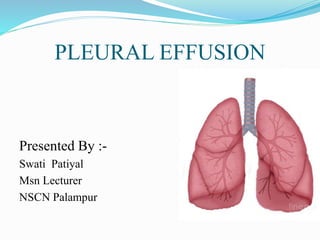
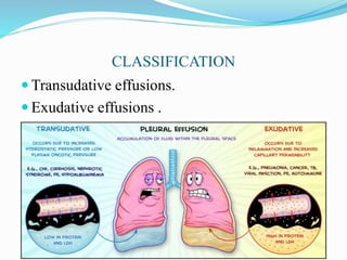
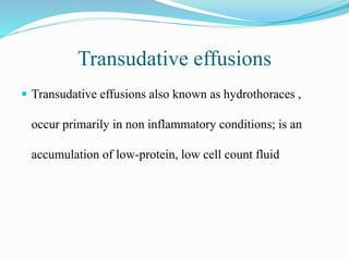
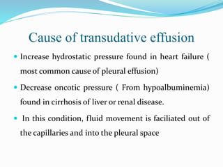
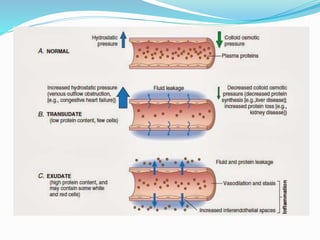
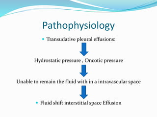
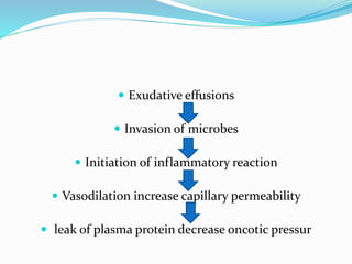
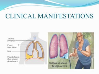
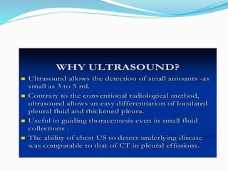
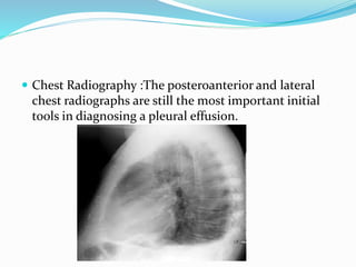
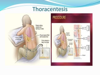
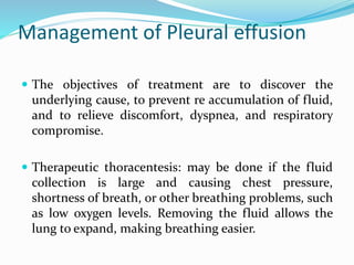
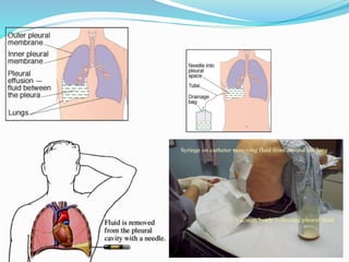
PLEURAL EFFUSION
Presented By :-
Swati Patiyal
Msn Lecturer
NSCN Palampur
Physiology of the Normal Lungs
The lungs are soft, spongy, cone shaped organs located
in the chest cavity.
They are separated by the mediastinum and the heart.
There are 3 lobes on the right lung and 2 lobes on the
left lung
 The lungs are supplied with blood via the pulmonary and
bronchial circulations.
• Pulmonary circulation: supplied from the pulmonary artery
and provides for gas exchange function of the lungs.
• Bronchial circulation: distributes blood to the conducting
airways and supporting structures of the lungs .
Layers of the lung
 • Parietal Pleura -Lines the thoracic cavity, including the
thoracic cage, mediastinum, and diaphragm.
 • Pleural space- Thin, transparent, serous membrane
which lines the thoracic cavity a potential space between
the parietal pleura and visceral pleura
 • Visceral pleura- Lines the entire surface of the lung.
PLEURA
Parietal pleura
cover the inner
surface of the
thoracic cavity,
including the
diaphragm, and ribs.
Visceral pleura
envelope all surfaces
of the lungs,
including the
interlobar fissures.
At the Hilum
where pulmonary vessels, bronchi, and
nerves enter the lung tissue, the parietal
pleura is continuous with the visceral pleura.
Introduction
 Pleural effusion, a collection of fluid in the pleural
space, is rarely a primary disease process but is
usually secondary to other diseases.
 The pleural space normally contains only about 10-20
ml of serous fluid.
Contd..
 Pleural fluid normally seeps continually into the
pleural space from the capillaries lining the parietal
pleura and is reabsorbed by the visceral pleural
capillaries and lymphatic system .
 Any condition that interferes with either secretion or
drainage of this fluid leads to pleural effusion
Definition
Pleural effusion is a collection of abnormal amount of
fluid in the pleural space.
OR
Presence of large amount of fluid in the pleural space
irrespective of the underlying causes
CLASSIFICATION
 Transudative effusions.
 Exudative effusions .
Transudative effusions
 Transudative effusions also known as hydrothoraces ,
occur primarily in non inflammatory conditions; is an
accumulation of low-protein, low cell count fluid
Cause of transudative effusion
 Increase hydrostatic pressure found in heart failure (
most common cause of pleural effusion)
 Decrease oncotic pressure ( From hypoalbuminemia)
found in cirrhosis of liver or renal disease.
 In this condition, fluid movement is faciliated out of
the capillaries and into the pleural space
Exudative effusions
 Exudative effusions occur in an area of inflammation;
is an accumulation of high-protein fluid.
CAUSE
 An exudative effusion results from increased capillary
permeability characteristic of inflammatory reaction.
 This types of effusion occurs secondary to conditions
such as pulmonary malignancies, pulmonary
infections and pulmonary embolization
Etiology
 Disseminated cancer (particularly lung and breast),
lymphoma
 Pleuro-pulmonary infections (pneumonia).
 Heart failure, cirrhosis, nephrotic syndrome
 Other conditions sarcoidosis, systemic lupus
erythematosus (SLE)
 Peritoneal dialysis
Pathophysiology
 Transudative pleural effusions:
Hydrostatic pressure , Oncotic pressure
Unable to remain the fluid with in a intravascular space
 Fluid shift interstitial space Effusion
 Exudative effusions
 Invasion of microbes
 Initiation of inflammatory reaction
 Vasodilation increase capillary permeability
 leak of plasma protein decrease oncotic pressur
CLINICAL MANIFESTATIONS
SYMPTOMS
 * key symptom -------> shortness of
breath
 Fluid filling the pleural space makes it hard
for the lungs to fully expand, causing the
patient to take many breaths so as to get
enough oxygen.
 .
* If parietal pleura is irritated -------> mild pain or
a sharp stabbing ,pleuritic type of pain
** Some patients will have a dry cough Occasionally
------> no symptoms at all.
 * This is more likely when the effusion results from:
 recent abdominal surgery, cancer, or tuberculosis.
Decreased or absent breath sounds
 Tapping on the chest will show stony dullness, and
decrease breath sound.
 Pneumonia causes fever, chills, and pleuritic chest
pain,
DIAGNOSTIC EVALUATION
 During a physical examination, the doctor will listen
to the sound of your breathing with a stethoscope and
may tap on your chest to listen for dullness.
 x ray
The fluid itself can be seen at the bottom of the lung
or lungs, hiding the normal lung structure.
If heart failure is present,
 the x-ray shadow of the heart will be enlarged

 Chest Radiography :The posteroanterior and lateral
chest radiographs are still the most important initial
tools in diagnosing a pleural effusion.
Computed Tomography: Cross-sectional computed
tomography (CT) It helps distinguish anatomic
compartments more clearly This modality is useful as
well in distinguishing empyema Normal Pleural effus
Thoracentesis
Management of Pleural effusion
 The objectives of treatment are to discover the
underlying cause, to prevent re accumulation of fluid,
and to relieve discomfort, dyspnea, and respiratory
compromise.
 Therapeutic thoracentesis: may be done if the fluid
collection is large and causing chest pressure,
shortness of breath, or other breathing problems, such
as low oxygen levels. Removing the fluid allows the
lung to expand, making breathing easier.
 General Treatment is aimed at underlying cause (heart
disease, infection).
 Thoracentesis is done to remove fluid, collect a
specimen, and relieve dyspnea
Complications
 Large effusion could lead to respiratory failure.
Nursing assessment
 Obtain history of previous pulmonary condition
 Assess patient for dyspnea and tachypnea
 Auscultate and percuss lungs for abnormalities
Nursing Intervention
 • 1. Ineffective breathing pattern related to decreased
lung expansion(accumulation of liquid), as evidenced
by dyspnea, changes in depth of breathing, accessory
muscle use.
 Interventions •
 Maintain a comfortable position is usually elevated
headboard
 • Given oxygen through a cannula (8mls)
 Assist with thoracentesis if indicated
 Maintain chest drainage as needed
 Provide care after pleurodesis.
 Monitor for excessive pain from the sclerosing agent, which
may cause hypoventilation.
 Administer prescribed analgesic.
 Assist patient undergoing instillation of intrapleural
lidocaine if pain relief is not forthcoming
 Administer oxygen as indicated by dyspnea and
hypoxemia.
 Observe patient's breathing pattern, oxygen saturation
 2. Acute Pain related to accumulation of fluid in the pleural
space and rubbing of thoracostomy tube to the lungs
 • Interventions • -
 The presence of pain, the scale and intensity of pain was
well assessed
• -The client taught about pain management and relaxation
with distraction
• -Chest tube secured to restrict movement and avoid
irritation
• -Given prescribed analgesics i.e diclofenac 75mg. 3
 3. Risk for nutrition impariment, less than body
requirement related to inability to ingest adequate
nutrients
 Interventions
 • -Patient relative i.e his father encouraged to give him
energy reaching food stuff together with energy
supplement so that he can get enough energy.
 • -Administer DNS as prescribed to the patient to
increase energy lost.
 4. Risk for fluid volume deficit related to chest tube
drainage.
 • Interventions
• -encourage the patient to drink enough water to
supplement the one lost by chest tube drainage
• -IV fluids & DNS to replace fluid lost in drainage
system monitored in 24hour
 5. Risk for infection related to the presence of fluid in
the pleural space and the incision site
 . • Interventions
 • -The patient dressed at the incision site when it is
wetted, probably after 2 to 3 days
• -Given antibiotics as prescribed i.e IV metronidazole
500mg 8 hourly, IV ceftiaxone 1gm.
References
 Chintamani, Lewis, Heitkemper, Dirksen, O’Brien and
Bucher. (2011). Lewis’s Medical Surgical Nursing:
Assessment and Management of Clinical Problems. (7th
Ed.). Mosby. P 595
Black, J.M., Hawks, J.H., & Annabelle, M.K. (2005). Medical-
Surgical Nursing-clinical management for positive
outcomes.(6th ed.). P 1631
Suzanne C. S., Brenda G. B., Janice L. H. , and Kerry H. C.
Brunner & Suddarth’s Textbook of Medical-Surgical
Nursing.(11th ed). 540
Lippincott Manual of Nursing Practice. (2010).William And
wiklins .ninth edition .302
Swati pal ppt

Swati pal ppt

  • 1.
    PLEURAL EFFUSION Presented By:- Swati Patiyal Msn Lecturer NSCN Palampur
  • 2.
    Physiology of theNormal Lungs The lungs are soft, spongy, cone shaped organs located in the chest cavity. They are separated by the mediastinum and the heart. There are 3 lobes on the right lung and 2 lobes on the left lung
  • 3.
     The lungsare supplied with blood via the pulmonary and bronchial circulations. • Pulmonary circulation: supplied from the pulmonary artery and provides for gas exchange function of the lungs. • Bronchial circulation: distributes blood to the conducting airways and supporting structures of the lungs .
  • 4.
    Layers of thelung  • Parietal Pleura -Lines the thoracic cavity, including the thoracic cage, mediastinum, and diaphragm.  • Pleural space- Thin, transparent, serous membrane which lines the thoracic cavity a potential space between the parietal pleura and visceral pleura  • Visceral pleura- Lines the entire surface of the lung.
  • 5.
  • 6.
    Parietal pleura cover theinner surface of the thoracic cavity, including the diaphragm, and ribs. Visceral pleura envelope all surfaces of the lungs, including the interlobar fissures. At the Hilum where pulmonary vessels, bronchi, and nerves enter the lung tissue, the parietal pleura is continuous with the visceral pleura.
  • 7.
    Introduction  Pleural effusion,a collection of fluid in the pleural space, is rarely a primary disease process but is usually secondary to other diseases.  The pleural space normally contains only about 10-20 ml of serous fluid.
  • 8.
    Contd..  Pleural fluidnormally seeps continually into the pleural space from the capillaries lining the parietal pleura and is reabsorbed by the visceral pleural capillaries and lymphatic system .  Any condition that interferes with either secretion or drainage of this fluid leads to pleural effusion
  • 9.
    Definition Pleural effusion isa collection of abnormal amount of fluid in the pleural space. OR Presence of large amount of fluid in the pleural space irrespective of the underlying causes
  • 10.
  • 11.
    Transudative effusions  Transudativeeffusions also known as hydrothoraces , occur primarily in non inflammatory conditions; is an accumulation of low-protein, low cell count fluid
  • 12.
    Cause of transudativeeffusion  Increase hydrostatic pressure found in heart failure ( most common cause of pleural effusion)  Decrease oncotic pressure ( From hypoalbuminemia) found in cirrhosis of liver or renal disease.  In this condition, fluid movement is faciliated out of the capillaries and into the pleural space
  • 13.
    Exudative effusions  Exudativeeffusions occur in an area of inflammation; is an accumulation of high-protein fluid.
  • 14.
    CAUSE  An exudativeeffusion results from increased capillary permeability characteristic of inflammatory reaction.  This types of effusion occurs secondary to conditions such as pulmonary malignancies, pulmonary infections and pulmonary embolization
  • 15.
    Etiology  Disseminated cancer(particularly lung and breast), lymphoma  Pleuro-pulmonary infections (pneumonia).  Heart failure, cirrhosis, nephrotic syndrome  Other conditions sarcoidosis, systemic lupus erythematosus (SLE)  Peritoneal dialysis
  • 17.
    Pathophysiology  Transudative pleuraleffusions: Hydrostatic pressure , Oncotic pressure Unable to remain the fluid with in a intravascular space  Fluid shift interstitial space Effusion
  • 18.
     Exudative effusions Invasion of microbes  Initiation of inflammatory reaction  Vasodilation increase capillary permeability  leak of plasma protein decrease oncotic pressur
  • 19.
  • 20.
    SYMPTOMS  * keysymptom -------> shortness of breath  Fluid filling the pleural space makes it hard for the lungs to fully expand, causing the patient to take many breaths so as to get enough oxygen.  .
  • 21.
    * If parietalpleura is irritated -------> mild pain or a sharp stabbing ,pleuritic type of pain ** Some patients will have a dry cough Occasionally ------> no symptoms at all.  * This is more likely when the effusion results from:  recent abdominal surgery, cancer, or tuberculosis. Decreased or absent breath sounds
  • 22.
     Tapping onthe chest will show stony dullness, and decrease breath sound.  Pneumonia causes fever, chills, and pleuritic chest pain,
  • 23.
    DIAGNOSTIC EVALUATION  Duringa physical examination, the doctor will listen to the sound of your breathing with a stethoscope and may tap on your chest to listen for dullness.  x ray The fluid itself can be seen at the bottom of the lung or lungs, hiding the normal lung structure. If heart failure is present,  the x-ray shadow of the heart will be enlarged 
  • 26.
     Chest Radiography:The posteroanterior and lateral chest radiographs are still the most important initial tools in diagnosing a pleural effusion.
  • 27.
    Computed Tomography: Cross-sectionalcomputed tomography (CT) It helps distinguish anatomic compartments more clearly This modality is useful as well in distinguishing empyema Normal Pleural effus
  • 28.
  • 29.
    Management of Pleuraleffusion  The objectives of treatment are to discover the underlying cause, to prevent re accumulation of fluid, and to relieve discomfort, dyspnea, and respiratory compromise.  Therapeutic thoracentesis: may be done if the fluid collection is large and causing chest pressure, shortness of breath, or other breathing problems, such as low oxygen levels. Removing the fluid allows the lung to expand, making breathing easier.
  • 30.
     General Treatmentis aimed at underlying cause (heart disease, infection).  Thoracentesis is done to remove fluid, collect a specimen, and relieve dyspnea
  • 32.
    Complications  Large effusioncould lead to respiratory failure.
  • 33.
    Nursing assessment  Obtainhistory of previous pulmonary condition  Assess patient for dyspnea and tachypnea  Auscultate and percuss lungs for abnormalities
  • 34.
    Nursing Intervention  •1. Ineffective breathing pattern related to decreased lung expansion(accumulation of liquid), as evidenced by dyspnea, changes in depth of breathing, accessory muscle use.  Interventions •  Maintain a comfortable position is usually elevated headboard  • Given oxygen through a cannula (8mls)
  • 35.
     Assist withthoracentesis if indicated  Maintain chest drainage as needed  Provide care after pleurodesis.  Monitor for excessive pain from the sclerosing agent, which may cause hypoventilation.  Administer prescribed analgesic.  Assist patient undergoing instillation of intrapleural lidocaine if pain relief is not forthcoming  Administer oxygen as indicated by dyspnea and hypoxemia.  Observe patient's breathing pattern, oxygen saturation
  • 36.
     2. AcutePain related to accumulation of fluid in the pleural space and rubbing of thoracostomy tube to the lungs  • Interventions • -  The presence of pain, the scale and intensity of pain was well assessed • -The client taught about pain management and relaxation with distraction • -Chest tube secured to restrict movement and avoid irritation • -Given prescribed analgesics i.e diclofenac 75mg. 3
  • 37.
     3. Riskfor nutrition impariment, less than body requirement related to inability to ingest adequate nutrients  Interventions  • -Patient relative i.e his father encouraged to give him energy reaching food stuff together with energy supplement so that he can get enough energy.  • -Administer DNS as prescribed to the patient to increase energy lost.
  • 38.
     4. Riskfor fluid volume deficit related to chest tube drainage.  • Interventions • -encourage the patient to drink enough water to supplement the one lost by chest tube drainage • -IV fluids & DNS to replace fluid lost in drainage system monitored in 24hour
  • 39.
     5. Riskfor infection related to the presence of fluid in the pleural space and the incision site  . • Interventions  • -The patient dressed at the incision site when it is wetted, probably after 2 to 3 days • -Given antibiotics as prescribed i.e IV metronidazole 500mg 8 hourly, IV ceftiaxone 1gm.
  • 40.
    References  Chintamani, Lewis,Heitkemper, Dirksen, O’Brien and Bucher. (2011). Lewis’s Medical Surgical Nursing: Assessment and Management of Clinical Problems. (7th Ed.). Mosby. P 595 Black, J.M., Hawks, J.H., & Annabelle, M.K. (2005). Medical- Surgical Nursing-clinical management for positive outcomes.(6th ed.). P 1631 Suzanne C. S., Brenda G. B., Janice L. H. , and Kerry H. C. Brunner & Suddarth’s Textbook of Medical-Surgical Nursing.(11th ed). 540 Lippincott Manual of Nursing Practice. (2010).William And wiklins .ninth edition .302