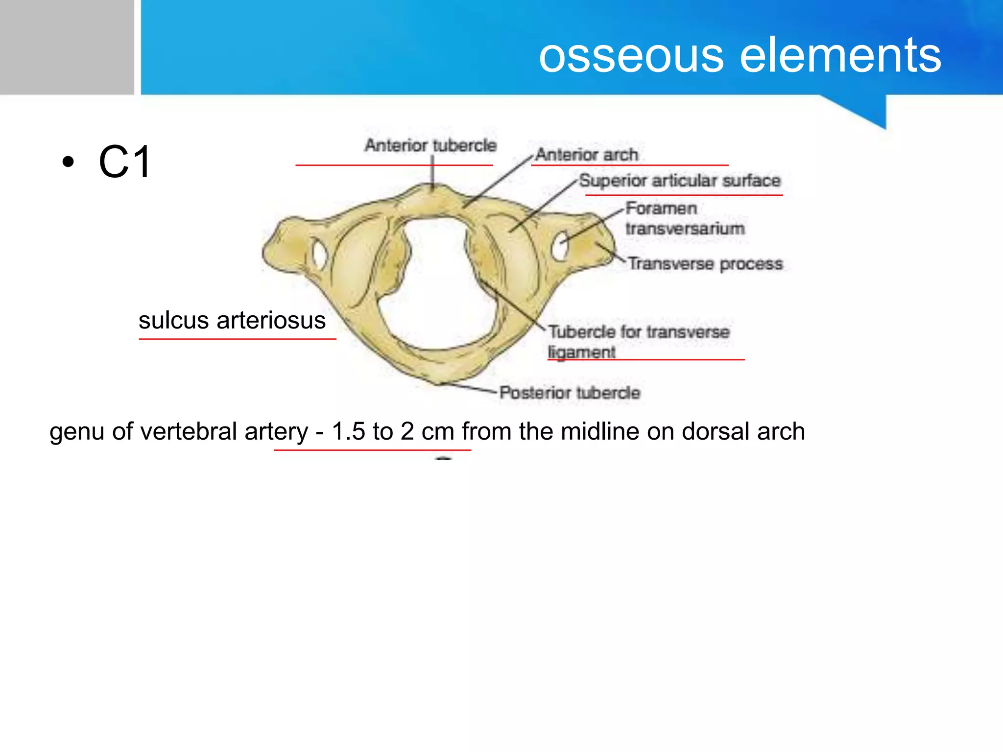
approach to cervical spine
dr himanshu soni
applied anatomy
• 7 vertebrae
• these segments are connected by an
extensive coplex of viscoelastic structures
to form a single flexible but constrained
column serving both neuroprotective and
structural function
• thorough understanding of the normal
relationshiops between these osseous,
discoligamentous and neural elements is
essential for safe and effective surgical
intervention
osseous elements
• C1
sulcus arteriosus
genu of vertebral artery - 1.5 to 2 cm from the midline on dorsal arch
• C2
– odoontoid process
– synovial joint
– apical ligament
– alar ligaments
– tr. AA ligament
– neck of the O. P.
• common site of fracture
– articular surfaces
– pars interartericularis
– sup and inf articulating surfaces - coronally offset
– forceful hyperextension, pars interartercularis breaks
• hangman's fracture
– foramen tranversarium
– C2 foramen is angulated 45 degrees laterally - VA is partially roofed by
artericular surface
• subaxial vertebra
– vertebral bodies
– uncovertebral joint - not a true joint
– transverse process
• foramen
• groove on superior surface - spinal
nerves
• tubercles - attachment of dorsal
musculature
– pedicles - connect the dorsal arch
angled medially between 38 to 48
degrees from saggital plane
– pedicles - range 6 to 6.5 mm
diameter
• thinnest along lateral wall, exposing
the risk of vertebral aftery damage
while screwig
• lateral mass column with articular surfaces
which are oriented obliquely on sagittal
plane
• affords axial load bearing
• oblique configuration restricts translation
• lateral mass serves as a common anchor
point for instrumentation
– rostral screw - protects nerve root
– lateral screw - protects the VA
discoligamentous structures
• disc space
– inferior endplate is concave causing taller disc
space at midbody
– discs constitute 20% of height
– height increases consecutively from C3-4 to
C7-T1 and space depth increases
– approx 16mm at C3 to 20mm at C7
– nucleus pulposus - 88% water at birth
– 64% at seventh decade
– nutrition by diffusion
– immune - isolated avascular space
– Sharpey fibers
• ALL
• PLL
• ligamentum flavum
• intertransverseligaments
– restrict lateral bending
• inter spinous ligaments
• nuchal ligament
– from occ protruberance to sp pr of C7
– thick elastic fibrous - resists hyperflexion
– forms posterior tension band
neural structures - cervical cord
• cylindrical, symmetrical
• ventral median fissure - anterior
spinal artery, true space
• posterior median sulcus -
seperated hemicords
• anterolateral sulcus -
emergence of ventral / motor
roots
• dorsolateral sulcus - dorsal /
sensory roots
• three columns : anterior, lateral
and posterior
• posterior column - medial
gracilis and lateral cuneatus
fasciculus
• cord segments approx same
level as the column segment
neural structures - spinal nerves
• anterolateral sulcus - 1-3 mm lateral to midline
– multiple rootlets
– allows intradural anastomosis causing atypical
distribution of radiculopathies
• dorsal rootlets
– proprioceptive large fibres run medially and
ventrally
– small pain fibres located laterally and dorsally
• both traverse the intervertebral foramen above
the same pedicle ( C5 through C4-5 foramen
above C5 pedicle )
• C1 has no DRG and hence no dermatome
intervertebral foramen
• dorsal roots more rostrally hence comprression affects
disproportionately
• exiting root - oblique with a ventrolateral trajectory
• at origin
– bounded ventromedially by the UV joint
– laterally, the nerve root and DRG abut the VA
– UV hypertrophy and lateral discs cause radiculopathy
– UV hypertrophy also distorts the VA causing secondary
compression on the exiting root or DRG
• dorsolateral wall
– superior and inf articular processes, hence facet hypertrophy
triggers radiculopathy
– basis of Spurling's maneuver
• the roots lie more caudally, abutting the subjacent pedicle
VASCULAR ANATOMY
• SPINAL CORD PERFUSION
• three principal arteries
– the anterior spinal artery
– paired posterio spinal arteries
• none of these vessels represent single
vessels that traverse teh entire length of
spinal cord
• they are anastomotic, interrupted,tudinal
vessels, drawing supply from numerous
segments.
• spinal cord perfusion is achieved by
– two indenpendent vasular systems :
• the centripetal ssytem
– runs along the perimeter of the spinal cord in
perimedullary plexus
– gives rise to arteries that penetrate radially into the cord
parenchyma
• the centrifugal system
– originates deep within the anterior median fissure from
branches of the ASA
– the centripetal system perfuses the white
matter
– centrifugal system perfuses the gray matter
• the ASA
– descending branches of bilateral intracrahial
vertebral arteries
– enters the midline ventral median fissure
– supplies ventral two thirds of the cord
– infarct causes dysfunction of
• the anterior columns
• spinothalamic tracts
• corticospinal tracts
• the PSAs
– intracranial vertebral arterires or teh PICAs
– run in the dorsolateral sulci
– form a peripheral anastomotic plexus
– perfuses the dorsal one third.
• cervical segmental vessels
– paired, arise from the verterberaltery and
branches of the cubclavian arteries
– perfuse the spinal roots and DRG before
entering the intervertebral foramen
– 3 branches
• the dural - perfuses spinal dura and nerve
root sleeve
• radicular - dura and ventral and dorsal
nerve roots
• medullary - anastomoses with the ASA
within one or two levels of origin
• VERTEBRAL ARTERY
– paired, arises from the subclavian artery, from the
innominate artery or from the aorta
– course is divided into four segments
– V1: origin to foramen transvarserium
• 87.5 % - at C6 level
• 5.4% - at C7 level
• 6.6 % - at C5 level
• 0.4% - at C4 level
– V2: traverses the FT, ends after C2 FT
• most vulnerable segment
• may have a tortuous course
• FT is closer to UV joint, warranting cautious drilling
– V3: until the VA penetrates the dura
– V4: intradural portion, ends at the VB junction
• muscular and osseous branches - each level
• anterior and posterior central arteries
– epidural plexus
– perfuses the vertebral bodies
• anterior and posterior ascending arteries
– at C2 level, anastomose to perfuse the
antlantoaxial complex
• collateral recruitment
– thyrocervical and costocervical in the lower spine
– ascending pharyngeal and occipital in the uppper
cervical spine
• odontoid process
– base - ascending branches of the VA
– tip - apical artery of the OP, branch from the
hypoglossal artery
• Venous anatomy
– The intrinsic spinal cord is drained
centrifugally by radially oriented veins that
empty into a circumferential venous plexus
called the vasa corona.
– The dorsal half of the spinal cord venous
drainage empties into this plexus, eventually
converging on the median dorsal longitudinal
vein,
– whereas the ventral half of the spinal cord
drainage empties into a comparable plexus,
eventually draining into the median ventral
Approach to cervical spine

Approach to cervical spine

  • 1.
    approach to cervicalspine dr himanshu soni
  • 2.
    applied anatomy • 7vertebrae • these segments are connected by an extensive coplex of viscoelastic structures to form a single flexible but constrained column serving both neuroprotective and structural function • thorough understanding of the normal relationshiops between these osseous, discoligamentous and neural elements is essential for safe and effective surgical intervention
  • 3.
    osseous elements • C1 sulcusarteriosus genu of vertebral artery - 1.5 to 2 cm from the midline on dorsal arch
  • 4.
    • C2 – odoontoidprocess – synovial joint – apical ligament – alar ligaments – tr. AA ligament – neck of the O. P. • common site of fracture – articular surfaces – pars interartericularis – sup and inf articulating surfaces - coronally offset – forceful hyperextension, pars interartercularis breaks • hangman's fracture – foramen tranversarium – C2 foramen is angulated 45 degrees laterally - VA is partially roofed by artericular surface
  • 9.
    • subaxial vertebra –vertebral bodies – uncovertebral joint - not a true joint – transverse process • foramen • groove on superior surface - spinal nerves • tubercles - attachment of dorsal musculature – pedicles - connect the dorsal arch angled medially between 38 to 48 degrees from saggital plane – pedicles - range 6 to 6.5 mm diameter • thinnest along lateral wall, exposing the risk of vertebral aftery damage while screwig
  • 11.
    • lateral masscolumn with articular surfaces which are oriented obliquely on sagittal plane • affords axial load bearing • oblique configuration restricts translation • lateral mass serves as a common anchor point for instrumentation – rostral screw - protects nerve root – lateral screw - protects the VA
  • 18.
    discoligamentous structures • discspace – inferior endplate is concave causing taller disc space at midbody – discs constitute 20% of height – height increases consecutively from C3-4 to C7-T1 and space depth increases – approx 16mm at C3 to 20mm at C7 – nucleus pulposus - 88% water at birth – 64% at seventh decade – nutrition by diffusion – immune - isolated avascular space – Sharpey fibers
  • 20.
    • ALL • PLL •ligamentum flavum • intertransverseligaments – restrict lateral bending • inter spinous ligaments • nuchal ligament – from occ protruberance to sp pr of C7 – thick elastic fibrous - resists hyperflexion – forms posterior tension band
  • 23.
    neural structures -cervical cord • cylindrical, symmetrical • ventral median fissure - anterior spinal artery, true space • posterior median sulcus - seperated hemicords • anterolateral sulcus - emergence of ventral / motor roots • dorsolateral sulcus - dorsal / sensory roots • three columns : anterior, lateral and posterior • posterior column - medial gracilis and lateral cuneatus fasciculus • cord segments approx same level as the column segment
  • 24.
    neural structures -spinal nerves • anterolateral sulcus - 1-3 mm lateral to midline – multiple rootlets – allows intradural anastomosis causing atypical distribution of radiculopathies • dorsal rootlets – proprioceptive large fibres run medially and ventrally – small pain fibres located laterally and dorsally • both traverse the intervertebral foramen above the same pedicle ( C5 through C4-5 foramen above C5 pedicle ) • C1 has no DRG and hence no dermatome
  • 25.
    intervertebral foramen • dorsalroots more rostrally hence comprression affects disproportionately • exiting root - oblique with a ventrolateral trajectory • at origin – bounded ventromedially by the UV joint – laterally, the nerve root and DRG abut the VA – UV hypertrophy and lateral discs cause radiculopathy – UV hypertrophy also distorts the VA causing secondary compression on the exiting root or DRG • dorsolateral wall – superior and inf articular processes, hence facet hypertrophy triggers radiculopathy – basis of Spurling's maneuver • the roots lie more caudally, abutting the subjacent pedicle
  • 28.
    VASCULAR ANATOMY • SPINALCORD PERFUSION • three principal arteries – the anterior spinal artery – paired posterio spinal arteries • none of these vessels represent single vessels that traverse teh entire length of spinal cord • they are anastomotic, interrupted,tudinal vessels, drawing supply from numerous segments.
  • 29.
    • spinal cordperfusion is achieved by – two indenpendent vasular systems : • the centripetal ssytem – runs along the perimeter of the spinal cord in perimedullary plexus – gives rise to arteries that penetrate radially into the cord parenchyma • the centrifugal system – originates deep within the anterior median fissure from branches of the ASA – the centripetal system perfuses the white matter – centrifugal system perfuses the gray matter
  • 30.
    • the ASA –descending branches of bilateral intracrahial vertebral arteries – enters the midline ventral median fissure – supplies ventral two thirds of the cord – infarct causes dysfunction of • the anterior columns • spinothalamic tracts • corticospinal tracts
  • 31.
    • the PSAs –intracranial vertebral arterires or teh PICAs – run in the dorsolateral sulci – form a peripheral anastomotic plexus – perfuses the dorsal one third.
  • 32.
    • cervical segmentalvessels – paired, arise from the verterberaltery and branches of the cubclavian arteries – perfuse the spinal roots and DRG before entering the intervertebral foramen – 3 branches • the dural - perfuses spinal dura and nerve root sleeve • radicular - dura and ventral and dorsal nerve roots • medullary - anastomoses with the ASA within one or two levels of origin
  • 33.
    • VERTEBRAL ARTERY –paired, arises from the subclavian artery, from the innominate artery or from the aorta – course is divided into four segments – V1: origin to foramen transvarserium • 87.5 % - at C6 level • 5.4% - at C7 level • 6.6 % - at C5 level • 0.4% - at C4 level – V2: traverses the FT, ends after C2 FT • most vulnerable segment • may have a tortuous course • FT is closer to UV joint, warranting cautious drilling
  • 34.
    – V3: untilthe VA penetrates the dura – V4: intradural portion, ends at the VB junction
  • 35.
    • muscular andosseous branches - each level • anterior and posterior central arteries – epidural plexus – perfuses the vertebral bodies • anterior and posterior ascending arteries – at C2 level, anastomose to perfuse the antlantoaxial complex • collateral recruitment – thyrocervical and costocervical in the lower spine – ascending pharyngeal and occipital in the uppper cervical spine
  • 36.
    • odontoid process –base - ascending branches of the VA – tip - apical artery of the OP, branch from the hypoglossal artery
  • 37.
    • Venous anatomy –The intrinsic spinal cord is drained centrifugally by radially oriented veins that empty into a circumferential venous plexus called the vasa corona. – The dorsal half of the spinal cord venous drainage empties into this plexus, eventually converging on the median dorsal longitudinal vein, – whereas the ventral half of the spinal cord drainage empties into a comparable plexus, eventually draining into the median ventral