Downloaded 1,422 times


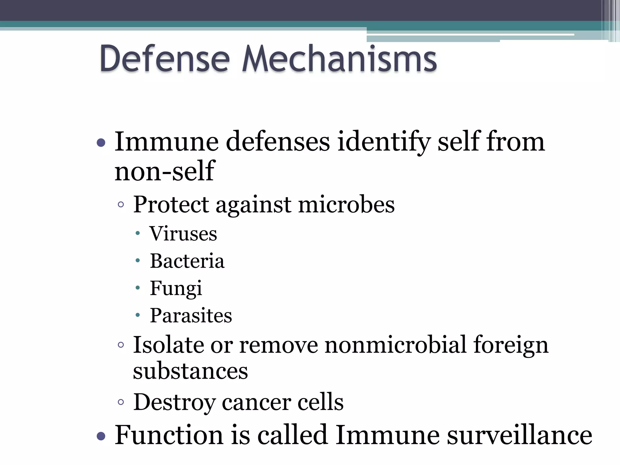
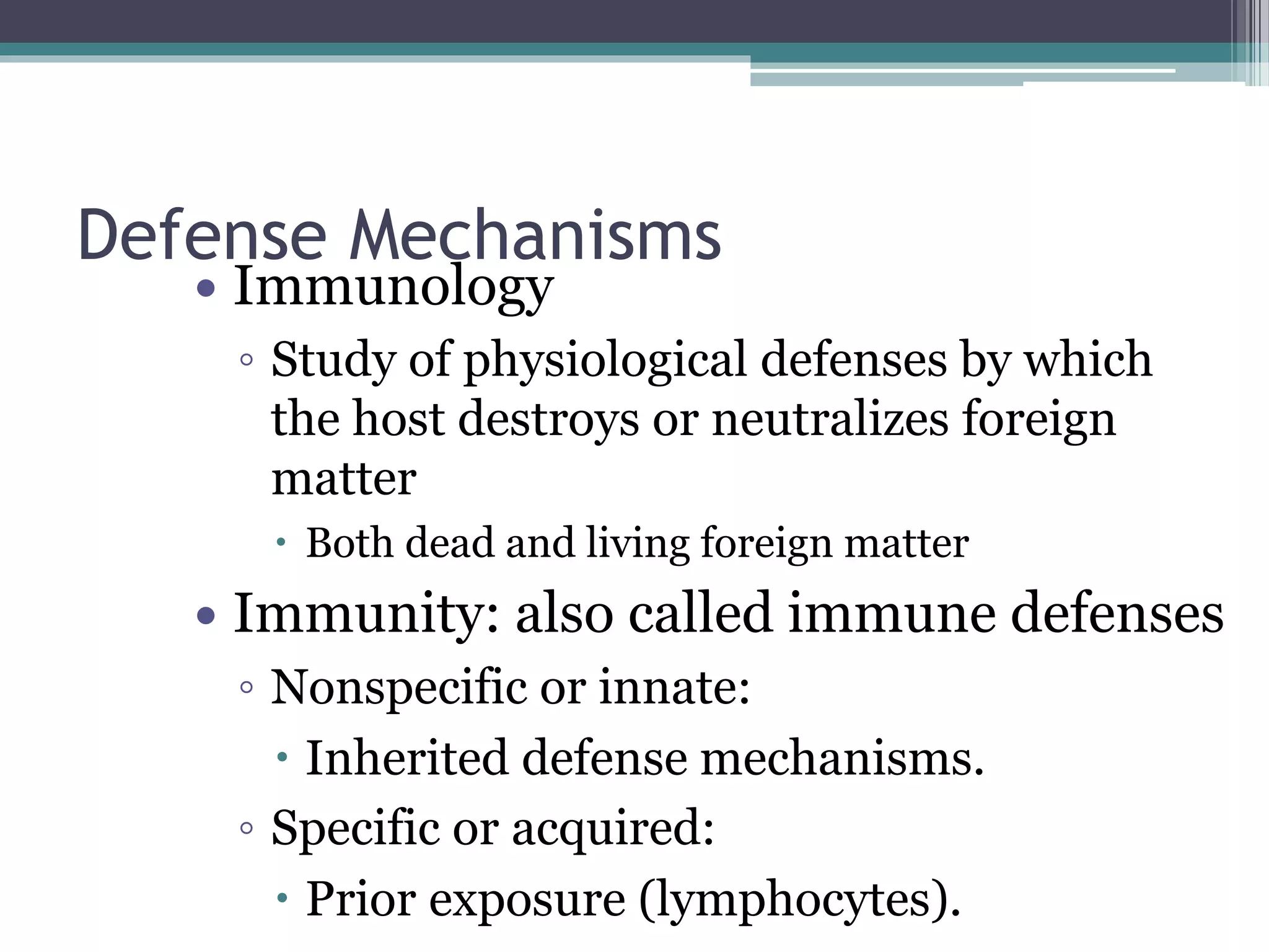


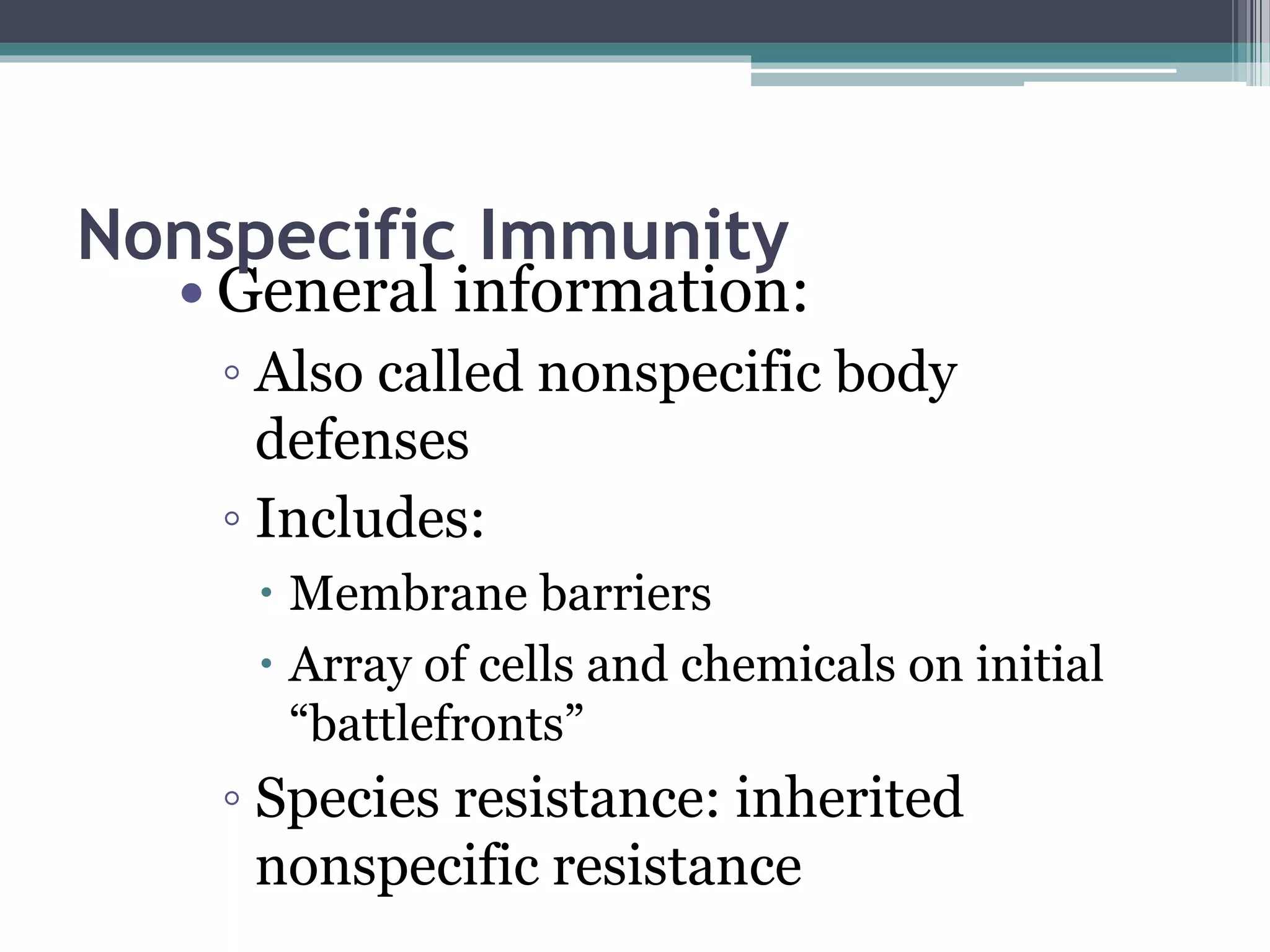

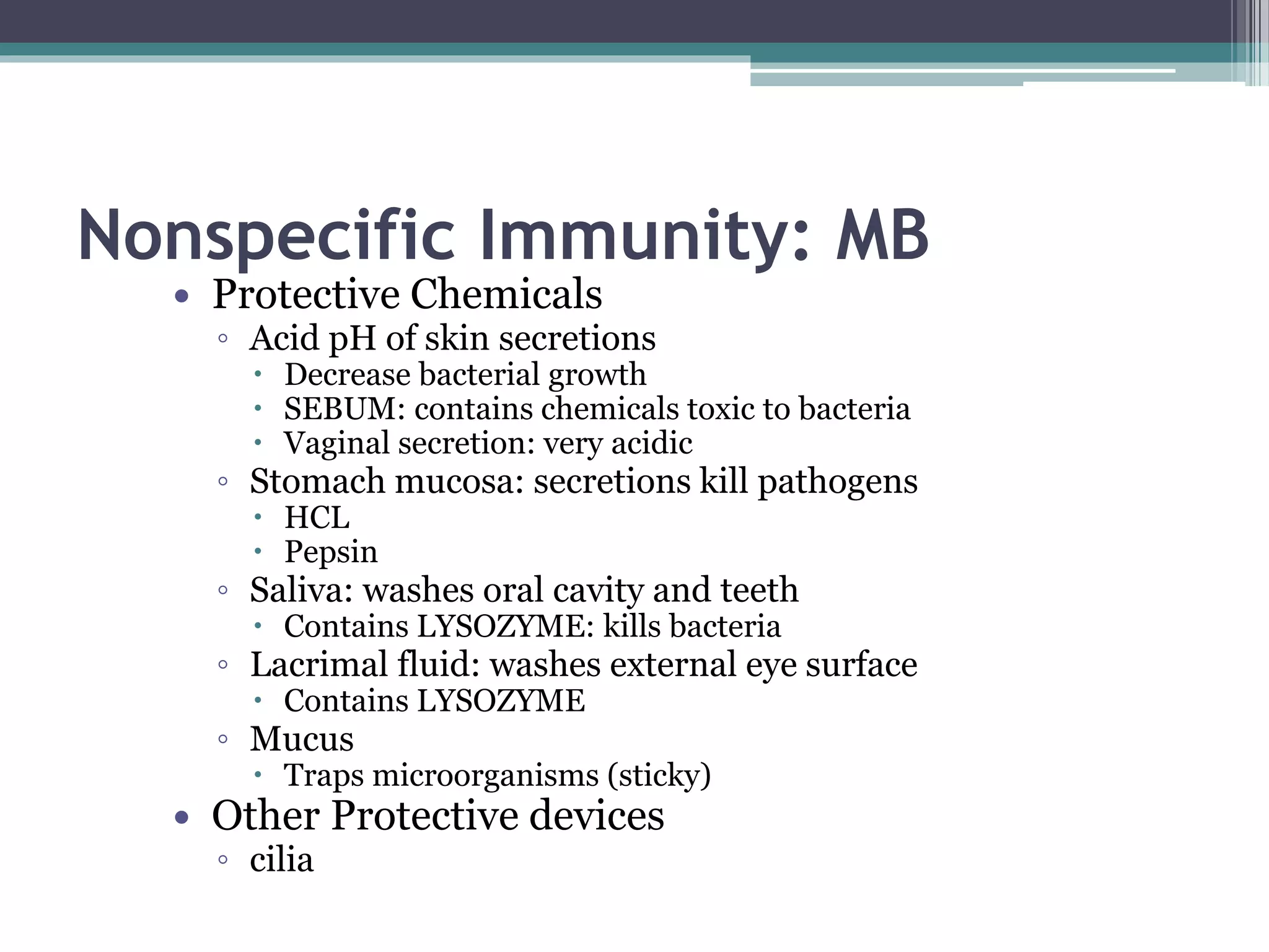
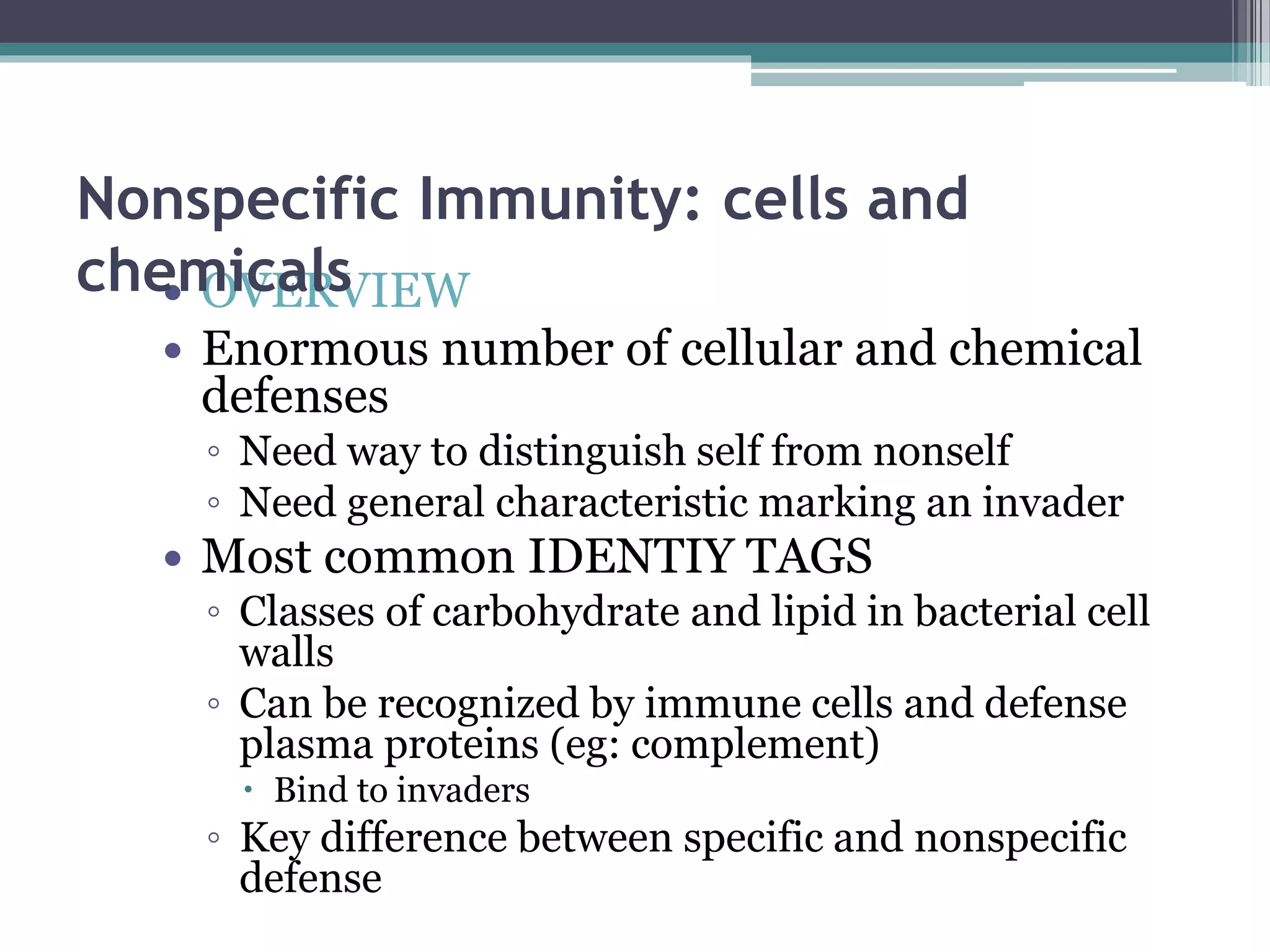


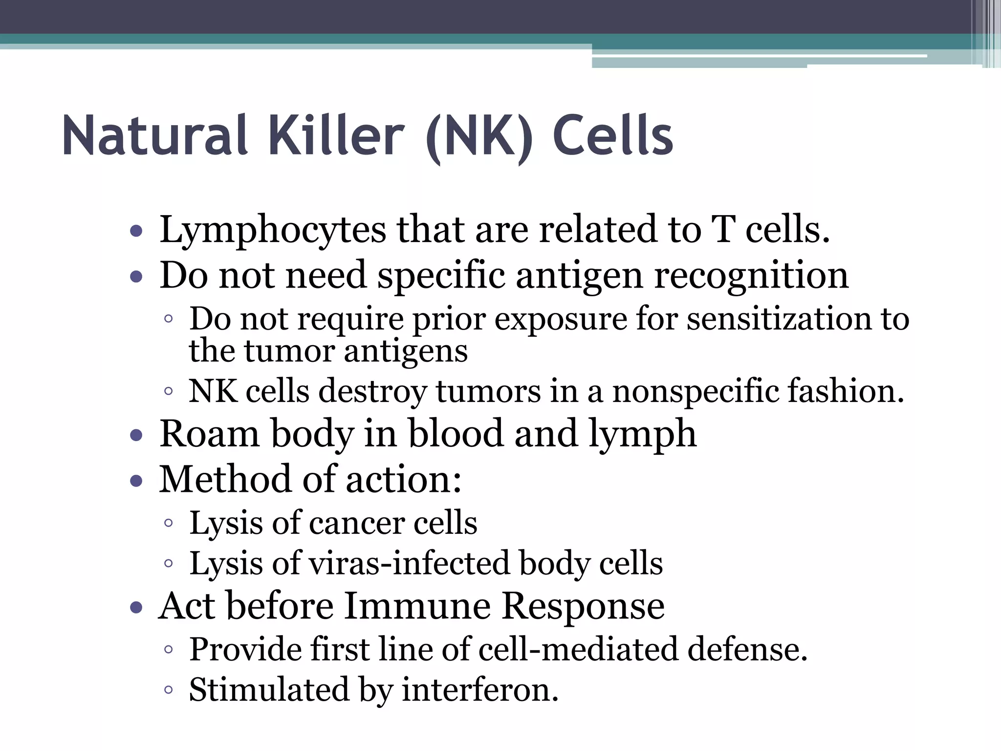


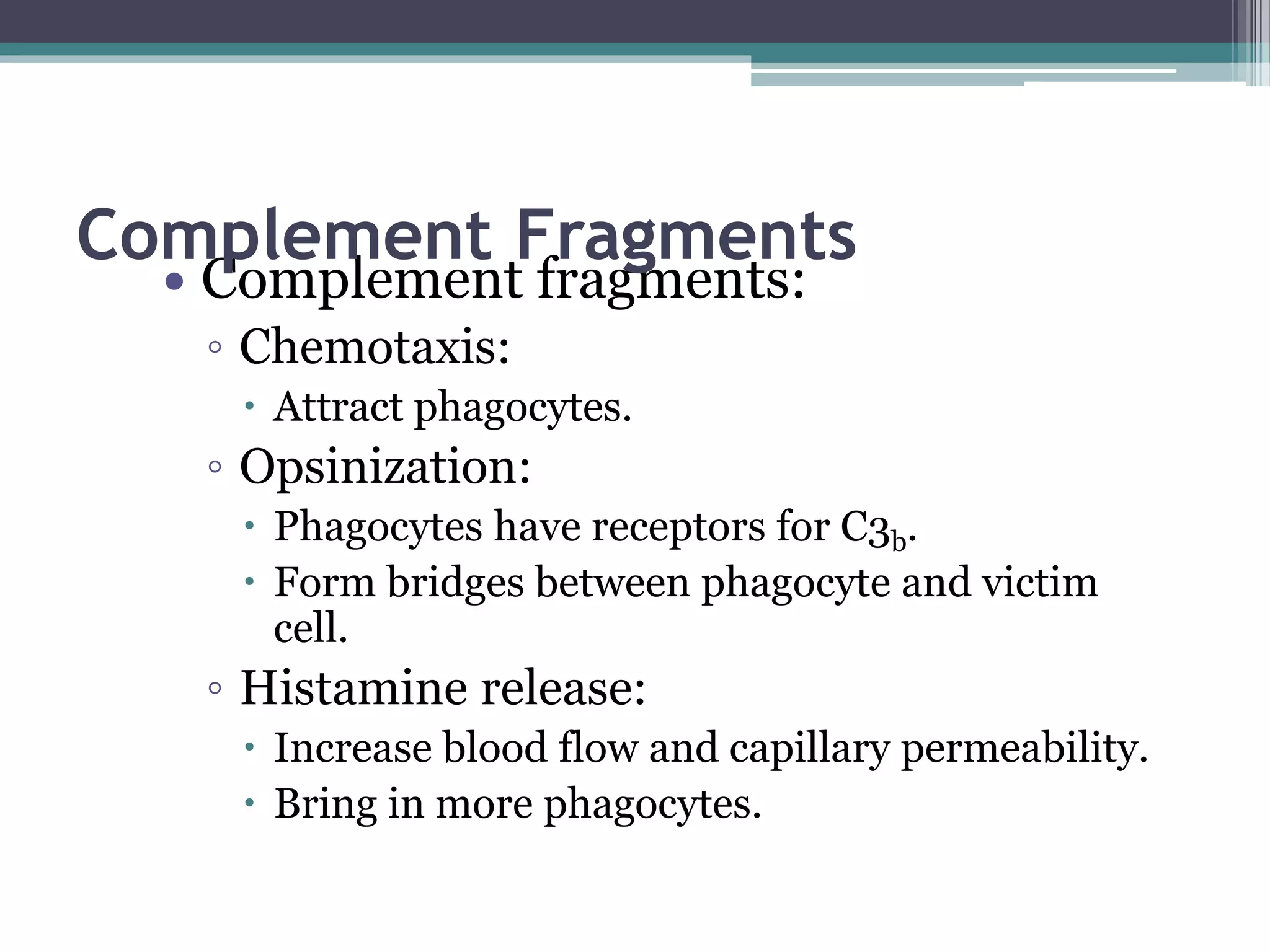

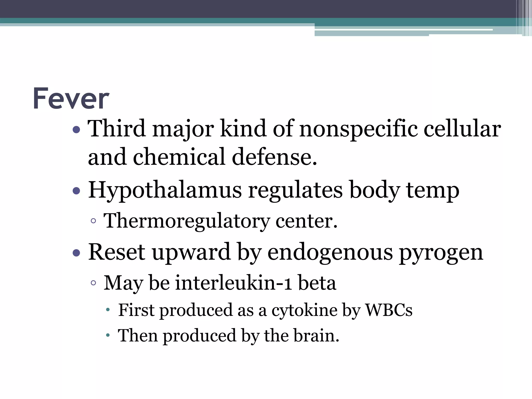


The immune system provides defense against pathogens. It includes cells that carry out immune functions and circulate in the body. The immune system distinguishes self from non-self and protects against microbes, viruses, bacteria, fungi and parasites. It also destroys cancer cells. Both nonspecific innate immunity and specific acquired immunity work together to provide protection. Nonspecific defenses include physical barriers, phagocytic cells, inflammation, complement proteins, and interferons that provide a first line of defense against pathogens.


