Downloaded 332 times
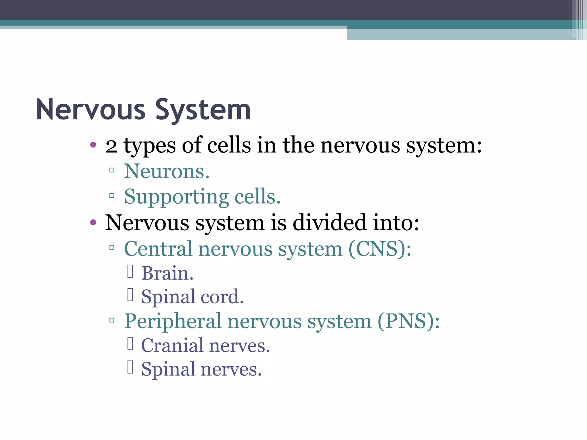
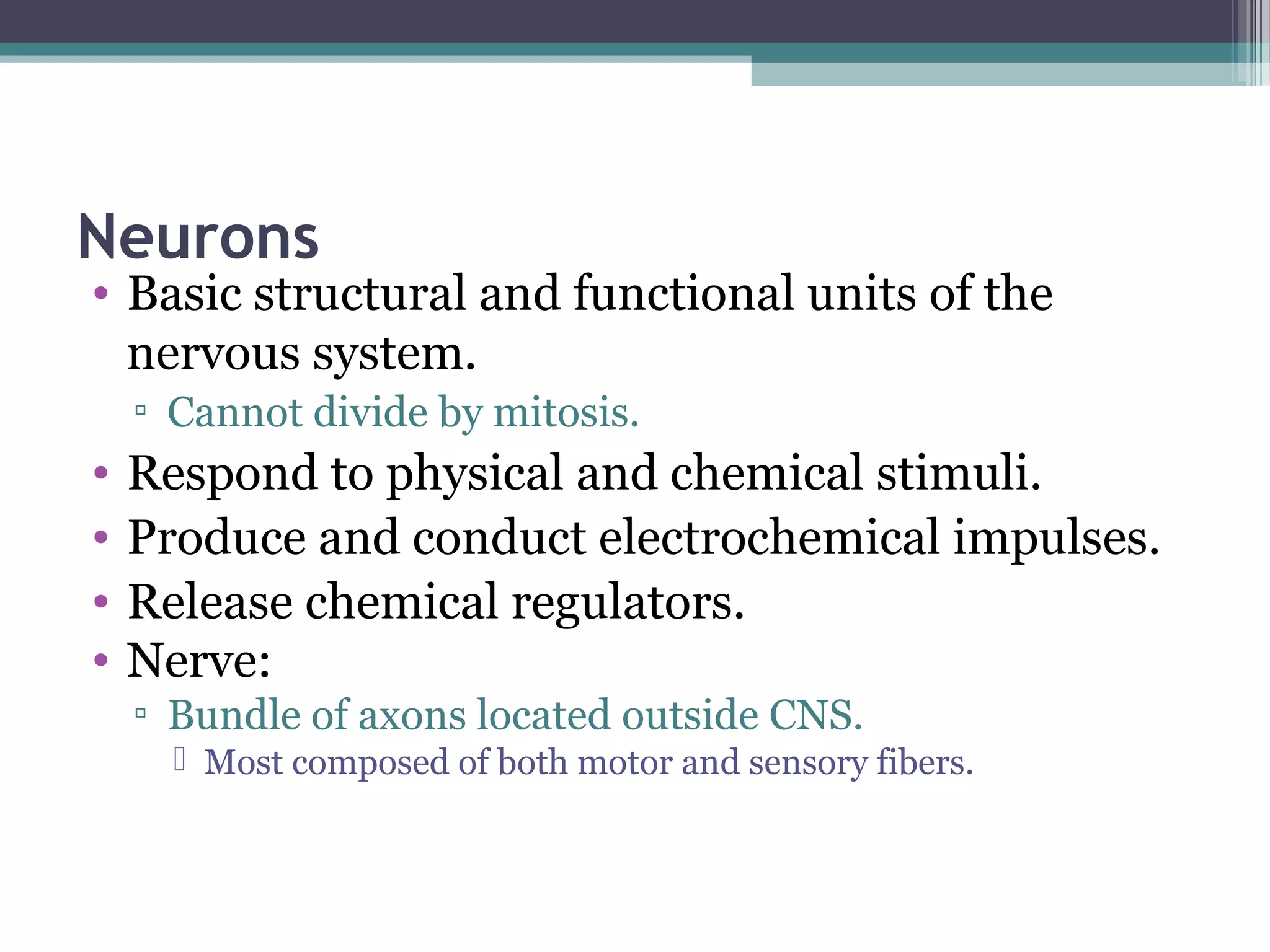
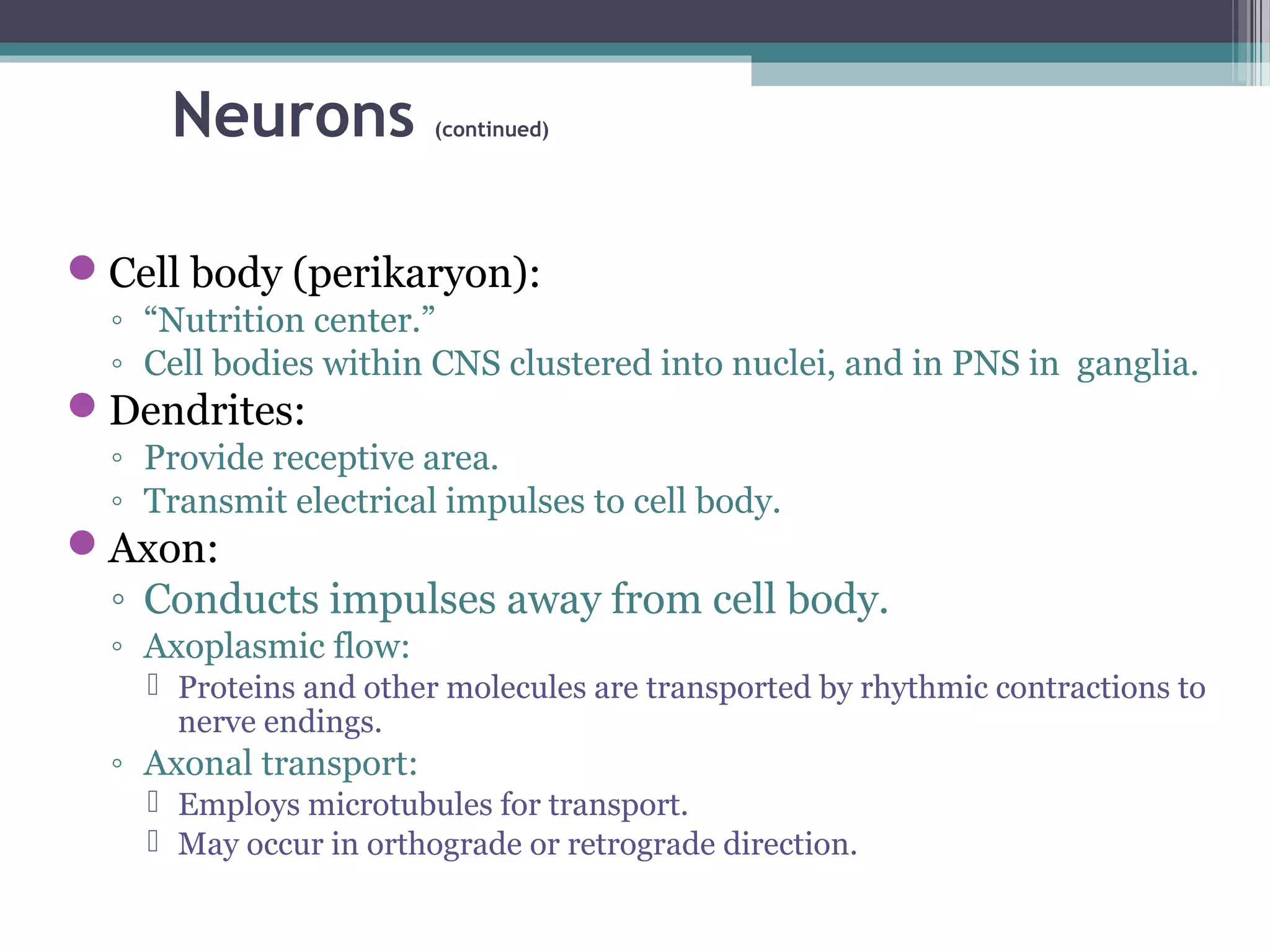
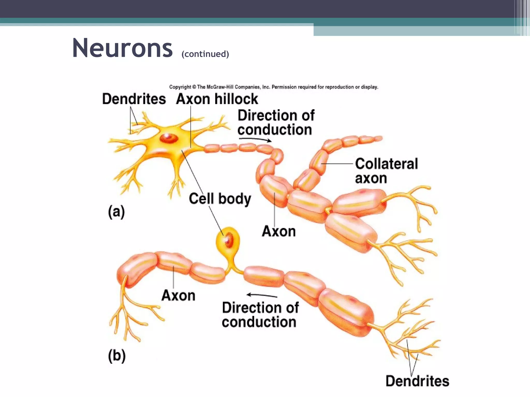
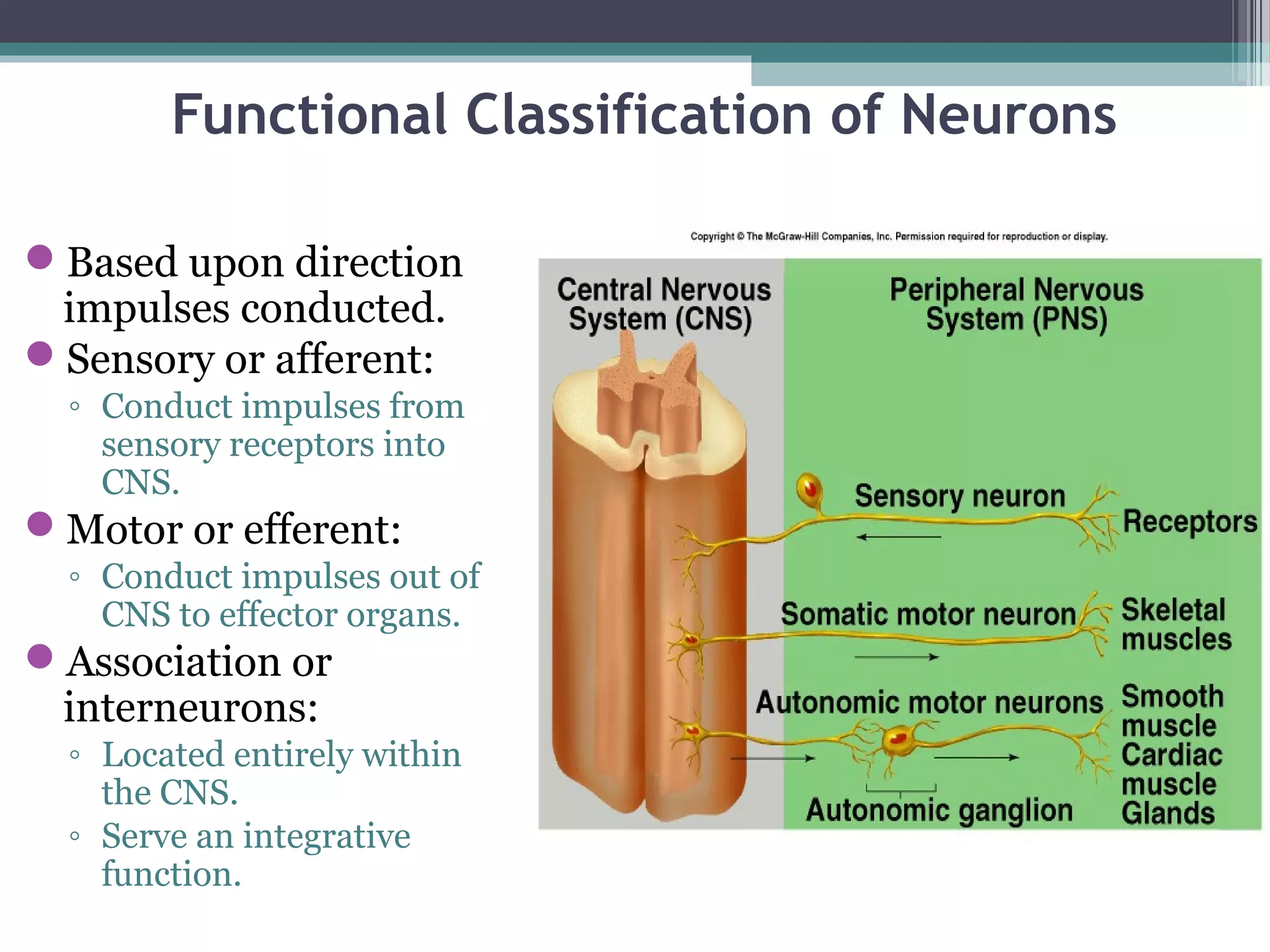
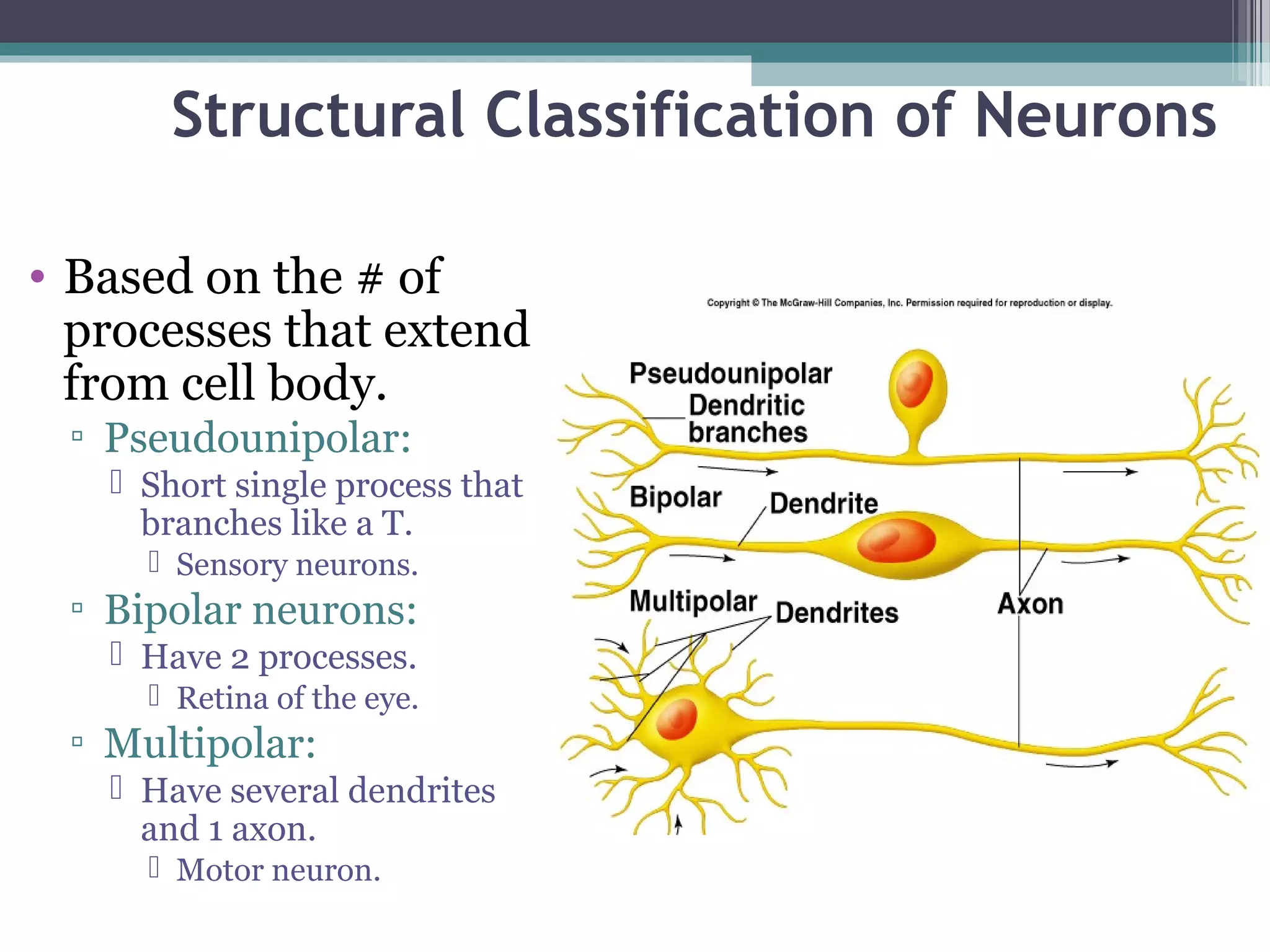
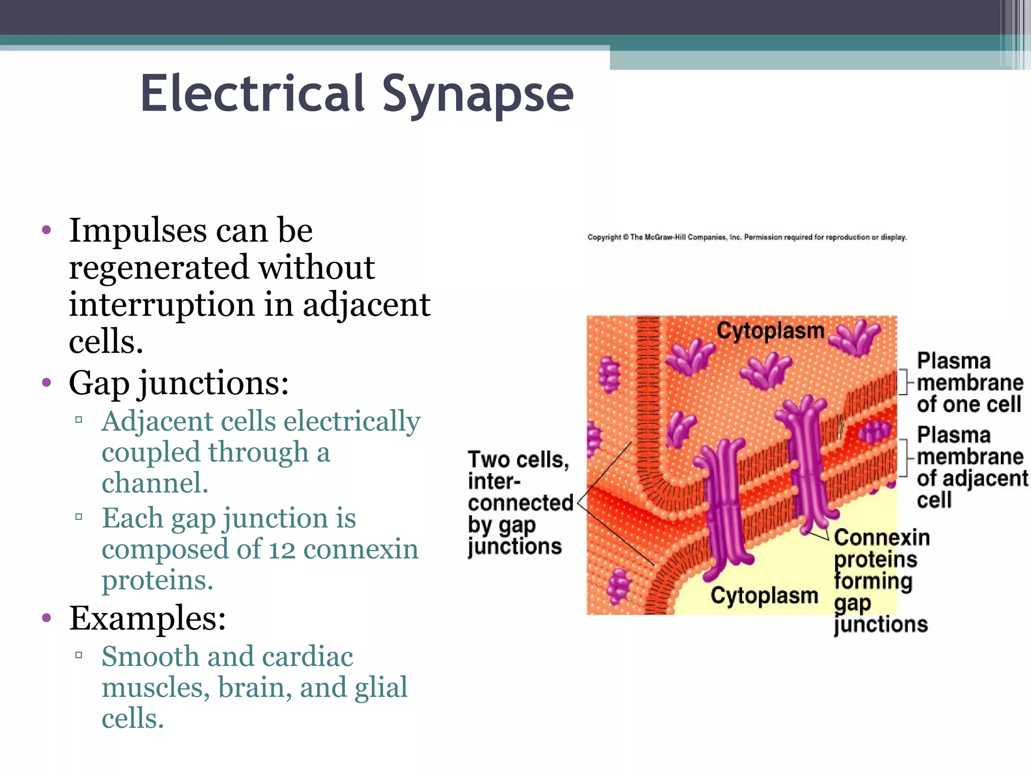
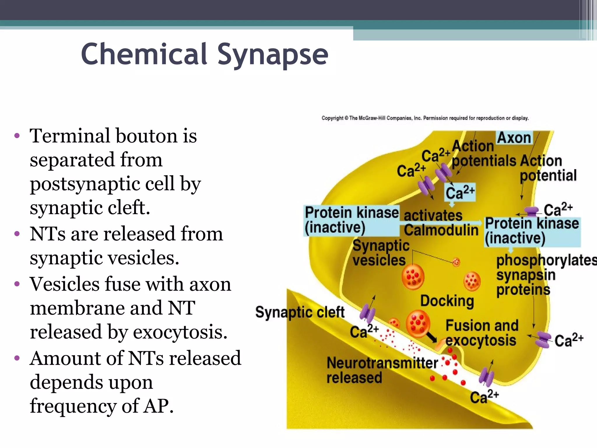
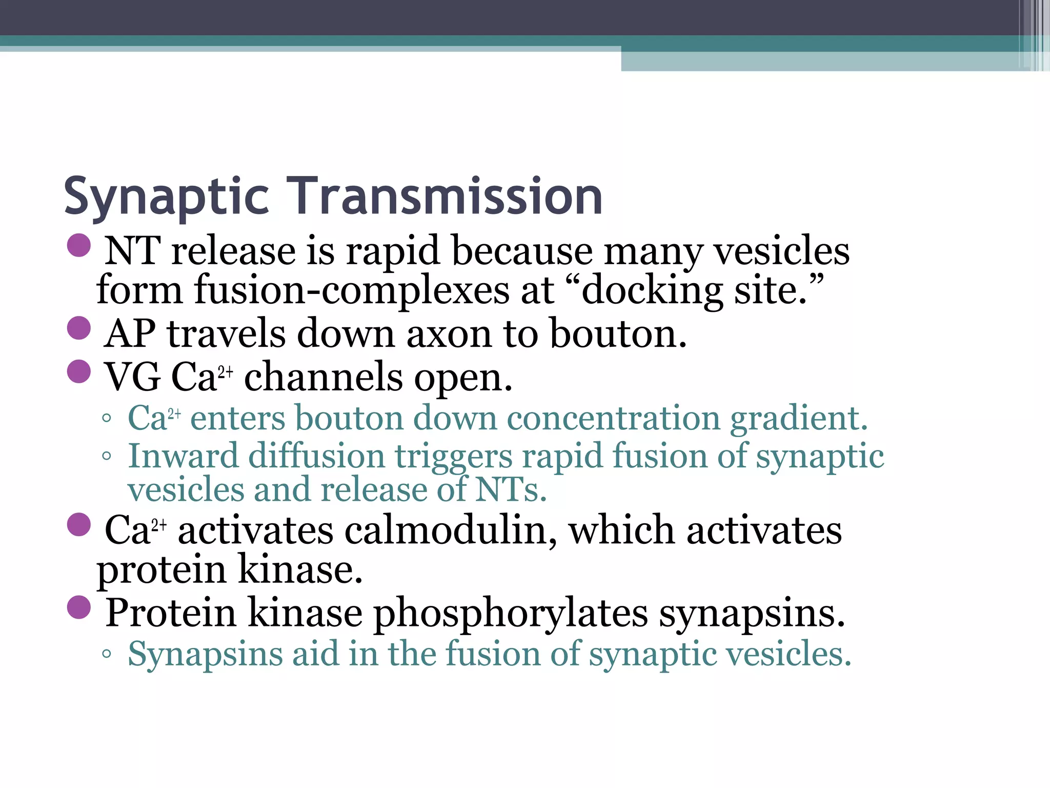
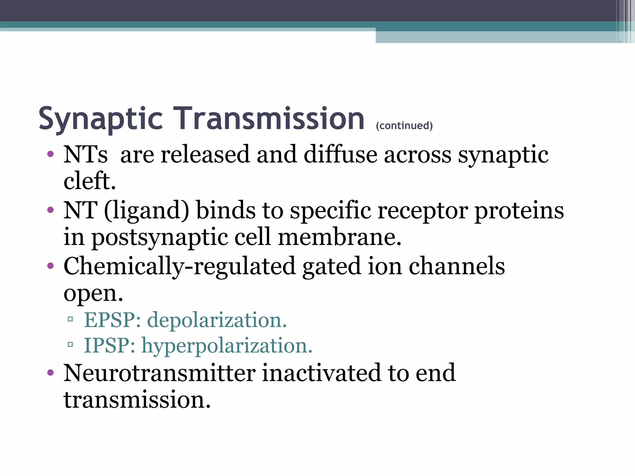
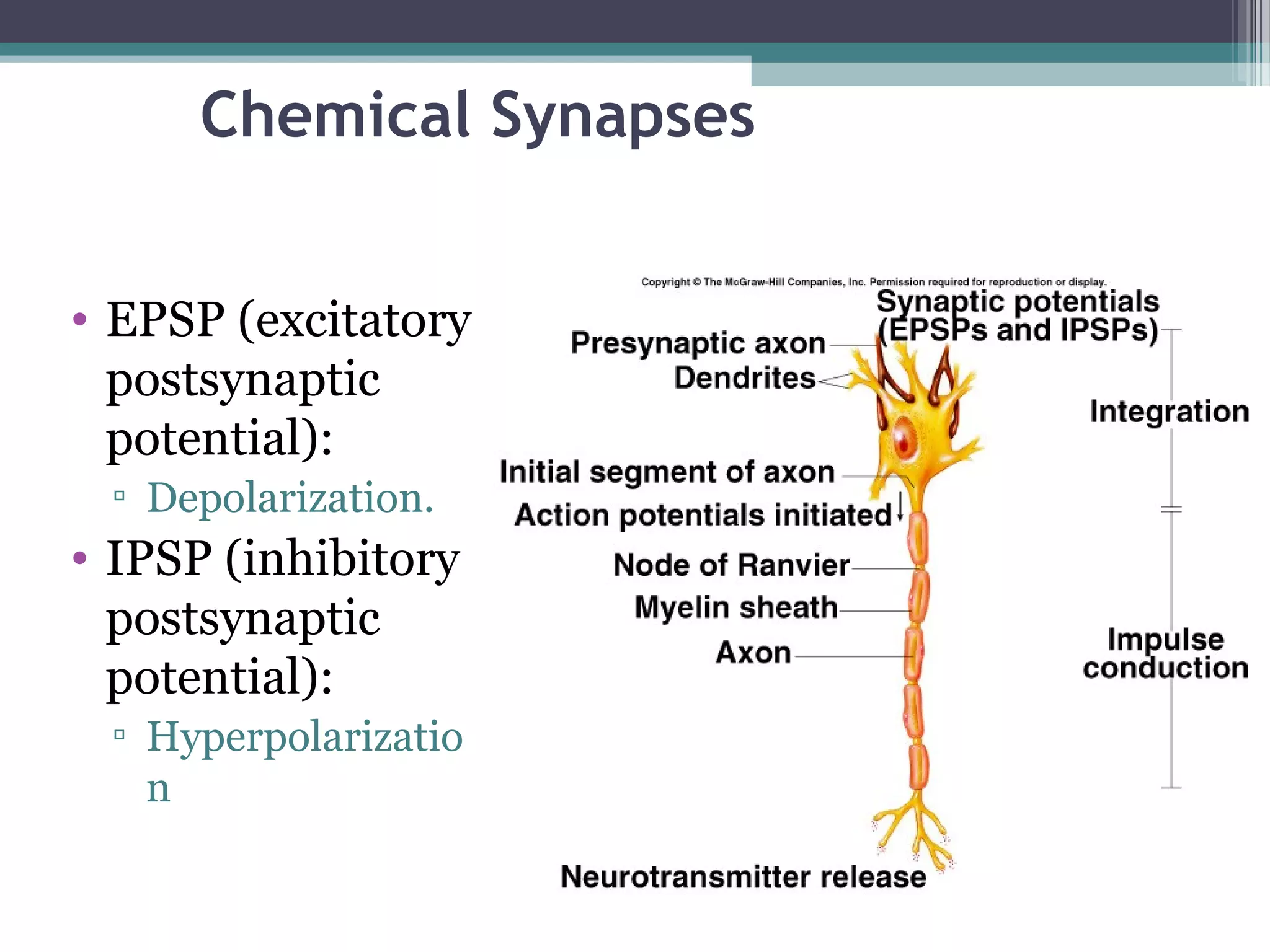
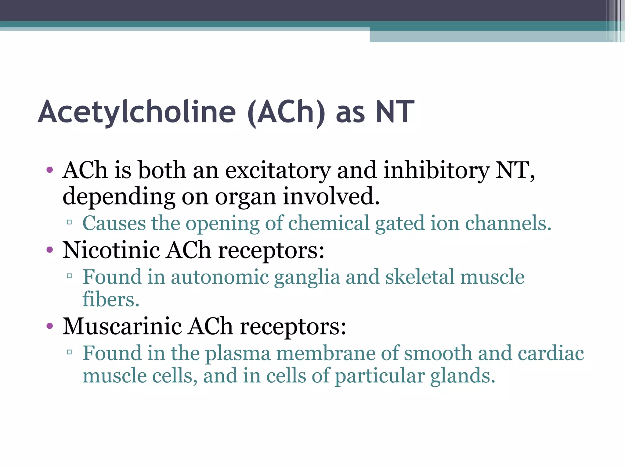
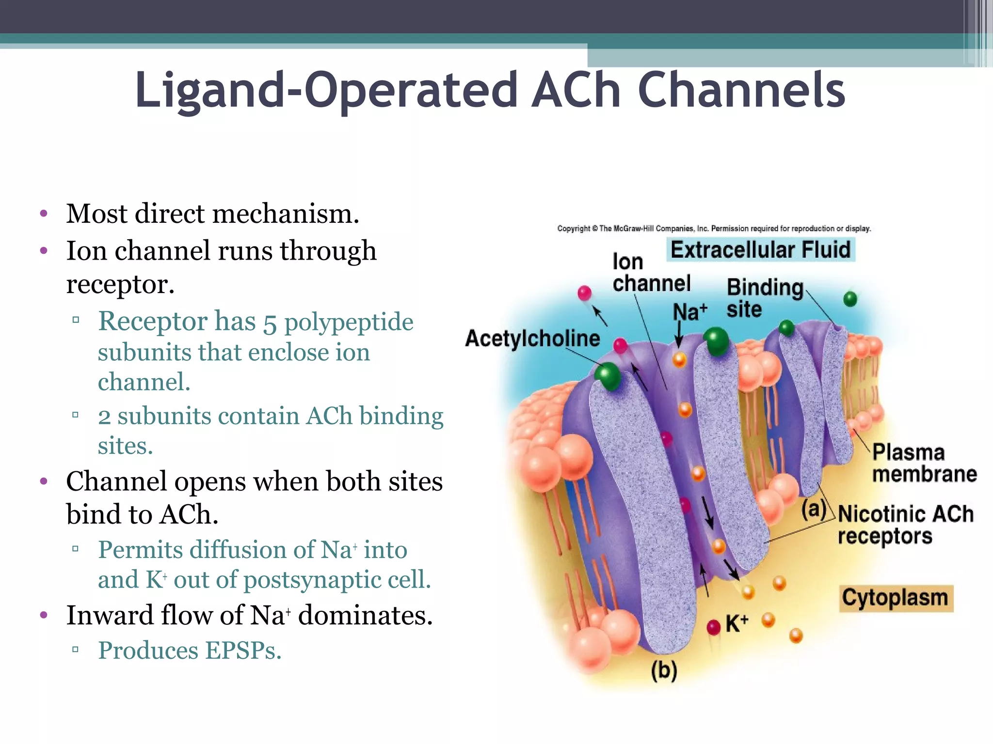
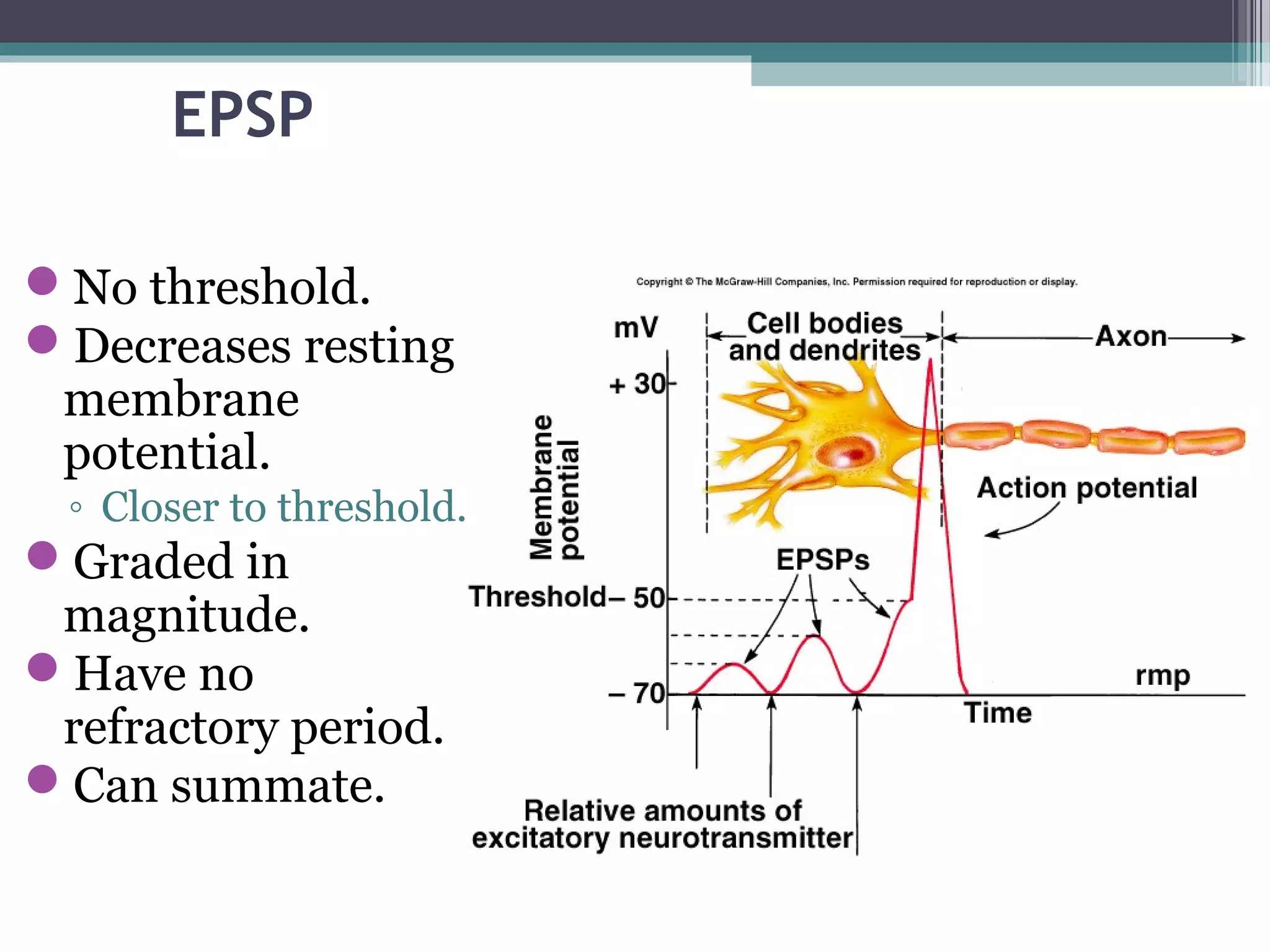
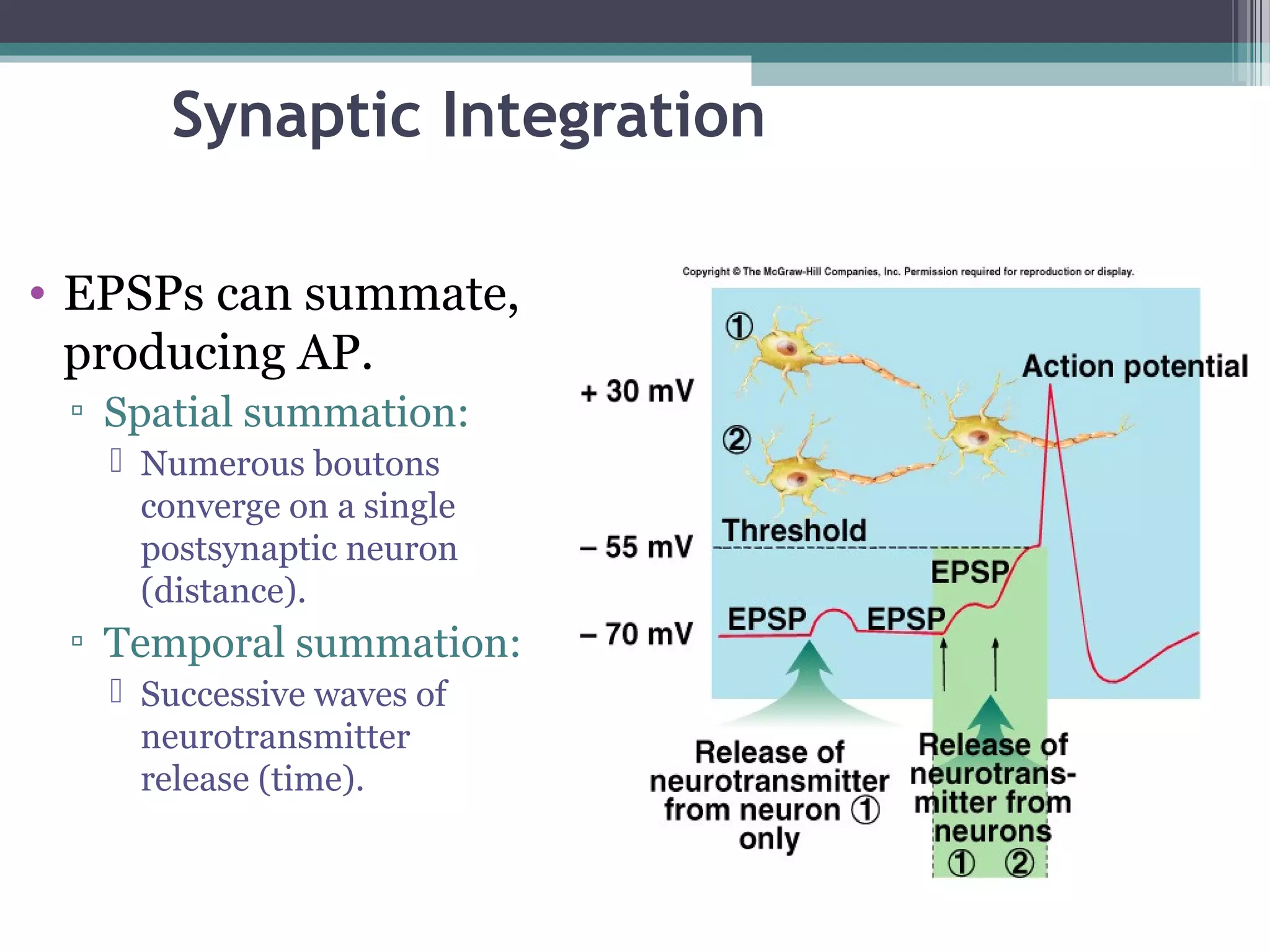

The nervous system contains two main cell types - neurons and supporting cells. Neurons transmit electrochemical signals and are divided into sensory, motor, and interneurons. Supporting cells aid neuron function. The nervous system is divided into the central nervous system (brain and spinal cord) and peripheral nervous system. Neurons contain a cell body, dendrites, and an axon. An action potential is generated when a threshold is reached due to ion flow through voltage-gated channels. At a synapse, a neurotransmitter is released and binds to receptors, generating an excitatory or inhibitory postsynaptic potential.


![Chapt10 Holes Lecture Animation[1]](https://cdn.slidesharecdn.com/ss_thumbnails/chapt10holeslectureanimation1-091122123853-phpapp02-thumbnail.jpg?width=640&height=640&fit=bounds)


