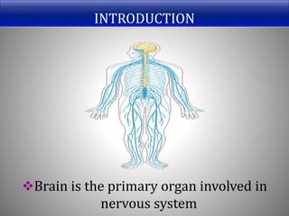
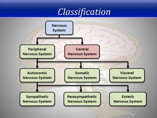
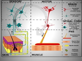
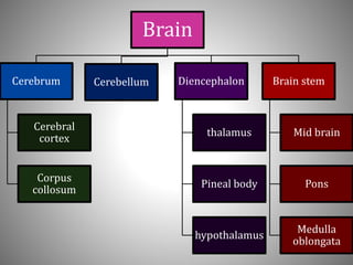
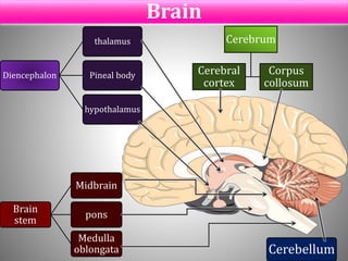
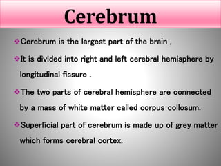
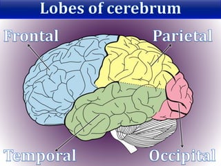
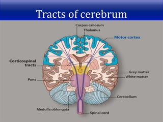
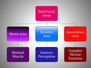
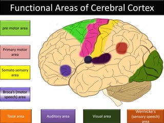
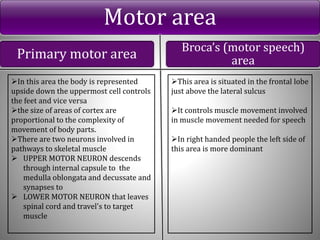
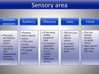
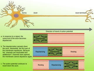
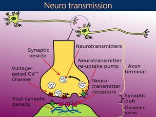
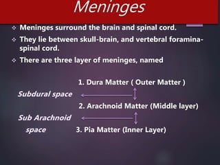
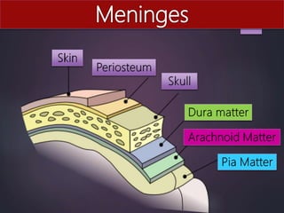
INTRODUCTION
Brain is the primary organ involved in
nervous system
Classification
Brain
Cerebrum
Cerebral
cortex
Corpus
collosum
Cerebellum Diencephalon
thalamus
Pineal body
hypothalamus
Brain stem
Mid brain
Pons
Medulla
oblongata
Brain
Cerebrum
Cerebral
cortex
Corpus
collosum
Diencephalon
thalamus
Pineal body
hypothalamus
Brain
stem
Midbrain
pons
Medulla
oblongata Cerebellum
Cerebrum
Cerebrum is the largest part of the brain ,
It is divided into right and left cerebral hemisphere by
longitudinal fissure .
The two parts of cerebral hemisphere are connected
by a mass of white matter called corpus collosum.
Superficial part of cerebrum is made up of grey matter
which forms cerebral cortex.
Tracts of cerebrum
Tracts
1. Association Tract- Connects different part of hemisphere.
Extending from one gyrus to another
2. Commissural tracts- It connects corresponding areas of
two cerebral hemisphere, e.g.- Corpus collosum
3. Projection tracts- It connects cerebral cortex with grey
matter with lower part of brain and spinal cord
4. Corticospinal Tracts- the motor fibres within internal
capsule forms the pyramidal tracts (corticospinal tracts)
Internal Capsule
Internal capsule is an important projection tract that lies
within the brain between the basal ganglia and thalamus
Fibers of internal capsule carries the nerve impulses passing
to and from cerebral cortex
Corticospinal tracts decussate at the medulla oblongata
Motor fibres that do not pass over internal capsule forms
extrapyramidal tracts
Basal Ganglia
There is a group of cell bodies deep within cerebral
hemisphere called nuclei.
Some of these nuclei forms basal ganglia which forms
extrapyramidal tracts.
They act as relay station that connects to many parts of
the brain.
They initiate complex movement.
Cerebral Cortex
Superficial part of cerebrum forms cerebral cortex
made up of grey matter
It is involved in higher order function such as learning ,
memory, thinking decision making
It is active in sensory perception such as pain,
temperature, touch, hearing, taste and smell
It is also involved in voluntary movement of skeletal
muscle
Functional
areas
Motor area
Skeletal
Muscle
Sensory
area
Sensory
Perception
Association
area
Complex
Mental
Function
Functional Areas of Cerebral Cortex
Wernicke‘s
(sensory speech)
area
Auditory areaTaste area
pre motor area
Somato sensory
area
Broca’s (motor
speech) area
Primary motor
area
Visual area
Motor area
Primary motor area Broca’s (motor speech)
area
In this area the body is represented
upside down the uppermost cell controls
the feet and vice versa
the size of areas of cortex are
proportional to the complexity of
movement of body parts.
There are two neurons involved in
pathways to skeletal muscle
 UPPER MOTOR NEURON descends
through internal capsule to the
medulla oblongata and decussate and
synapses to
 LOWER MOTOR NEURON that leaves
spinal cord and travel's to target
muscle
This area is situated in the frontal lobe
just above the lateral sulcus
It controls muscle movement involved
in muscle movement needed for speech
In right handed people the left side of
this area is more dominant
Somato
sensory
• This area
lies behind
central
sulcus
• It receive
signals of
pain
temperature
pressure
and touch .
Auditory
•Present
below lateral
sulcus
• This area
receive
impulse from
8th cranial
nerve
Olfactory
• It lies deep
within
temporal
lobe
• it recieves
impulses
from nose
via 1st
cranial
nerve
(olfactory
nerve )
taste
• This lies just
above
lateral
sulcus
• It receive
impulses
from taste
buds
visual
• This lies
behind
parieto
occipital
sulcus
• Optic nerves
pass from
this area .
Sensory area
Association areas
Premotor
area
• The neurons
coordinate
movement
initiated by
primary
motor cortex.
Prefrontal
area
• It is a large
area
• Developed
more highly
in humans
• Intellectual
function are
controlled
here.
Sensory
speech area
• This area is
associated
with
language
perception.
Parieto
occipital
area
• This area is
involved in
spatial
awareness
language
interpretatio
n and naming
of object.
Thalamus
• They are two
masses of grey
and white matter
• The thalamus
relays and
redistributes
impulses from
most part of the
brain to the
cerebral cortex
Hypothalamus
• Hypothalamus
consist of nuclei
• It is linked to the
posterior lobe of
the pituitary
gland by complex
system of blood
vessels
Diencephalon
Mid brain
• It is situated
around cerebral
aqueduct
• It consist of nuclei
and nerve fibres
(tracts)
• These tracts
connect cerebrum
with lower part of
the brain and with
the spinal cord
• They help in visual
and auditory
reflexes
Pons
• It is situated in
front of cerebellum
pons contains
white matter that
forms bridge
between the 2
hemisphere of
cerebellum
• Pons act as relay
station and some
of the part forms
apnoustic and
pneumotoxic
centres
Medulla oblongata
• It is the most
interior portion of
brain stem and is
continues with
spinal cord
• Outer structure of
medulla are
composed of white
matter and grey
matter centrally
Features of Medulla oblongata
• In this motor nerves descend from motor
area in the cerebrum to the spinal cord in
the corticospinal tracts
Decussation (crossing)
• Crossing over of sensory nervesSensory decussation
• This centre controls the rate and force of
cardiac contraction, it controls blood
pressure. It has Vasomotor centres
Cardio Vascular
Centre
• This area control rate of respiration,
these centres are connected with
Intercostal nerves which control
intercostal muscle
Respiratory System
•Certain irritants present in stomach or
respiratory tract stimulate the Reflex
Centres ( vomiting, gagging, coughing and
sneezing)
Reflex centres
cerebellum
 The cerebellum is situated behind the pons and
immediately below posterior portion of cerebrum
 It occupies posterior cranial fossa
 Grey matter forms outer portion and white matter
lies deep
Functions of Cerebellum
Cerebellum controls and coordinates the
movement of various group of muscle
Maintenance of posture, balance and
equilibrium
They also have a role in language processing
Grey matter and White matter
 GREY MATTER  WHITE MATTER
Grey matter is formed by nerve cell
body, dendrites and terminal
knobs
Grey matter is grey in color because
of grey nuclei
Grey matter is involved in learning
and complex function
Grey matter is responsible for
cognitive function
White matter is formed only by axons
White matter is white in color because
of myelin
White matter is involved
in insulation of neuron
White matter transfers impulses to
and from grey matter
Cell bodies form the grey matter of the
nervous system
They are present at the periphery of the
brain and in center of spinal cord
In CNS cell bodies are called nuclei
In PNS cell bodies are called ganglia
Dendrites form parts of synapses in motor
neuron
Dendrites form sensory receptors in
sensory receptors
Axons are extensions of cell bodies and are
found deep in brain called tracts.
Group of axons in spinal cord are called
nerve or nerve fibers.
They form the white matter of the nervous
system.
Oligodendrocytes are found in clusters around nerve cell
bodies in grey matter.
They have supportive function.
They are present adjacent to and along length of myelinated
nerve fibers.
They aid in transmission of impulses
These cells form Ventricle of brain
These cells also form central canal in spinal chord
Choroid plexus inside ventricles are formed by Ependymal
cells
Choroid plexus is a vascular area surrounded by Ependymal
cells
Microglia
They are present mainly in area of blood vessels
These cells are derived from monocyte
They have phagocytic action in area of inflammation
and cell destruction
Action Potential
 Action potential is defined as transmission of nerve impulses.
 Action potential arises due to movement of ions across nerve cell
membrane.
 K+(potassium) is present inside the membrane(intracellular cation)
 Na+ (sodium) is present outside the membrane (extracellular cation )
 During stimulation the permeability of these membrane changes and
Na+ ions flood in causing depolarization
 In resting stage the nerve cell membrane is polarised due to different
charges present in and outside the membrane, this stage is called
resting potential.
 Inside the nerve cell membrane the the net charge is negative due to
presence of other negative charged ions
Immediately after repolarization occurs i.e
immediately after entry of Na+ ions into the
membrane the k+ ions flood out that returns
the initial state of the mebrane
Meninges
 Meninges surround the brain and spinal cord.
 They lie between skull-brain, and vertebral foramina-
spinal cord.
 There are three layer of meninges, named
1. Dura Matter ( Outer Matter )
2. Arachnoid Matter (Middle layer)
3. Pia Matter (Inner Layer)
Dura matter
 Cerebral dura matter consist of dense fibrous
tissue.
 Dura matter covers periosteum of skull and is
outer most layer of meninges .
 Dura matter provide a protective covering of
brain
 Dura matter forms a potential space anterior to
cerebram called falx cerebri
 Dura matter between cerebellar hemisphere forms falx
cerebelli
 Dura matter between cerebrum and cerebellum to form
tentorium cerebelli
 Spinal dura forms a sheath round spinal cord extending
from foramen magnum to second sacral vertibrae .
 It encloses filum terminale and fuses with periosteum
of coccyx .
Arachnoid matter
 It is separated from the dura matter by the subdural
space .
 They from the pia matter by the subaracnoid space
containing cerebrospinal fluid .
 It continues to the spinal cord and ends by merging
with the dura matter ; at the level of the 2nd sacral
vertebra.
Pia matter
 This is a delicate layer of connective tissue containing many minute
blood vessels .
 It adheres to the brain completely covering the convolutions and
dipping into each fissure .
 It continues downwards surrounding the spinal cord .
 Beyond the end of cord it continues as the filum terminale pierces
the arachnoid tube and goes on ,with the dura matter , to fuse with
the periosteum of the coccyx.
Ventricles
Ventricles are irregular shaped cavities
Lateral
ventricles
3rd ventricle 4th ventricle
Lateral ventricles
 There are two ventricles right and left.
 They are present within cerebral hemispheres, one on
each side of medial plane.
 Lateral ventricles are separated by septum lucidum.
 Lateral ventricles communicate with third ventricle via
interventricular foramina.
The third ventricle
 Third ventricle is situated below lateral ventricle
and between two parts of thalamus.
 It communicates with fourth ventricle via cerebral
aqueduct.
Fourth ventricle
 Fourth ventricle is a diamond shaped cavity
 It is situated below and behind the third ventricle between the
cerebellum and pons.
 It is continuous with central canal of spinal cord.
 It communicates with sub-arachanoid space by foramina
present in roof.
 CSF enters sub-arachanoid space by these foraminas.
CSF (Cerebrospinal Fluid)
 It is secreted by ependymal cells of choroid plexus
 CSF can enter Blood vessels through tiny diverticula
present in sub-arachnoid space when pressure of CSF
increases within the space
 But when blood pressure increases the arachnoid villi
collapses and prevents the blood entering the sub-
arachnoid space
Nervous system ppt
Nervous system ppt
Nervous system ppt

Nervous system ppt

  • 2.
    INTRODUCTION Brain is theprimary organ involved in nervous system
  • 3.
  • 5.
  • 6.
  • 7.
    Cerebrum Cerebrum is thelargest part of the brain , It is divided into right and left cerebral hemisphere by longitudinal fissure . The two parts of cerebral hemisphere are connected by a mass of white matter called corpus collosum. Superficial part of cerebrum is made up of grey matter which forms cerebral cortex.
  • 9.
  • 10.
    Tracts 1. Association Tract-Connects different part of hemisphere. Extending from one gyrus to another 2. Commissural tracts- It connects corresponding areas of two cerebral hemisphere, e.g.- Corpus collosum 3. Projection tracts- It connects cerebral cortex with grey matter with lower part of brain and spinal cord 4. Corticospinal Tracts- the motor fibres within internal capsule forms the pyramidal tracts (corticospinal tracts)
  • 11.
    Internal Capsule Internal capsuleis an important projection tract that lies within the brain between the basal ganglia and thalamus Fibers of internal capsule carries the nerve impulses passing to and from cerebral cortex Corticospinal tracts decussate at the medulla oblongata Motor fibres that do not pass over internal capsule forms extrapyramidal tracts
  • 12.
    Basal Ganglia There isa group of cell bodies deep within cerebral hemisphere called nuclei. Some of these nuclei forms basal ganglia which forms extrapyramidal tracts. They act as relay station that connects to many parts of the brain. They initiate complex movement.
  • 13.
    Cerebral Cortex Superficial partof cerebrum forms cerebral cortex made up of grey matter It is involved in higher order function such as learning , memory, thinking decision making It is active in sensory perception such as pain, temperature, touch, hearing, taste and smell It is also involved in voluntary movement of skeletal muscle
  • 14.
  • 15.
    Functional Areas ofCerebral Cortex Wernicke‘s (sensory speech) area Auditory areaTaste area pre motor area Somato sensory area Broca’s (motor speech) area Primary motor area Visual area
  • 16.
    Motor area Primary motorarea Broca’s (motor speech) area In this area the body is represented upside down the uppermost cell controls the feet and vice versa the size of areas of cortex are proportional to the complexity of movement of body parts. There are two neurons involved in pathways to skeletal muscle  UPPER MOTOR NEURON descends through internal capsule to the medulla oblongata and decussate and synapses to  LOWER MOTOR NEURON that leaves spinal cord and travel's to target muscle This area is situated in the frontal lobe just above the lateral sulcus It controls muscle movement involved in muscle movement needed for speech In right handed people the left side of this area is more dominant
  • 17.
    Somato sensory • This area liesbehind central sulcus • It receive signals of pain temperature pressure and touch . Auditory •Present below lateral sulcus • This area receive impulse from 8th cranial nerve Olfactory • It lies deep within temporal lobe • it recieves impulses from nose via 1st cranial nerve (olfactory nerve ) taste • This lies just above lateral sulcus • It receive impulses from taste buds visual • This lies behind parieto occipital sulcus • Optic nerves pass from this area . Sensory area
  • 18.
    Association areas Premotor area • Theneurons coordinate movement initiated by primary motor cortex. Prefrontal area • It is a large area • Developed more highly in humans • Intellectual function are controlled here. Sensory speech area • This area is associated with language perception. Parieto occipital area • This area is involved in spatial awareness language interpretatio n and naming of object.
  • 19.
    Thalamus • They aretwo masses of grey and white matter • The thalamus relays and redistributes impulses from most part of the brain to the cerebral cortex Hypothalamus • Hypothalamus consist of nuclei • It is linked to the posterior lobe of the pituitary gland by complex system of blood vessels Diencephalon
  • 20.
    Mid brain • Itis situated around cerebral aqueduct • It consist of nuclei and nerve fibres (tracts) • These tracts connect cerebrum with lower part of the brain and with the spinal cord • They help in visual and auditory reflexes Pons • It is situated in front of cerebellum pons contains white matter that forms bridge between the 2 hemisphere of cerebellum • Pons act as relay station and some of the part forms apnoustic and pneumotoxic centres Medulla oblongata • It is the most interior portion of brain stem and is continues with spinal cord • Outer structure of medulla are composed of white matter and grey matter centrally
  • 21.
    Features of Medullaoblongata • In this motor nerves descend from motor area in the cerebrum to the spinal cord in the corticospinal tracts Decussation (crossing) • Crossing over of sensory nervesSensory decussation • This centre controls the rate and force of cardiac contraction, it controls blood pressure. It has Vasomotor centres Cardio Vascular Centre • This area control rate of respiration, these centres are connected with Intercostal nerves which control intercostal muscle Respiratory System •Certain irritants present in stomach or respiratory tract stimulate the Reflex Centres ( vomiting, gagging, coughing and sneezing) Reflex centres
  • 22.
    cerebellum  The cerebellumis situated behind the pons and immediately below posterior portion of cerebrum  It occupies posterior cranial fossa  Grey matter forms outer portion and white matter lies deep
  • 23.
    Functions of Cerebellum Cerebellumcontrols and coordinates the movement of various group of muscle Maintenance of posture, balance and equilibrium They also have a role in language processing
  • 24.
    Grey matter andWhite matter  GREY MATTER  WHITE MATTER Grey matter is formed by nerve cell body, dendrites and terminal knobs Grey matter is grey in color because of grey nuclei Grey matter is involved in learning and complex function Grey matter is responsible for cognitive function White matter is formed only by axons White matter is white in color because of myelin White matter is involved in insulation of neuron White matter transfers impulses to and from grey matter
  • 26.
    Cell bodies formthe grey matter of the nervous system They are present at the periphery of the brain and in center of spinal cord In CNS cell bodies are called nuclei In PNS cell bodies are called ganglia
  • 27.
    Dendrites form partsof synapses in motor neuron Dendrites form sensory receptors in sensory receptors
  • 28.
    Axons are extensionsof cell bodies and are found deep in brain called tracts. Group of axons in spinal cord are called nerve or nerve fibers. They form the white matter of the nervous system.
  • 30.
    Oligodendrocytes are foundin clusters around nerve cell bodies in grey matter. They have supportive function. They are present adjacent to and along length of myelinated nerve fibers. They aid in transmission of impulses
  • 31.
    These cells formVentricle of brain These cells also form central canal in spinal chord Choroid plexus inside ventricles are formed by Ependymal cells Choroid plexus is a vascular area surrounded by Ependymal cells
  • 32.
    Microglia They are presentmainly in area of blood vessels These cells are derived from monocyte They have phagocytic action in area of inflammation and cell destruction
  • 34.
    Action Potential  Actionpotential is defined as transmission of nerve impulses.  Action potential arises due to movement of ions across nerve cell membrane.  K+(potassium) is present inside the membrane(intracellular cation)  Na+ (sodium) is present outside the membrane (extracellular cation )
  • 35.
     During stimulationthe permeability of these membrane changes and Na+ ions flood in causing depolarization  In resting stage the nerve cell membrane is polarised due to different charges present in and outside the membrane, this stage is called resting potential.  Inside the nerve cell membrane the the net charge is negative due to presence of other negative charged ions
  • 36.
    Immediately after repolarizationoccurs i.e immediately after entry of Na+ ions into the membrane the k+ ions flood out that returns the initial state of the mebrane
  • 39.
    Meninges  Meninges surroundthe brain and spinal cord.  They lie between skull-brain, and vertebral foramina- spinal cord.  There are three layer of meninges, named 1. Dura Matter ( Outer Matter ) 2. Arachnoid Matter (Middle layer) 3. Pia Matter (Inner Layer)
  • 41.
    Dura matter  Cerebraldura matter consist of dense fibrous tissue.  Dura matter covers periosteum of skull and is outer most layer of meninges .  Dura matter provide a protective covering of brain
  • 42.
     Dura matterforms a potential space anterior to cerebram called falx cerebri  Dura matter between cerebellar hemisphere forms falx cerebelli  Dura matter between cerebrum and cerebellum to form tentorium cerebelli  Spinal dura forms a sheath round spinal cord extending from foramen magnum to second sacral vertibrae .  It encloses filum terminale and fuses with periosteum of coccyx .
  • 43.
    Arachnoid matter  Itis separated from the dura matter by the subdural space .  They from the pia matter by the subaracnoid space containing cerebrospinal fluid .  It continues to the spinal cord and ends by merging with the dura matter ; at the level of the 2nd sacral vertebra.
  • 44.
    Pia matter  Thisis a delicate layer of connective tissue containing many minute blood vessels .  It adheres to the brain completely covering the convolutions and dipping into each fissure .  It continues downwards surrounding the spinal cord .  Beyond the end of cord it continues as the filum terminale pierces the arachnoid tube and goes on ,with the dura matter , to fuse with the periosteum of the coccyx.
  • 45.
    Ventricles Ventricles are irregularshaped cavities Lateral ventricles 3rd ventricle 4th ventricle
  • 47.
    Lateral ventricles  Thereare two ventricles right and left.  They are present within cerebral hemispheres, one on each side of medial plane.  Lateral ventricles are separated by septum lucidum.  Lateral ventricles communicate with third ventricle via interventricular foramina.
  • 48.
    The third ventricle Third ventricle is situated below lateral ventricle and between two parts of thalamus.  It communicates with fourth ventricle via cerebral aqueduct.
  • 49.
    Fourth ventricle  Fourthventricle is a diamond shaped cavity  It is situated below and behind the third ventricle between the cerebellum and pons.  It is continuous with central canal of spinal cord.  It communicates with sub-arachanoid space by foramina present in roof.  CSF enters sub-arachanoid space by these foraminas.
  • 50.
    CSF (Cerebrospinal Fluid) It is secreted by ependymal cells of choroid plexus  CSF can enter Blood vessels through tiny diverticula present in sub-arachnoid space when pressure of CSF increases within the space  But when blood pressure increases the arachnoid villi collapses and prevents the blood entering the sub- arachnoid space