
Recommended
More Related Content
What's hot
What's hot (20)
Similar to anatomyofcerebellum-181202085620.pdf
Similar to anatomyofcerebellum-181202085620.pdf (20)
Recently uploaded
Recently uploaded (20)
anatomyofcerebellum-181202085620.pdf
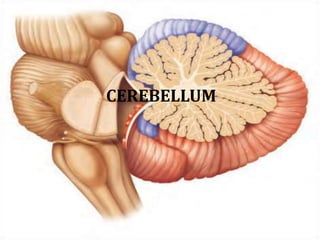
- 1. CEREBELLUM
- 2. GROSS ANATOMY OF CEREBELLUM Location: The term cerebellum is from “latin meaning” the little brain.It is a part of the hindbrain situated in the posterior cranial fossa. It is also present behind the pons and medulla ablongata, seperated from two structures by the cavity of fourth ventricle. It is covered by tentorium cerebelli and is connected to brain stem by three cerebellar peduncles. In adults the weight ratio between cerebellum and cerebrum is 1:10,Infants 1:20
- 3. Consists of two laterally, large hemisphere which are united by midline vermis. Cerebellar surface is divided by numerous curve transverse fissures giving it a laminated appearance One conspicious fissure “horizontal fissure” extends around dorsolateral border of each hemisphere from middle cerebellar peduncle to vallecula, seperating superior and inferior surface
- 4. The deepest fissure in the vermis is primary fissure, which curves ventrolaterally in the superior surface of the cerebellum to meet horizontal fissure. Primary fissure divides the cerebellum into anterior and posterior lobe.
- 5. Vermis • The cerebellar vermis (Latin for worm) is located in the medial, cortico-nuclear zone of the cerebellum, residing in the posterior fossa of the cranium. The primary fissure in the vermis curves ventrolaterally to the superior surface of the cerebellum, dividing it into anterior and posterior lobes. Functionally, the vermis is associated with bodily posture and locomotion.
- 7. Cerebellar Hemispheres • Each hemisphere consists of lobes separated by deep and distinct fissures. • The anterior lobe and posterior lobe govern subconscious aspects of skeletal muscle movements. • The flocculonodular lobe on the inferior surface contributes to equilibrium and balance.
- 8. Cerebral Nuclei • Dentate • Emboliform • Festigial • Globose
- 9. Dentate Nucleus The dentate nucleus is a cluster of neurons, or nerve cells, in the central nervous system that has a dentate – tooth-like or serrated – edge. It is located within the deep white matter of each cerebellar hemisphere, and it is the largest single structure linking the cerebellum to the rest of the brain.
- 10. Emboliform Nucleus The emboliform nucleus (or anterior interposed nucleus) is a deep cerebellar nucleus that lies immediately to the medial side of the nucleus dentatus, and partly covering its hilum.
- 11. Fastigial Nucleus • The fastigial nucleus is located in the cerebellum. It is one of the four deep cerebellar nuclei and is grey matter embedded in the white matter of the cerebellum. • It refers specifically to the concentration of gray matter nearest to the middle line at the anterior end of the superior vermis, and immediately over the roof of the fourth ventricle, from which it is separated by a thin layer of white matter
- 12. Globose Nucleus • The globose nucleus is one of the deep cerebellar nuclei. It is located medial to the emboliform nucleus and lateral to the fastigial nucleus. This nucleus contains primarily large and small multipolar neurons.
- 13. Fourth ventricle Arbor vitae cerebelli Arbor vitae •In latin “ tree of life” it is the white matter of the white matter of cerebellum. •It is so called because of the tree like appearance. •It brings sensory and motor sensation to and from cerebellum.
- 14. Cerebellar peduncles • Cerebellar peduncles connect the cerebellum to the brain stem. • Superior cerebellar peduncle is a paired structure of white matter that connects the cerebellum to the midbrain. • Inferior cerebellar peduncle is a thick rope- like strand that occupies the upper part of the posterior district of the medulla oblongata. • Middle cerebellar peduncles connect the cerebellum to the pons and are composed entirely of centripetal fibers.
- 15. The cerebellum is connected to Brain stem by three peduncles Superior cerebellar peduncle Midbr ain Middle cerebellar peduncle Pon s Inferior cerebellar peduncle Medulla ablongata
- 16. Peduncles of the cerebellum
- 17. LOBES OF CEREBELLUM Anatomical Anterior lobe Posterior lobe Flocculonodular lobe Superior surface
- 18. Archicerebellum Posterior lobe (Vestibular part) • It is formed of the flocculo- nodular lobe + associated fastigial nuclei, lying on inf. Surface in front of postero- lateral fissure. • Embryologically, it is the oldest part of cerebellum. • It receives afferent Fibres. From vestibular apparatus of internal ear Via vestibulo-cerebellar tracts. • It is concerned with equlibrium
- 19. Archicerebellum …….contd. It has connections with vestibular & reticular nuclei of brain stem through the inferior cerebellar peduncle. Afferent vestibular Fibres. Pass from vestibular nuclei in pons & medulla to the cortex of ipsilateral flocculo-nodular lobe. Efferent cortical (purkinje cell) Fibres. Project to fastigial nucleus (It is one of the four deep cerebellar nuclei and is grey matter embedded in the white matter of the cerebellum), which projects to vestibular nuclei & reticular formation. It affects the locomotror system bilaterally via descending vestibulo- spinal & reticulo-spinal tracts. Sub – divisions Vermis:- Nodulus Hemispheres:- Fluocculus and parafuocculus
- 20. Paleocerebellum (spinal part) • It is formed of midline vermis + surrounding paravermis + globose & emboliform nuclei. • It receives afferent proprio-ceptive impulses from Ms.& tendons Via spino- cerebellar tracts (dorsal & ventral) mainly. • It sends efferents to red nucleus of midbrain. (The red nucleus or nucleus ruber is a structure in the rostral midbrain involved in motor coordination. It is pale pink in color; the color is believed to be due to iron, which is present in the red nucleus in at least two different forms : hemoglobin and ferritin) • It is concerned with muscle tone
- 21. It is concerned with muscle tone & posture. Afferents spinal Fibres consist of dorsal & ventral spino-cerebellar tract from muscle, joint & cutaneous receptors to enter the cortex of ipsilateral vermis & para vermis Via inferior & superior cerebellar peduncles . Efferents cortical fibres pass to globose & emboliform nuclei, then Via sup. C. peduncle to contra- lateral red nucleus of midbrain to give rise descending rubro-spinal tract (motor control pathway) Sub – divisions Vermis:- Lingula, Columen Hemispheres:- Central Lobule and anterior quadrang lobule
- 22. Neo-cerebellum (cerebral part) • It is the remaining largest part of cerebellum. • It includes the most 2- cerebellar hemispheres + dendate nuclei. • It receives afferent impulses from the cerebral cortex+pons Via cerebro- ponto- cerebellar pathway. • It sends efferents to Ventro lateral nucleus of thalamus. • It controls voluntary movements (muscle coordination).
- 23. It is concerned with muscular coordination. It receives afferents from cerebral cortex involved in planning of movement- to pontine nuclei ,cross to opposite side Via middle Cerebellar peduncle to end in lateral parts of cerebellum (cerebro-ponto- cerebellar tract). Neo-cerebellar efferents project to dendate nucleus,which in turn projects to contra-lateral red nucleus & ventral lateral nucleus of thalamus ,then to motor cortex of frontal lobe, giving rise descending cortico-spinal & cortico-bulbar pathways. Efferents of dentate nucleus form a major part of Superior cerebellar peduncle. Sub – divisions Vermis:- Declive, Folium, tuber, pyramis, uvula Hemispheres:- Simple lobule, superior semilunar lobule, superior semilunar lobule, Gracile lobule ,Biventral lobule, Tonsil
- 24. Cerebellar Cortex • Molecular Layer • Purkinje Cell Layer • Granular Layer
- 25. CEREBELLUM CORTEX 1. Molecular Layer a. Stellate Cell:- taurine (inhibitory) afferent: parallel fiber efferent: Purkinje cell dendrite b. Basket Cell :- GABA (gamma-aminobutyric acid)(inhibitory) (Inhibit or reduce the activity of the neurons or nerve cells) afferent: parallel fiber efferent: Purkinje cell soma c. Parallel Fiber granule cell axon d. Purkinje Cell Dendrite
- 26. 2. Purkinje Cell Layer Purkinje Cell • 15,000,000 in number • GABA (inhibitory) • afferent: – parallel fiber – climbing fiber – stellate cell – basket cell • efferent: – deep cortical nuclei Bergman’s glial cell
- 28. Purkinje cells flaskshaped cell, single layered Dendrites –Molecular layer –Profuse branching –Dendritic spines Axon – synapse with deep cerebellar nucleus – basket & stellate cells – vestibular nuclei
- 29. 3.Granular Layer Granular Cell • 50,000,000,000 in number • glutamic acid (excitatory) • afferent: mossy fiber • efferent: Purkinje cell axon, basket cell, stellate cell Golgi cell GABA (inhibitory) • afferent: parallel fiber, mossy fiber rosette • efferent: granule cell dendrite
- 30. Climbing fibres - from inferior olivary complex - direct action on individual Purkinje cell -powerful , sharply localised -- Basket cells, stellate cells, Golgi cells act as inhibitory interneurons. Mossy fibres -from spinal cord / brain stem centres -indirect action on Purkinje cells via granule cells -diffuse ( thousands of Punkinje cells may be excited )
- 31. White matter of the cerebellum Consists of three types of nerve fibres in the white matter A. Axons of purkinje cells The only axons to leave cerebellar cortex to end in deep cerebellar nuclei specially dendate nucleus. B. Mossy fibres They end in the granular layer. C. Climbing fibres They end in the molecular layer
- 32. White matter of cerebellum The internal circuitry of cerebellum Do not leave the cerebellum, interconnect different regions of cerebellum. Some connect the same side. Some connect the two cerebellar hemisphere The cerebellar efferent via middle cerebellar • Peduncle(MCP) and inferior cerebellar peduncle (ICP) The cerebellar afferent via superior cerebellar • Peduncle(SCP) and from fastigial from inferior cerebellar peduncle(ICP)
- 33. Intrinsic pathway • Afferent pathways to cerebellar cortex excite Purkinje cells. • Basket, stellate and Golgi cells regulate Purkinje cell activity • Efferent pathways from the cerebellar cortex originate from Purkinje cells -
- 36. Cerebellar afferent pathway From cerebral cortex cortico-ponto-cerebellar fibres cerebro-olivo-cerebellar fibres cerebro- reticulo- cerebellar fibres From spinal cord anterior spinocerebellar tract posterior spinocerebellar tract cuneocerebellar tract From vestibular nucleus vestibulocerebellar tract [ flocculonodular lobe ] From other areas red nucleus, tectum
- 37. Afferent pathway origin Destination via Corticopontocerebellar Frontal, parietal, temporal, occipital Pontine nuclei & mossy fibres to cerebellar cortex Cerebroolivocerebellar climb fibres to cerebellar cortex Cerebroreticulocerebellar Sensorimotor areas Reticular formation Ant spinocerebellar Muscle spindles,tendons, joints Mossy fibres to cerebellar cortex Post spinocerebellar Cuneocerebellar Vestibular nerve Utricle, saccule,semicircular canals Mossy fibres to cortex of FN node others Red nuc, tectum cerebellar cortex
- 38. Cerebellar efferent pathways • Axons of Purkinje cells synapse with the cerebellar nuclei. • Axons of the neurones form the efferent pathways Connect with • Red nucleus • Thalamus • Vestibular nuclei • Reticular formation
- 39. Cerebellar cortex…..contd. Synaptic Glomerulus • Afferent terminals on granular layer Mossy Fiber Rosette – afferent fibers except inferior olivary input – 2/3 of medullary center Granular Cell Dendrite – main afferent input Golgi Cell Axon – synapse on granule cell dendrite – GABA (inhibitory) Surrounded by Astrocyte
- 40. Fibres entering and leaving through cerebellar peduncles Superior cerebellar peduncle A. Fibres entering the cerebellum 1. Ventral spino-cerebellar tract 2. Rostral spino-cerebellar tract 3. Tecto-cerebellar fibres 4. Rubro-cerebellar fibres 5. Trigemino-cerebellar fibres 6. Hypothalamo-cerebellar fibres 7. Coerulo-cerebellar fibres B. Fibres leaving the cerebellum 1. Cerebello-rubral fibres 2. Cerebello-thalamic fibres 3. Cerebello-reticular fibres 4. Cerebello-olivary fibres 5. Cerebello-nuclear fibres 6. Some fibres to hypothalamus and thalamus Superior cerebellar pedunc
- 41. Middle cerebellar peduncle Pontocerebellar fibres Inferior cerebellar peduncle A. Fibres entering cerebellum 1. Posterior spino cerebellar tract 2. Cuneo-cerebellar tract 3. Olivo-cerebellar fibres 4. Reticulo-cerebellar fibres 5. Vestibulo-cerebellar fibres 6. Anterior external arcuate fibres 7. Fibres of striae medullaries 8. Trigemino-cerebellar fibres B. Fibres Leaving the cerebellum 1. Cerebello-olivary fibres 2. Cerebello-vestibular fibres 3. Cerebello spinal and cerebello reticular fibres Middle cerebellar pedunc Inferior cerebellar peduncle
- 42. Functions • Regulates posture and postural activities • Muscular co-ordination and balance • Detection and correction of motor output from cortex • Controls timing of motor activities • Maintenance of body posture,equlibrium and eye movements. • cerebellum may also have non-motor functions such as cognition (acquisition of knowledge) and language processing.
- 43. • Damage to the cerebellum can result in a loss of ability to coordinate muscular movements, a condition called ataxia . • Cerebellar syndrome : muscular hypertonia, intensional tremors, nystagmus, unsteady gait etc.
- 44. Balance
- 45. Motor skills
- 46. BLOOD SUPPLY OF BRAIN • Internal Carotid Artery:- – Ant. cerebral artery – Middle cerebral artery • Vertebral Artery – Anterior spinal artery – Posterior spinal artery – Posterior and inferior cerebellar artery – Medullary artery – Meningial branches
- 47. Basilar artery branches • Superior, Inferior & anterior cerebellar artery • Pontine branches • Labyrinthine artery
- 51. Circle of Willis • The circle of Willis (also called Willis' circle, loop of Willis, cerebral arterial circle, and Willis polygon) is a circulatory anastomosis that supplies blood to the brain and surrounding structures.
- 52. The circle of Willis is a part of the cerebral circulation and is composed of the following arteries Anterior cerebral artery (left and right) forms anterolateral part Anterior communicating artery-which connects right and left Anterior cerebral artery and forms the anterior part. Internal carotid artery (left and right)-lateral part Posterior cerebral artery (left and right) Posterior communicating artery (left and right) – gives the link between internal carotid and posterior cerebaral Circle completed at the bifurcation of basilar artery The middle cerebral arteries, supplying the brain, are not considered part of the circle,
- 53. Functional Importance • Equalizes the pressure of the blood flow to the two sides of brain. • The arterial anastamosis provides an alternative route
- 54. Applied anatomy Berry Aneurysm • Localized dilatation on one of the arteries of circle of Willis due to congenital muscular weakness.
- 55. Venous drainage • Cerebral veins and finally drain in to dural venous sinuses
- 56. Ataxia: incoordination of movement - decomposition of movement - dysmetria, past-pointing - dysdiadochokinesia - rebound phenomenon of Holmes - gait ataxia, truncal ataxia, titubation Intention Tremor Hypotonia, Nystagmus Archicerebellar Lesion: medulloblastoma Paleocerebellar Lesion: gait disturbance Neocerebellar Lesion: hypotonia, ataxia, tremor Syndromes
- 57. Cerebellar Ataxia Ataxic gait and position: Left cerebellar tumor a. Sways to the right in standing position b. Steady on the right leg c. Unsteady on the left leg d. ataxic gait
- 58. Cerebellar Medulloblastoma Cerebellar tumors on vermis - Truncal Ataxia - Frequent Falling
- 59. THANK YOU