Downloaded 1,421 times


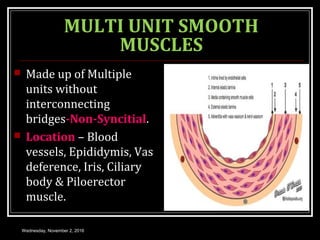


Dr. Nilesh Kate's document provides an overview of smooth muscle physiology. It discusses the functional anatomy and organization of smooth muscle, including that it is non-striated, involuntary muscle that exists in bundles. It describes the two types of smooth muscle - single unit and multi unit - and their characteristics. The document outlines the structure of smooth muscle fibers and covers the processes of excitability, contraction, and relaxation. It explains excitation and inhibition of smooth muscle can occur through nerves, hormones, pacemakers, stretching or temperature changes. In summary, the document provides a comprehensive review of smooth muscle types, organization, function and physiology.


![Smooth muscle and Cardiac Muscle [Compatibility Mode].pdf](https://cdn.slidesharecdn.com/ss_thumbnails/smoothmuscleandcardiacmusclecompatibilitymode-241026115752-31dc00ea-thumbnail.jpg?width=640&height=640&fit=bounds)