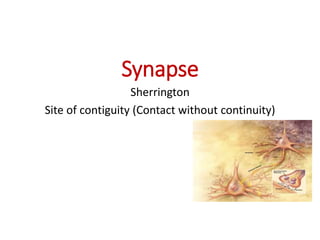
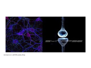
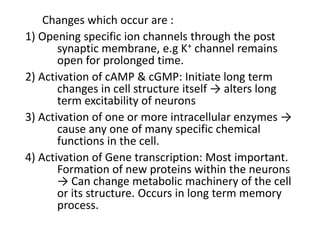
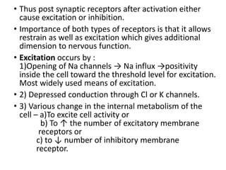
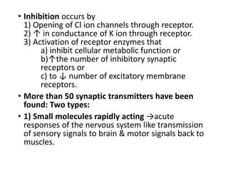
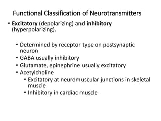
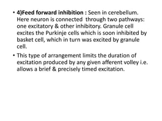
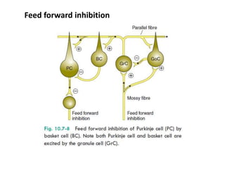
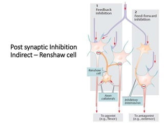
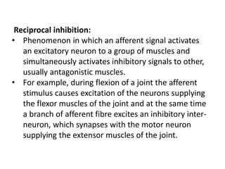
Downloaded 100 times


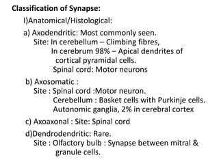
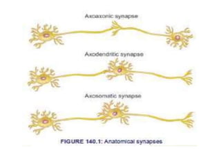
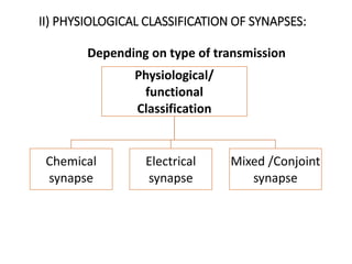

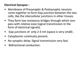
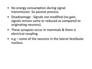
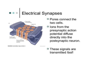
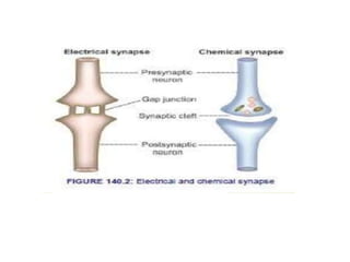
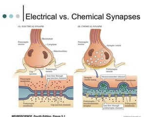
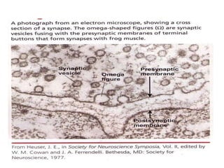
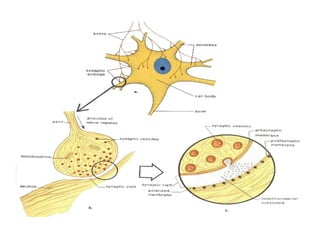

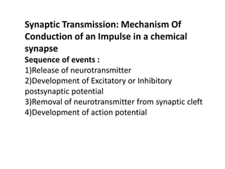

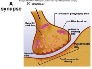
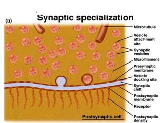
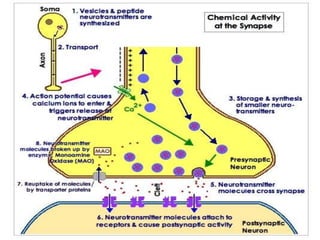
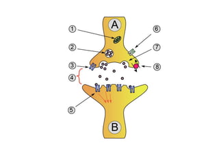
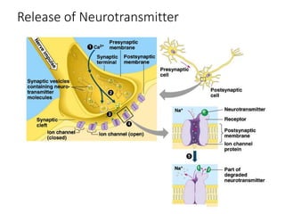
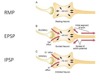
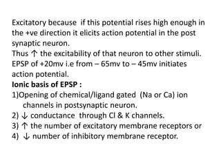
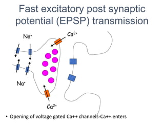
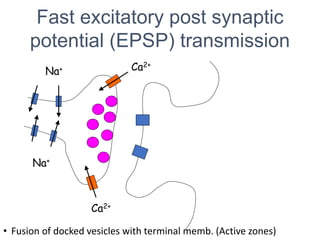
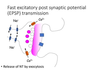
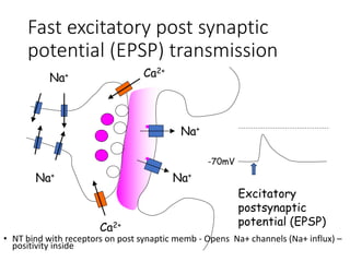
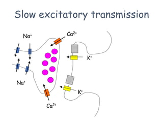
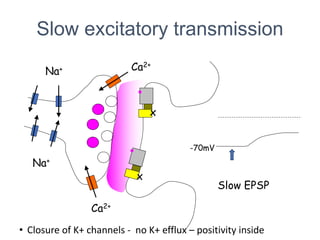
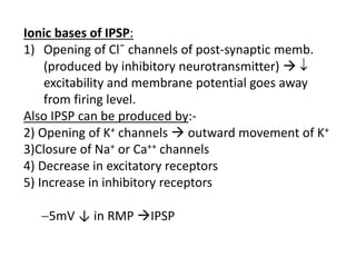
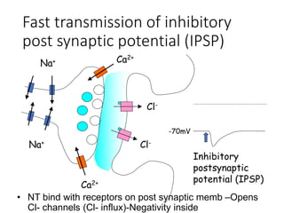
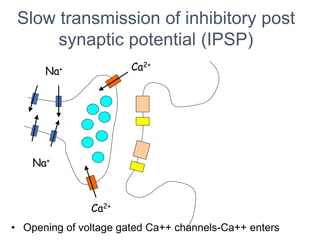
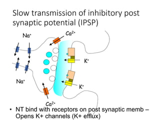
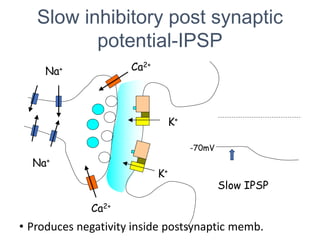
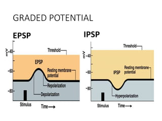

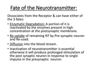
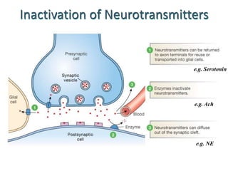
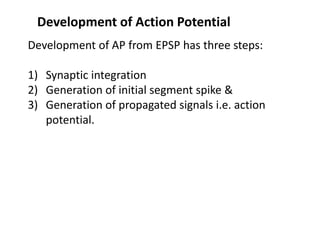

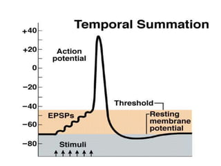
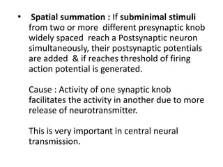
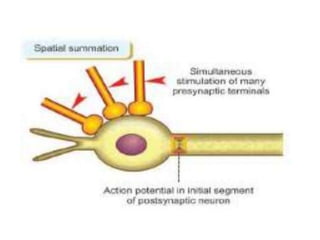
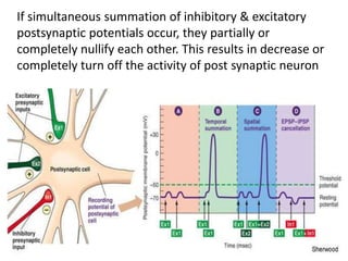


The document provides an extensive overview of synapses, detailing their structure and function in neural communication. It classifies synapses into anatomical and physiological categories, discusses the mechanisms behind chemical and electrical transmission, and describes the role of neurotransmitters in excitatory and inhibitory postsynaptic potentials. The content elaborates on the processes involved in synaptic transmission and the biochemical interactions that take place between presynaptic and postsynaptic neurons.


