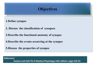
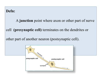
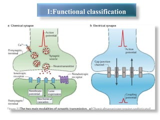
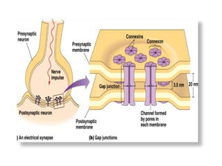
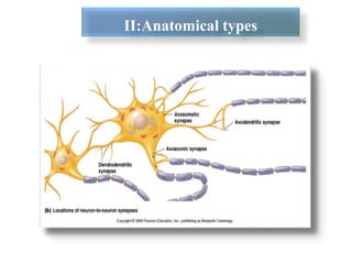
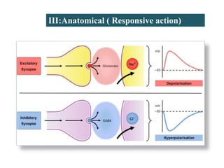
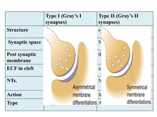
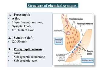
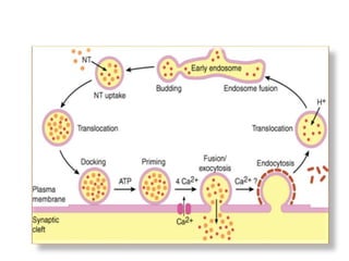
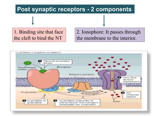
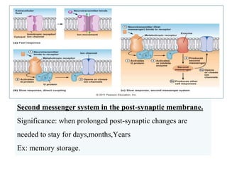
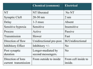
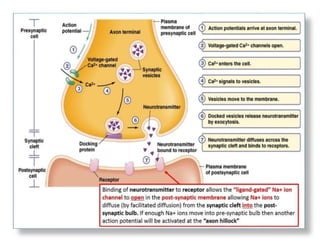
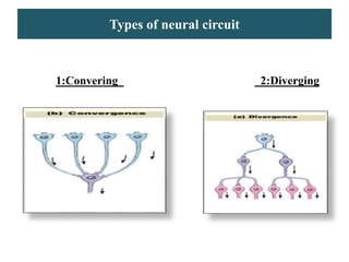
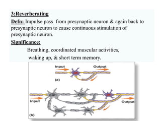
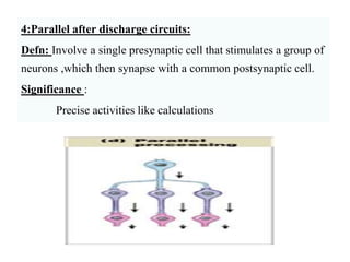
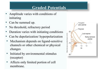
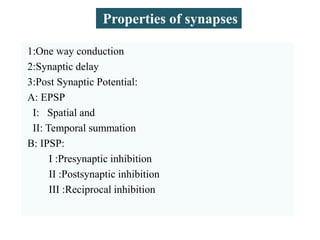
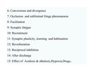
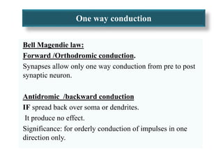
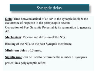
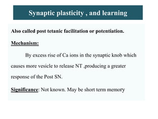
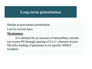
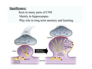
Lecture13
Synapses & their transmission
Dr Shamshad
Majamaah University
Objectives
1.Define synapse
2. Discuss the classification of synapses
3.Describe the functional anatomy of synapse
4.Describe the events occurring at the synapse
5.Discuss the properties of synapse
Reference:
Guyton and Hall-TB of Medical Physiology 13th edition: page 543-55
Defn:
A junction point where axon or other part of nerve
cell (presynaptic cell) terminates on the dendrites or
other part of another neuron (postsynaptic cell).
I:Functional classification
II:Anatomical types
III:Anatomical ( Responsive action)
Type I (Gray’s I
synapses)
Type II (Gray’s II
synapses)
Structure Asymmetric symmetric
Synaptic space wide Narrow
Post synaptic
membrane
thickened thin
ECF in cleft present absent
NTs. NT:5HT,Glutamate,
NE,E,DOPA
NT GABA,Glycine
Action Excitatory Inhibitory
Type Axo-dendritic Axo-somatic
Structure of chemical synapse
1. Presynaptic
‱ A flat,
‱ 20-ÎŒm2 membrane area,
‱ Synaptic knob ,
‱ tuft, bulb of axon
2. Synaptic cleft
‱ (20-30 nm)
3. Postsynaptic neuron
‱ Grid
‱ Sub synaptic membrane,
‱ Sub synaptic web.
Post synaptic receptors - 2 components
1. Binding site that face
the cleft to bind the NT
2. Ionophore: It passes through
the membrane to the interior.
Ion channels
Channels Cation channels Anion channels
Ions Na+ (most common) ClÂŻ (mainly)
Other ions K+,Ca2
+
Action Opening of Na+ channels
increase membrane
potential in positive
direction toward
threshold level of
excitation  (+) neuron.
Opening of ClÂŻ channels 
diffusion of negative charges
into the membrane decrease
membrane potential making
it more negative away
from threshold level  (-)
neuron.
Second messenger system in the post-synaptic membrane.
Significance: when prolonged post-synaptic changes are
needed to stay for days,months,Years
Ex: memory storage.
Chemical (common) Electrical
NT NT liberated No NT
Synaptic Cleft 20-30 nm 2 nm
Delay 1-3 msec Absent
Sensitive hypoxia Sensitive Insensitive
Process Active Passive
Transmission Slower Fast
Direction of flow Unidirectional pre-post. Bi/Unidirectional
Inhibitory Effect Inhibitory +/- No
Post synaptic
duration
Longer-mediated by
second messengers.
No
Direction of Ions
current transmission
From outside to inside From cell inside to
inside.
https://media.sciencephoto.com/image/k0039491/p
review/K0039491-
Synaptic_transmission,_animation.mp4
https://media.sciencephoto.com/image/k004
4312/preview/K0044312-
Electrical_synapse,_animation.mp4
Types of neural circuit
1:Convering 2:Diverging
3:Reverberating
Defn: Impulse pass from presynaptic neuron & again back to
presynaptic neuron to cause continuous stimulation of
presynaptic neuron.
Significance:
Breathing, coordinated muscular activities,
waking up, & short term memory.
4:Parallel after discharge circuits:
Defn: Involve a single presynaptic cell that stimulates a group of
neurons ,which then synapse with a common postsynaptic cell.
Significance :
Precise activities like calculations
Graded Potentials
‱ Amplitude varies with conditions of
initiating
‱ Can be summed up.
‱ No threshold, refractory period
‱ Duration varies with initiating conditions
‱ Can be depolarization/ hyperpolarization
‱ Mechanism depends on ligand-sensitive
channels or other chemical or physical
changes
‱ Initiated by environmental stimulus
(receptor)
‱ Affects only limited portion of cell
membrane.
1:One way conduction
2:Synaptic delay
3:Post Synaptic Potential:
A: EPSP
I: Spatial and
II: Temporal summation
B: IPSP:
I :Presynaptic inhibition
II :Postsynaptic inhibition
III :Reciprocal inhibition
Properties of synapses
6: Conversions and divergence
7: Occlusion and subliminal fringe phenomenon
8: Facilitation
9: Synaptic fatigue
10: Recruitment
11: Synaptic plasticity ,learning and habituation
12: Reverberation
13: Reciprocal inhibition
14: After discharge
15: Effect of Acidosis & alkalosis,Hypoxia,Drugs..
One way conduction
Bell Magendie law:
Forward /Orthodromic conduction.
Synapses allow only one way conduction from pre to post
synaptic neuron.
Antidromic /backward conduction
IF spread back over soma or dendrites.
It produce no effect.
Significance: for orderly conduction of impulses in one
direction only.
Synaptic delay
Defn: Time between arrival of an AP to the synaptic knob & the
occurrence of response in the postsynaptic neuron.
Generation of Post Synaptic Potential & its summation to generate
AP.
Mechanism: Release and diffusion of the NTs.
Binding of the NTs. to the post Synaptic membrane.
Minimum delay : 0.5 msec.
Significance: can be used to determine the number of synapses
present in a polysynaptic reflex.
EPSP IPSP
Cause by binding of the
excitatory NT & makes
post SM generate an AP.
by the inhibitory NT &
makes Post SM membrane
less likely to generate an AP
Ions
Flow
+vly ions. –vly ions.
Leads to Depolarization.
Brings the Post SM
towards threshold.
Hyperpolarization.
Takes the post SM away
from threshold.
Effect
on Post
SM
Excited.
Facilitated the firing of
an AP on Post SM.
Less excited.
Lowers the firing of an AP
on the post SM.
NTs &
ions
involved
Glutamate or Na+
aspartate ions
Glycine and GABA Cl-.
Temporal (Time):
When repeated stimuli applied in short duration.
Spatial (Space):
When post synaptic membrane receives impulses
simultaneously from large number of presynaptic terminals.
Inhibition
Postsynaptic : Direct by releasing inhibitory NT due to refractory
period. By development of IPSP .
Golgi-tendon inhibition
Presynaptic: Release of GABA opens K+ or Cl channels—leads to
diffusion of K ions or Cl ions .
Hyperpolarization of post synaptic membrane by inhibitory.
Location: In spinal alpha motor neuron
Neuron inhibit those neuron which excite it
Significance:
It serves to limit excitability of motor neurons.
Feedback (Renshaw cell inhibition)
Neuron connected through 2 pathways Excitatory and inhibitory.
It allows brief and precisely timed excitation.
EX : Deep neural circuit of cerebellum
Significance :
For restriction over neurons and muscles to react properly
and appropriately
Feed forward inhibition.
Fatigue
Defn:
Progressive decline in rate of discharge of the Post SN,
Following intense prolonged stimulation of the PreSN .
If sever can lead to compete stop called synaptic block
Temporary in nature.
Cause:
Fatigue is mainly due to exhaustion of Nm substances .
Due to lack of time to resynthesize and reuptake of NT.
Significance:
Protects CNS from over excitability.
Dale law
Defn:
Only one type of NT is released at one synapse.
Either excitatory or Inhibitory.
Occlusion
Defn:
Response to stimulation of 2 presynaptic neurons is
less than sum total of the response obtained when
stimulated separately.
Synaptic plasticity , and learning
Defn:
Synaptic transmission can be increased or decreased
on the basis of past experience
Defn:
When presynaptic axon is stimulated with several
consecutive individual stimuli each evokes larger post synaptic
potential than previous stimuli
Post tetanic potentiation:
Synaptic plasticity , and learning
Also called post tetanic facilitation or potentiation.
Mechanism:
By excess rise of Ca ions in the synaptic knob which
causes more vesicle to release NT ,producing a greater
response of the Post SN.
Significance: Not known. May be short term memory
Long term potentiation
Similar to post tetanic potentiation.
Last for several days.
Mechanism:
It is initiated by an increase of intracellular calcium
ion in post SN through opening of Ca 2+ channels in post
SM after binding of glutamate to its specific NMDA
receptors
Significance:
Seen in many parts of CNS
Mainly in hippocampus
Play role in long term memory and learning.

Lect 13

  • 1.
    Lecture13 Synapses & theirtransmission Dr Shamshad Majamaah University
  • 2.
    Objectives 1.Define synapse 2. Discussthe classification of synapses 3.Describe the functional anatomy of synapse 4.Describe the events occurring at the synapse 5.Discuss the properties of synapse Reference: Guyton and Hall-TB of Medical Physiology 13th edition: page 543-55
  • 3.
    Defn: A junction pointwhere axon or other part of nerve cell (presynaptic cell) terminates on the dendrites or other part of another neuron (postsynaptic cell).
  • 4.
  • 6.
  • 7.
  • 8.
    Type I (Gray’sI synapses) Type II (Gray’s II synapses) Structure Asymmetric symmetric Synaptic space wide Narrow Post synaptic membrane thickened thin ECF in cleft present absent NTs. NT:5HT,Glutamate, NE,E,DOPA NT GABA,Glycine Action Excitatory Inhibitory Type Axo-dendritic Axo-somatic
  • 9.
    Structure of chemicalsynapse 1. Presynaptic ‱ A flat, ‱ 20-ÎŒm2 membrane area, ‱ Synaptic knob , ‱ tuft, bulb of axon 2. Synaptic cleft ‱ (20-30 nm) 3. Postsynaptic neuron ‱ Grid ‱ Sub synaptic membrane, ‱ Sub synaptic web.
  • 11.
    Post synaptic receptors- 2 components 1. Binding site that face the cleft to bind the NT 2. Ionophore: It passes through the membrane to the interior.
  • 12.
    Ion channels Channels Cationchannels Anion channels Ions Na+ (most common) ClÂŻ (mainly) Other ions K+,Ca2 + Action Opening of Na+ channels increase membrane potential in positive direction toward threshold level of excitation  (+) neuron. Opening of ClÂŻ channels  diffusion of negative charges into the membrane decrease membrane potential making it more negative away from threshold level  (-) neuron.
  • 13.
    Second messenger systemin the post-synaptic membrane. Significance: when prolonged post-synaptic changes are needed to stay for days,months,Years Ex: memory storage.
  • 14.
    Chemical (common) Electrical NTNT liberated No NT Synaptic Cleft 20-30 nm 2 nm Delay 1-3 msec Absent Sensitive hypoxia Sensitive Insensitive Process Active Passive Transmission Slower Fast Direction of flow Unidirectional pre-post. Bi/Unidirectional Inhibitory Effect Inhibitory +/- No Post synaptic duration Longer-mediated by second messengers. No Direction of Ions current transmission From outside to inside From cell inside to inside.
  • 15.
  • 17.
    Types of neuralcircuit 1:Convering 2:Diverging
  • 18.
    3:Reverberating Defn: Impulse passfrom presynaptic neuron & again back to presynaptic neuron to cause continuous stimulation of presynaptic neuron. Significance: Breathing, coordinated muscular activities, waking up, & short term memory.
  • 19.
    4:Parallel after dischargecircuits: Defn: Involve a single presynaptic cell that stimulates a group of neurons ,which then synapse with a common postsynaptic cell. Significance : Precise activities like calculations
  • 20.
    Graded Potentials ‱ Amplitudevaries with conditions of initiating ‱ Can be summed up. ‱ No threshold, refractory period ‱ Duration varies with initiating conditions ‱ Can be depolarization/ hyperpolarization ‱ Mechanism depends on ligand-sensitive channels or other chemical or physical changes ‱ Initiated by environmental stimulus (receptor) ‱ Affects only limited portion of cell membrane.
  • 21.
    1:One way conduction 2:Synapticdelay 3:Post Synaptic Potential: A: EPSP I: Spatial and II: Temporal summation B: IPSP: I :Presynaptic inhibition II :Postsynaptic inhibition III :Reciprocal inhibition Properties of synapses
  • 22.
    6: Conversions anddivergence 7: Occlusion and subliminal fringe phenomenon 8: Facilitation 9: Synaptic fatigue 10: Recruitment 11: Synaptic plasticity ,learning and habituation 12: Reverberation 13: Reciprocal inhibition 14: After discharge 15: Effect of Acidosis & alkalosis,Hypoxia,Drugs..
  • 23.
    One way conduction BellMagendie law: Forward /Orthodromic conduction. Synapses allow only one way conduction from pre to post synaptic neuron. Antidromic /backward conduction IF spread back over soma or dendrites. It produce no effect. Significance: for orderly conduction of impulses in one direction only.
  • 24.
    Synaptic delay Defn: Timebetween arrival of an AP to the synaptic knob & the occurrence of response in the postsynaptic neuron. Generation of Post Synaptic Potential & its summation to generate AP. Mechanism: Release and diffusion of the NTs. Binding of the NTs. to the post Synaptic membrane. Minimum delay : 0.5 msec. Significance: can be used to determine the number of synapses present in a polysynaptic reflex.
  • 25.
    EPSP IPSP Cause bybinding of the excitatory NT & makes post SM generate an AP. by the inhibitory NT & makes Post SM membrane less likely to generate an AP Ions Flow +vly ions. –vly ions. Leads to Depolarization. Brings the Post SM towards threshold. Hyperpolarization. Takes the post SM away from threshold. Effect on Post SM Excited. Facilitated the firing of an AP on Post SM. Less excited. Lowers the firing of an AP on the post SM. NTs & ions involved Glutamate or Na+ aspartate ions Glycine and GABA Cl-.
  • 26.
    Temporal (Time): When repeatedstimuli applied in short duration. Spatial (Space): When post synaptic membrane receives impulses simultaneously from large number of presynaptic terminals.
  • 27.
    Inhibition Postsynaptic : Directby releasing inhibitory NT due to refractory period. By development of IPSP . Golgi-tendon inhibition Presynaptic: Release of GABA opens K+ or Cl channels—leads to diffusion of K ions or Cl ions . Hyperpolarization of post synaptic membrane by inhibitory.
  • 28.
    Location: In spinalalpha motor neuron Neuron inhibit those neuron which excite it Significance: It serves to limit excitability of motor neurons. Feedback (Renshaw cell inhibition)
  • 29.
    Neuron connected through2 pathways Excitatory and inhibitory. It allows brief and precisely timed excitation. EX : Deep neural circuit of cerebellum Significance : For restriction over neurons and muscles to react properly and appropriately Feed forward inhibition.
  • 30.
    Fatigue Defn: Progressive decline inrate of discharge of the Post SN, Following intense prolonged stimulation of the PreSN . If sever can lead to compete stop called synaptic block Temporary in nature. Cause: Fatigue is mainly due to exhaustion of Nm substances . Due to lack of time to resynthesize and reuptake of NT. Significance: Protects CNS from over excitability.
  • 31.
    Dale law Defn: Only onetype of NT is released at one synapse. Either excitatory or Inhibitory. Occlusion Defn: Response to stimulation of 2 presynaptic neurons is less than sum total of the response obtained when stimulated separately.
  • 32.
    Synaptic plasticity ,and learning Defn: Synaptic transmission can be increased or decreased on the basis of past experience Defn: When presynaptic axon is stimulated with several consecutive individual stimuli each evokes larger post synaptic potential than previous stimuli Post tetanic potentiation:
  • 33.
    Synaptic plasticity ,and learning Also called post tetanic facilitation or potentiation. Mechanism: By excess rise of Ca ions in the synaptic knob which causes more vesicle to release NT ,producing a greater response of the Post SN. Significance: Not known. May be short term memory
  • 34.
    Long term potentiation Similarto post tetanic potentiation. Last for several days. Mechanism: It is initiated by an increase of intracellular calcium ion in post SN through opening of Ca 2+ channels in post SM after binding of glutamate to its specific NMDA receptors
  • 35.
    Significance: Seen in manyparts of CNS Mainly in hippocampus Play role in long term memory and learning.

Editor's Notes

  • #6 Channels: Connexon Diameter is 2nm 6 subunits: connexin
  • #8 Axodendritic Axosomatic Marked thickening slight thickening which is not continous Wider 30nm 20nm Filled with dense extrcellular tissue no such tissue is present Sites CC , C LIMBING FIBRES A.N.S, BASKET CELLS 1. SPHERICAL-E Small-molecule NT (synthesized in the cytoplasm of the terminal buttons and packed into vesicles by the Golgi complex) 2. HORIZONTAL/FLAT-I (synthesized in the soma by ribosomes and packed into vesicles by the soma’s Golgi complex and then moved down to the terminals by microtubules) Clear vesicles acetylcholine) Dense-core vesicles(catecholamine) Small-molecule transmitters (synthesized in the cytoplasm of the terminal buttons and packed into vessicles by the Golgi complex) Large-molecule (peptide) transmitters (synthesized in the soma by ribosomes and packed into vessicles by the soma’s Golgi complex and then moved down to the terminals by microtubules)
  • #10 A chemical synapse consists Chemical synapses between neurons are generally small, often less than 1 ÎŒm in diameter, which means that their detailed structure can be seen only with an electron microscope; light microscope, brain synapses are usually visible only as swellings along or at the termination of the axons . . Synapses are polarized, which means that their two apposed sides have different structures. This polarity reflects the fact that most synapses transmit information in one direction but not in the other (we will see that some rare exceptions do exist). The presynaptic side contains numerous clear vesicles, 40 to 50 nm in diameter, that appear empty when viewed by transmission electron microscopy. Synaptic termini may also contain large (100 to 200 nm in diameter), dense-core secretory granules that are morphologically quite similar to the secretory granules of endocrine cells. These granules contain neuropeptides, that is, peptides or small proteins that act as neurotransmitters and for which receptors exist in the postsynaptic membranes . Endocrine hormones such as adrenocorticotropic hormone, vasoactive intestinal polypeptide, and cholecystokinin are found in dense-core secretory granules present in the terminals of certain central and peripheral neurons. The clear synaptic vesicles (i.e., not the dense-core granules) are anchored and shifted about by a dense network of cytoskeletal proteins. Some vesicles are clustered close to the part of the presynaptic membrane that apposes the synaptic contact; these vesicle attachment sites are called active zones. Synaptic vesicles are lined up several deep along the active zones, which are probably the regions of actual exocytosis. The number of active zones per synapse varies greatly (active zones . Most synapses in the central nervous system (CNS) have relatively few active zones, often only 1 but occasionally as many as 10 or 20 (versus the hundreds in the neuromuscular junction). If we could view the presynaptic face of an active zone from the perspective of a synaptic vesicle, we would see filaments and particles projecting from the presynaptic membrane, often forming a regular hexagonal arrangement called a presynaptic grid. Specific points along the grid are thought to be the vesicle release sites. Unlike the clear synaptic vesicles containing non-peptide transmitters, dense-core secretory granules are distributed randomly throughout the cytoplasm of the synaptic terminus. They are not concentrated at the presynaptic density, and they do not appear to release their contents at the active zone. Although the molecular pathways that control exocytosis of the neuronal dense-core granules are still being elucidated, it appears that a rise in [Ca2+]i is a primary stimulus.
  • #21  An action potential consists of a temporary change in a neuron’s electrical potential The neuron must recover to Resting Potential