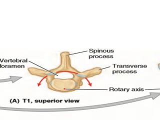
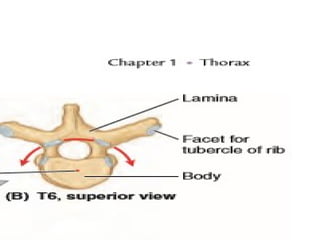
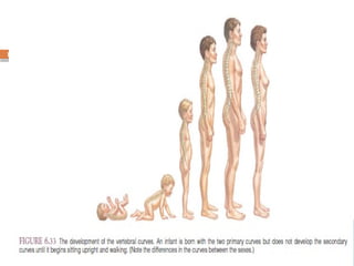
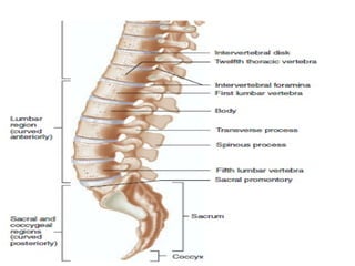
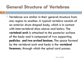
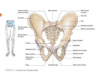
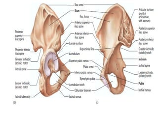
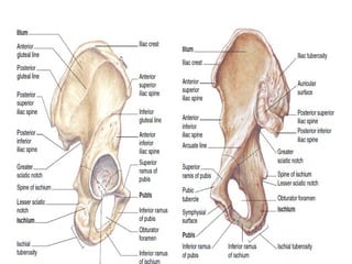
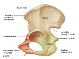
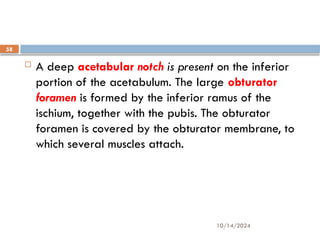
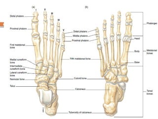
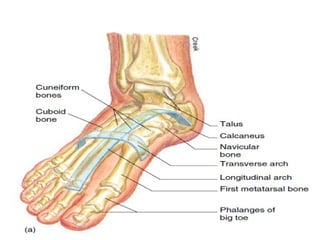
10/14/2024
1
VERTEBRAL COLUMN
10/14/2024
2
VERTEBRAL COLUMN
 The vertebral column consists of a series of irregular
bones called vertebrae, separated from each other
by fibrocartilaginous intervertebral discs. Vertebrae
enclose and protect the spinal cord,
10/14/2024
3
10/14/2024
4
10/14/2024
5
 support the skull and allow for its movement,
articulate with the rib cage, and provide for the
attachment of trunk muscles. The intervertebral discs
lend flexibility to the vertebral column and absorb
vertical shock.
10/14/2024
6
 The vertebral column (“backbone”) and the spinal
cord of the nervous system constitute the spinal
column. The vertebral column has four functions:
1) to support the head and upper extremities while
permitting freedom of movement
2) to provide attachment for various muscles, ribs, and
visceral organs; and
3) to protect the spinal cord and permit passage of
the spinal nerves.
10/14/2024
7
 The vertebral column is typically composed of 33
individual vertebrae, some of which are fused.
There are 7 cervical, 12 thoracic, 5 lumbar, 3 to 5
fused sacral, and 4 or 5 fused coccygeal
vertebrae;
10/14/2024
8
 the adult vertebral column is composed of a total of
26 movable parts. Vertebrae are separated by
fibrocartilaginous intervertebral discs and are
secured to each other by interlockin Between the
vertebrae are openings called intervertebral
foramina that allow passage of spinal nerves
processes and binding ligaments.
10/14/2024
9
 When viewed from the side, four curvatures of the
vertebral column can be identified. The cervical,
thoracic, and lumbar curves are identified by the
type of include. The pelvic curve (sacral curve) is
formed by the shape of the sacrum and coccyx
10/14/2024
10
 The four vertebral curves are not present in an
infant. The cervical curve begins to develop at
about 3 months as the baby begins holding up its
head. The lumbar curve develops as a child begins
to walk. The thoracic and pelvic curves are called
primary curves because they retain the shape of the
fetus. The cervical and lumbar curves are called
secondary curves because they are modifications of
the fetal shape.
10/14/2024
11
10/14/2024
12
10/14/2024
13
10/14/2024
14
General Structure of Vertebrae
 Vertebrae are similar in their general structure from
one region to another. A typical vertebra consists of
an anterior drum shaped body, which is in contact
with intervertebral discs above and below. The
vertebral arch is attached to the posterior surface
of the body and is composed of two supporting
pedicles and two arched laminae. The space formed
by the vertebral arch and body is the vertebral
foramen, through which the spinal cord passes.
10/14/2024
15
 Between the pedicles of adjacent vertebrae are the
intervertebral foramina, through which spinal
nerves emerge as they branch off the spinal cord.
Seven processes arise from the vertebral arch of a
typical vertebrae: the spinous process, two
transverse processes, two superior articular
processes, and two inferior articular processes.
10/14/2024
16
10/14/2024
17
The Appendicular Skeleton
 Pectoral Girdle
 Two scapulae and two clavicles make up the pectoral
(shoulder) girdle. It is not a complete girdle, having
only an anterior attachment to the axial skeleton,
via the sternoclavicular joint at the sternum.
10/14/2024
18
Conti…..
 the pectoral girdle has a wide range of movement.
Because it is not weight-bearing, it is structurally
more delicate than the pelvic girdle. The primary
function of the pectoral girdle is to provide
attachment areas for the numerous muscles that
move the shoulder and elbow joints
10/14/2024
19
 Clavicle: The slender S-shaped clavicle connects the upper
extremity to the axial skeleton and holds the shoulder joint
away from the trunk to permit freedom of movement. The
articulation of the medial sternal extremity of the clavicle to
the manubrium of the sternum is referred to as the
sternoclavicular joint. The lateral acromial extremity of the
clavicle articulates with the acromion of the scapula. This
articulation is referred to as the acromioclavicular joint. A
conoid tubercle is present on the acromial extremity of the
clavicle, and a costal tuberosity is present on the inferior
surface of the sternal extremity. Both processes serve as
attachments for ligaments.
10/14/2024
20
 Scapula: The scapula is a large, triangular flat bone
on the posterior side of the rib cage, overlying ribs
2 through 7. The spine of the scapula is a
prominent diagonal bony ridge seen on the
posterior surface. The spine strengthens the scapula,
making it more resistant to bending
10/14/2024
21
 Above the spine is the supraspinous fossa, and
below the spine is the infraspinous fossa. The spine
broadens toward the shoulder as the acromion
process.
 Inferior to the acromion is a shallow depression, the
glenoid cavity, into which the head of the humerus
fits. The coracoid process is a thick upward
projection lying superior and anterior to the glenoid
cavity.
10/14/2024
22
 On the anterior surface of the scapula is a slightly
concave area known as the subscapular fossa.
 The scapula has three borders delimited by three
angles. The superior edge is called the superior
border. The medial border is nearest to the
vertebral column, and the lateral border is directed
toward the arm.
10/14/2024
23
 The superior angle is located between the superior
and medial borders; the inferior angle, at the
junction of the medial and lateral borders; and the
lateral angle, at the junction of the superior and
lateral borders. It is at the lateral angle that the
scapula articulates with the head of the humerus.
Along the superior border, a distinct depression
called the scapular notch is a passageway for the
suprascapular nerve.
10/14/2024
24
10/14/2024
25
 Brachium (Arm):The brachium extends from the
shoulder to the elbow. arm refers only to this
portion of the upper limb. The brachium contains a
single bone Humerus
 The humerus: is the longest bone of the upper
extremity. It consists of a proximal head, which
articulates with the glenoid cavity of the scapula; a
body (“shaft”); and a distal end, which is modified
to articulate with the two bones forearm.
10/14/2024
26
 Surrounding the margin of the head is a slightly
indented groove denoting the anatomical neck.
The surgical neck, the constriction just below the
head, is a frequent fracture site. The greater
tubercle is a large knob on the lateral proximal
portion of the humerus. The lesser tubercle is
slightly anterior to the greater tubercle and is
separated from the greater by an intertubercular
groove.
10/14/2024
27
10/14/2024
28
The tendon of the long head of the biceps brachii
muscle passes through this groove. Along the lateral
mid region of the body of the humerus is a
roughened area, the deltoid tuberosity, for the
attachment of the deltoid muscle. Small openings in
the body are called nutrient foramina.
10/14/2024
29
 The humeral condyle on the distal end of the
humerus has two articular surfaces. The capitulum
is the lateral rounded part that articulates with the
radius. The trochlea is the pulleylike medial part
that articulates with the ulna.
10/14/2024
30
 is the pulleylike medial part that articulates with the
ulna. On either side above the condyle are the
lateral and medial epicondyles. The large medial
epicondyle protects the ulnar nerve that passes
posteriorly through the ulnar sulcus. It is popularly
known as the “funny bone” because striking the
elbow on the edge of a table, for example,
stimulates the ulnar nerve and produces a tingling
sensation.
10/14/2024
31
 The coronoid fossa is a depression above the
trochlea on the anterior surface. The olecranon
fossa is a depression on the distal posterior surface.
Both fossae are adapted to work with the ulna
during movement of the forearm.
10/14/2024
32
10/14/2024
33
 Antebrachium (Forearm):
 The skeletal structures of the antebrachium are the
ulna on the medial side and the radius on the lateral
(thumb) side The ulna is more firmly connected to
the humerus than the radius, and it is longer than the
radius. The radius, however, contributes more
significantly to the articulation at the wrist joint than
does the ulna.
10/14/2024
34
Ulna
The proximal end of the ulna articulates with the
humerus and radius. A distinct depression, the
trochlear notch, articulates with the trochlea of the
humerus. The coronoid process forms the anterior
lip of the trochlear notch, and the olecranon forms
the posterior portion. Lateral and inferior to the
coronoid process is the radial notch, which
accommodates the head of the radius.
10/14/2024
35
 On the tapered distal end of the ulna is a knobbed
portion, the head, and a knoblike projection, the
styloid process. The ulna articulates at both ends
with the radius.
10/14/2024
36
Radius
 The radius consists of a body with a small proximal
end and a large distal end. A proximal disc-shaped
head articulates with the capitulum of the humerus
and the radial notch of the ulna. The prominent
tuberosity of radius (radial tuberosity), for
attachment of the biceps brachii muscle, is located
on the medial side of the body, just below the head.
On the distal end of the radius is a double-faceted
surface for articulation with the proximal carpal
bones.
10/14/2024
37
 The distal end of the radius also has a styloid
process on the lateral tip and an ulnar notch on the
medial side that receives the distal end of the ulna.
The styloid processes on the ulna and radius
provide lateral and medial stability for articulation
at the wrist.
10/14/2024
38
10/14/2024
39
Manus (Hand)
 The hand contains 27 bones, grouped into the
carpus, metacarpus, and phalanges
 Carpus: The carpus, or wrist, contains eight carpal
bones arranged in two transverse rows of four
bones each. The proximal row, naming from the
lateral (thumb) to the medial side, consists of the
scaphoid (navicular), lunate, triquetrum and
pisiform. The pisiform forms in a tendon as a
sesamoid bone.
10/14/2024
40
 The distal row, from lateral to medial, consists of the
trapezium (greater multangular), trapezoid (lesser
multangular), capitate, and hamate. The scaphoid
and lunate of the proximal row articulate with the
distal end of the radius.
10/14/2024
41
Metacarpus
 The metacarpus, or palm of the hand, contains five
metacarpal bones. Each metacarpal bone consists
of a proximal base, a body, and a distal head that
is rounded for articulation with the base of each
proximal phalanx. The heads of the metacarpal
bones are distally located and form the knuckles of
a clenched fist.
10/14/2024
42
Phalanges
 The 14 phalanges are the bones of the digits. A
single finger bone is called a phalanx. The
phalanges of the fingers are arranged in a
proximal row, a middle row, and a distal row. The
thumb, or pollex (adjective, pollicis), lacks a middle
phalanx. The digits are sequentially numbered I to
V starting with the thumb—the lateral side, in
reference to anatomical position.
10/14/2024
43
PELVIC GIRDLE
AND LOWER EXTREMITY
 The structure of the pelvic girdle and lower
extremities is adaptive for support and locomotion.
Extensive processes and surface features on certain
bones of the pelvic girdle and lower extremities
accommodate massive muscles used in body
movement and in maintaining posture.
10/14/2024
44
Pelvic Girdle
 The pelvic girdle is formed by two ossa coxae
united anteriorly at the symphysis pubis . It is
attached posteriorly to the sacrum of the vertebral
column. The sacrum, a bone of the axial skeleton,
The deep, basin like structure formed by the ossa
coxae, together with the sacrum and coccyx, is
called the pelvis.
10/14/2024
45
 The pelvis is divided into a greater (false) pelvis
and a lesser (true) pelvis. These two components
are divided by the pelvic brim, a curved bony rim
passing inferiorly from the sacral promontory to the
upper margin of the symphysis pubis. The greater
pelvis is the expanded portion of the pelvis, superior
to the pelvic brim. The pelvic brim not only divides
the two portions but surrounds the pelvic inlet of the
lesser pelvis. The lower circumference of the lesser
pelvis bounds the pelvic outlet.
10/14/2024
46
 The pelvic girdle and its associated ligaments
support the weight of the body from the vertebral
column. The pelvic girdle also supports and protects
the lower viscera, including the urinary bladder, the
reproductive organs, and in a pregnant woman, the
developing fetus.
10/14/2024
47
10/14/2024
48
 Each os coxae (“hipbone”) actually consists of three
separate bones: the ilium, the ischium, and the pubis.
These bones are fused together in the adult. On the
lateral surface of the os coxae, where the three
bones ossify, is a large circular depression, the
acetabulum which receives the head of the femur.
Although both ossa coxae are single bones in the
adult, the three components of each one are
considered separately for descriptive purposes
10/14/2024
49
10/14/2024
50
Ilium
 The ilium is the uppermost and largest of the three
pelvic bones. It has a crest and four angles, or
spines—important surface landmarks that serve for
muscle attachment. The iliac crest forms the
prominence of the hip. This crest terminates
anteriorly as the anterior superior iliac spine. Just
below this spine is the anterior inferior iliac spine.
10/14/2024
51
 The posterior termination of the iliac crest is the
posterior superior iliac spine, and just below this is
the posterior inferior iliac spine.
10/14/2024
52
 Below the posterior inferior iliac spine is the greater
sciatic notch, through which the sciatic nerve
passes. On the medial surface of the ilium is the
roughened auricular surface, which articulates with
the sacrum. The iliac fossa is the smooth, concave
surface on the anterior portion of the ilium. The
iliacus muscle originates from this fossa.
10/14/2024
53
 The iliac tuberosity, for the attachment of the
sacroiliac ligament, is positioned posterior to the
iliac fossa. Three roughened ridges are present on
the gluteal surface of the posterior aspect of the
ilium. These ridges, which serve to attach the gluteal
muscles, are the inferior, anterior, and posterior
gluteal lines
10/14/2024
54
10/14/2024
D
55
10/14/2024
56
Ischium
 The ischium is the posteroinferior bone of the os
coxae. This bone has several distinguishing features.
The spine of the ischium is the projection
immediately posterior and inferior to the greater
sciatic notch of the ilium. Inferior to this spine is the
lesser sciatic notch of the ischium. The ischial
tuberosity is the bony projection that supports the
weight of the body in the sitting position.
10/14/2024
57
10/14/2024
58
 A deep acetabular notch is present on the inferior
portion of the acetabulum. The large obturator
foramen is formed by the inferior ramus of the
ischium, together with the pubis. The obturator
foramen is covered by the obturator membrane, to
which several muscles attach.
10/14/2024
59
Pubis
 The pubis is the anterior bone of the os coxae. It
consists of a superior ramus and an inferior ramus
that support the body of the pubis. The body
contributes to the formation of the symphysis pubis
—the joint between the two ossa coxae. At the
lateral end of the anterior border of the body is
the pubic tubercle, one of the attachments for the
inguinal ligament.
10/14/2024
60
10/14/2024
61
Thigh
 The femur is the only bone of the thigh.
 Femur:
 The is the longest, heaviest, strongest bone in the body.
The proximal rounded head of the femur articulates with
the acetabulum of the os coxae. A roughened shallow
pit, the fovea capitis femoris, is present in the lower
center of the head of the femur. The fovea capitis
femoris provides the point of attachment for the
ligamentum capitis femoris, which helps to support the
head of the femur against the acetabulum.
10/14/2024
62
Conti…..
 It also provides the site for the entry of an artery
into the head of the femur. The constricted region
supporting the head is called the neck and is a
common site for fractures in the elderly.
10/14/2024
63
Conti…..
 The body of the femur has a slight medial curve to bring the
knee joint in line with the body’s plane of gravity. The
degree of curvature is greater in the female because of the
wider pelvis. The body of the femur has several distinguishing
features for muscle attachment. On the proximolateral side
of the body is the greater trochanter, and on the medial side
is the lesser trochanter. On the anterior side, between the
trochanters, is the intertrochanteric line. On the posterior
side, between the trochanters, is the intertrochanteric crest.
The linea aspera is a roughened vertical ridge on the
posterior surface of the body of the femur.
10/14/2024
64
 The distal end of the femur is expanded for
articulation with the tibia. The medial and lateral
condyles are the articular processes for this joint.
The shallow depression between the condyles on the
posterior aspect is called the intercondylar fossa.
The patellar surface is located between the condyles
on the anterior side. Above the condyles on the
lateral and medial sides are the epicondyles, which
serve for ligament and tendon attachment.
10/14/2024
65
10/14/2024
66
10/14/2024
67
10/14/2024
68
patella
The patella ( “kneecap”) is a large, triangular
sesamoid bone positioned on the anterior side of
the distal femur. It develops in response to strain in
the patellar tendon. It has a broad base and an
inferiorly pointed apex. Articular facets on the
articular surface of the patella articulate with the
medial and lateral condyles of the femur.
10/14/2024
69
 The functions of the patella are to protect the knee
joint and to strengthen the patellar tendon. It also
increases the leverage of the quadriceps femoris
muscle as it extends (straightens) the knee joint.
10/14/2024
70
Leg
 Technically speaking, leg refers only to that portion
of the lower limb between the knee and foot. The
tibia and fibula are the bones of the leg. The tibia is
the larger and more medial of the two.
10/14/2024
71
Tibia
 The tibia ( “shinbone”) articulates proximally with
the femur at the knee joint and distally with the
talus of the ankle. It also articulates both proximally
and distally with the fibula. Two slightly concave
surfaces on the proximal end of the tibia, the
medial and lateral condyles articulate with the
condyles of the femur.
10/14/2024
72
 The condyles are separated by a slight upward
 projection called the intercondylar eminence,
which provides attachment for the cruciate
ligaments of the knee joint. The tibial tuberosity, for
attachment of the patellar ligament, is located on
the proximoanterior part of the body of the tibia.
The anterior crest, commonly called the “shin,” is a
sharp ridge along the anterior surface of the body.
10/14/2024
73
 The medial malleolus is a prominent medial knob
of bone located on the distomedial end of the tibia.
A fibular notch, for articulation with the fibula, is
located on the distolateral end. In that the tibia is
the weight-bearing bone of the leg, it is much
larger than the fibula.
10/14/2024
74
Fibula
 The fibula is a long, slender bone that is more
important for muscle attachment than for support.
The head of the fibula articulates with the
proximolateral end of the tibia. The distal end has
a prominent knob called the lateral malleolus.
10/14/2024
75
10/14/2024
76
Pes (Foot)
 The foot contains 26 bones, grouped into the tarsus,
metatarsus, and phalanges
 Tarsus: There are seven tarsal bones. The most
superior in position is the talus, which articulates
with the tibia and fibula to form the ankle joint. The
calcaneus is the largest of the tarsal bones and
provides skeletal support for the heel of the foot.
10/14/2024
77
 It has a large posterior extension, called the
tuberosity of the calcaneus, for the attachment of
the calf muscles. Anterior to the talus is the block-
shaped navicular bone. The remaining four tarsal
bones form a distal series that articulate with the
metatarsal bones. They are, from the medial to the
lateral side, the medial, intermediate, and lateral
cuneiform bones and the cuboid bone.
10/14/2024
78
Metatarsus: The metatarsal bones and
phalanges are similar in name and number to the
metacarpals and phalanges of the hand. They
differ in shape, however, because of their load-
bearing role. The metatarsal bones are numbered I
to V, starting with the medial (great toe) side of the
foot. The first metatarsal bone is larger than the
others because of its major role in supporting body
weight.
10/14/2024
79
 The metatarsal bones each have a base, body, and
head. The proximal bases of the first, second, and
third metatarsals articulate proximally with the
cuneiform bones. The heads of the metatarsals
articulate distally with the proximal phalanges. The
proximal joints are called tarsometatarsal joints, and
the distal joints are called metatarsophalangeal
joints. The ball of the foot is formed by the heads of
the first two metatarsal bones.
10/14/2024
80
 Phalanges: The 14 phalanges are the skeletal
elements of the toes. As with the fingers of the
hand, the phalanges of the toes are arranged in a
proximal row, a middle row, and a distal row. The
great toe, or hallux has only a proximal and a distal
phalanx.
10/14/2024
81
10/14/2024
82
 Arches of the Foot: The foot has two arches. They
are formed by the structure and arrangement of
the bones and maintained by ligaments and tendons
The arches are not rigid; they “give” when weight is
placed on the foot, and they spring back as the
weight is lifted.
10/14/2024
83
 The longitudinal arch is divided into medial and
lateral parts. The medial part is the more elevated
of the two. The talus is keystone of the medial part,
which originates at the calcaneus, rises at the talus,
and descends to the first three metatarsal bones.
The shallower lateral part consists of the calcaneus,
cuboid, and fourth and fifth metatarsal bones. The
cuboid is the keystone bone of this arch
10/14/2024
84
 The transverse arch extends across the width of the
foot and is formed by the calcaneus, navicular, and
cuboid bones posteriorly and the bases of all five
metatarsal bones anteriorly. A weakening of the
ligaments and tendons of the foot may cause the
arches to “fall”—a condition known as pes planus,
or, more commonly, “flatfoot.”
10/14/2024
85

Veretebrea upper limb and lower limb.pptx

  • 1.
  • 2.
    10/14/2024 2 VERTEBRAL COLUMN  Thevertebral column consists of a series of irregular bones called vertebrae, separated from each other by fibrocartilaginous intervertebral discs. Vertebrae enclose and protect the spinal cord,
  • 3.
  • 4.
  • 5.
    10/14/2024 5  support theskull and allow for its movement, articulate with the rib cage, and provide for the attachment of trunk muscles. The intervertebral discs lend flexibility to the vertebral column and absorb vertical shock.
  • 6.
    10/14/2024 6  The vertebralcolumn (“backbone”) and the spinal cord of the nervous system constitute the spinal column. The vertebral column has four functions: 1) to support the head and upper extremities while permitting freedom of movement 2) to provide attachment for various muscles, ribs, and visceral organs; and 3) to protect the spinal cord and permit passage of the spinal nerves.
  • 7.
    10/14/2024 7  The vertebralcolumn is typically composed of 33 individual vertebrae, some of which are fused. There are 7 cervical, 12 thoracic, 5 lumbar, 3 to 5 fused sacral, and 4 or 5 fused coccygeal vertebrae;
  • 8.
    10/14/2024 8  the adultvertebral column is composed of a total of 26 movable parts. Vertebrae are separated by fibrocartilaginous intervertebral discs and are secured to each other by interlockin Between the vertebrae are openings called intervertebral foramina that allow passage of spinal nerves processes and binding ligaments.
  • 9.
    10/14/2024 9  When viewedfrom the side, four curvatures of the vertebral column can be identified. The cervical, thoracic, and lumbar curves are identified by the type of include. The pelvic curve (sacral curve) is formed by the shape of the sacrum and coccyx
  • 10.
    10/14/2024 10  The fourvertebral curves are not present in an infant. The cervical curve begins to develop at about 3 months as the baby begins holding up its head. The lumbar curve develops as a child begins to walk. The thoracic and pelvic curves are called primary curves because they retain the shape of the fetus. The cervical and lumbar curves are called secondary curves because they are modifications of the fetal shape.
  • 11.
  • 12.
  • 13.
  • 14.
    10/14/2024 14 General Structure ofVertebrae  Vertebrae are similar in their general structure from one region to another. A typical vertebra consists of an anterior drum shaped body, which is in contact with intervertebral discs above and below. The vertebral arch is attached to the posterior surface of the body and is composed of two supporting pedicles and two arched laminae. The space formed by the vertebral arch and body is the vertebral foramen, through which the spinal cord passes.
  • 15.
    10/14/2024 15  Between thepedicles of adjacent vertebrae are the intervertebral foramina, through which spinal nerves emerge as they branch off the spinal cord. Seven processes arise from the vertebral arch of a typical vertebrae: the spinous process, two transverse processes, two superior articular processes, and two inferior articular processes.
  • 16.
  • 17.
    10/14/2024 17 The Appendicular Skeleton Pectoral Girdle  Two scapulae and two clavicles make up the pectoral (shoulder) girdle. It is not a complete girdle, having only an anterior attachment to the axial skeleton, via the sternoclavicular joint at the sternum.
  • 18.
    10/14/2024 18 Conti…..  the pectoralgirdle has a wide range of movement. Because it is not weight-bearing, it is structurally more delicate than the pelvic girdle. The primary function of the pectoral girdle is to provide attachment areas for the numerous muscles that move the shoulder and elbow joints
  • 19.
    10/14/2024 19  Clavicle: Theslender S-shaped clavicle connects the upper extremity to the axial skeleton and holds the shoulder joint away from the trunk to permit freedom of movement. The articulation of the medial sternal extremity of the clavicle to the manubrium of the sternum is referred to as the sternoclavicular joint. The lateral acromial extremity of the clavicle articulates with the acromion of the scapula. This articulation is referred to as the acromioclavicular joint. A conoid tubercle is present on the acromial extremity of the clavicle, and a costal tuberosity is present on the inferior surface of the sternal extremity. Both processes serve as attachments for ligaments.
  • 20.
    10/14/2024 20  Scapula: Thescapula is a large, triangular flat bone on the posterior side of the rib cage, overlying ribs 2 through 7. The spine of the scapula is a prominent diagonal bony ridge seen on the posterior surface. The spine strengthens the scapula, making it more resistant to bending
  • 21.
    10/14/2024 21  Above thespine is the supraspinous fossa, and below the spine is the infraspinous fossa. The spine broadens toward the shoulder as the acromion process.  Inferior to the acromion is a shallow depression, the glenoid cavity, into which the head of the humerus fits. The coracoid process is a thick upward projection lying superior and anterior to the glenoid cavity.
  • 22.
    10/14/2024 22  On theanterior surface of the scapula is a slightly concave area known as the subscapular fossa.  The scapula has three borders delimited by three angles. The superior edge is called the superior border. The medial border is nearest to the vertebral column, and the lateral border is directed toward the arm.
  • 23.
    10/14/2024 23  The superiorangle is located between the superior and medial borders; the inferior angle, at the junction of the medial and lateral borders; and the lateral angle, at the junction of the superior and lateral borders. It is at the lateral angle that the scapula articulates with the head of the humerus. Along the superior border, a distinct depression called the scapular notch is a passageway for the suprascapular nerve.
  • 24.
  • 25.
    10/14/2024 25  Brachium (Arm):Thebrachium extends from the shoulder to the elbow. arm refers only to this portion of the upper limb. The brachium contains a single bone Humerus  The humerus: is the longest bone of the upper extremity. It consists of a proximal head, which articulates with the glenoid cavity of the scapula; a body (“shaft”); and a distal end, which is modified to articulate with the two bones forearm.
  • 26.
    10/14/2024 26  Surrounding themargin of the head is a slightly indented groove denoting the anatomical neck. The surgical neck, the constriction just below the head, is a frequent fracture site. The greater tubercle is a large knob on the lateral proximal portion of the humerus. The lesser tubercle is slightly anterior to the greater tubercle and is separated from the greater by an intertubercular groove.
  • 27.
  • 28.
    10/14/2024 28 The tendon ofthe long head of the biceps brachii muscle passes through this groove. Along the lateral mid region of the body of the humerus is a roughened area, the deltoid tuberosity, for the attachment of the deltoid muscle. Small openings in the body are called nutrient foramina.
  • 29.
    10/14/2024 29  The humeralcondyle on the distal end of the humerus has two articular surfaces. The capitulum is the lateral rounded part that articulates with the radius. The trochlea is the pulleylike medial part that articulates with the ulna.
  • 30.
    10/14/2024 30  is thepulleylike medial part that articulates with the ulna. On either side above the condyle are the lateral and medial epicondyles. The large medial epicondyle protects the ulnar nerve that passes posteriorly through the ulnar sulcus. It is popularly known as the “funny bone” because striking the elbow on the edge of a table, for example, stimulates the ulnar nerve and produces a tingling sensation.
  • 31.
    10/14/2024 31  The coronoidfossa is a depression above the trochlea on the anterior surface. The olecranon fossa is a depression on the distal posterior surface. Both fossae are adapted to work with the ulna during movement of the forearm.
  • 32.
  • 33.
    10/14/2024 33  Antebrachium (Forearm): The skeletal structures of the antebrachium are the ulna on the medial side and the radius on the lateral (thumb) side The ulna is more firmly connected to the humerus than the radius, and it is longer than the radius. The radius, however, contributes more significantly to the articulation at the wrist joint than does the ulna.
  • 34.
    10/14/2024 34 Ulna The proximal endof the ulna articulates with the humerus and radius. A distinct depression, the trochlear notch, articulates with the trochlea of the humerus. The coronoid process forms the anterior lip of the trochlear notch, and the olecranon forms the posterior portion. Lateral and inferior to the coronoid process is the radial notch, which accommodates the head of the radius.
  • 35.
    10/14/2024 35  On thetapered distal end of the ulna is a knobbed portion, the head, and a knoblike projection, the styloid process. The ulna articulates at both ends with the radius.
  • 36.
    10/14/2024 36 Radius  The radiusconsists of a body with a small proximal end and a large distal end. A proximal disc-shaped head articulates with the capitulum of the humerus and the radial notch of the ulna. The prominent tuberosity of radius (radial tuberosity), for attachment of the biceps brachii muscle, is located on the medial side of the body, just below the head. On the distal end of the radius is a double-faceted surface for articulation with the proximal carpal bones.
  • 37.
    10/14/2024 37  The distalend of the radius also has a styloid process on the lateral tip and an ulnar notch on the medial side that receives the distal end of the ulna. The styloid processes on the ulna and radius provide lateral and medial stability for articulation at the wrist.
  • 38.
  • 39.
    10/14/2024 39 Manus (Hand)  Thehand contains 27 bones, grouped into the carpus, metacarpus, and phalanges  Carpus: The carpus, or wrist, contains eight carpal bones arranged in two transverse rows of four bones each. The proximal row, naming from the lateral (thumb) to the medial side, consists of the scaphoid (navicular), lunate, triquetrum and pisiform. The pisiform forms in a tendon as a sesamoid bone.
  • 40.
    10/14/2024 40  The distalrow, from lateral to medial, consists of the trapezium (greater multangular), trapezoid (lesser multangular), capitate, and hamate. The scaphoid and lunate of the proximal row articulate with the distal end of the radius.
  • 41.
    10/14/2024 41 Metacarpus  The metacarpus,or palm of the hand, contains five metacarpal bones. Each metacarpal bone consists of a proximal base, a body, and a distal head that is rounded for articulation with the base of each proximal phalanx. The heads of the metacarpal bones are distally located and form the knuckles of a clenched fist.
  • 42.
    10/14/2024 42 Phalanges  The 14phalanges are the bones of the digits. A single finger bone is called a phalanx. The phalanges of the fingers are arranged in a proximal row, a middle row, and a distal row. The thumb, or pollex (adjective, pollicis), lacks a middle phalanx. The digits are sequentially numbered I to V starting with the thumb—the lateral side, in reference to anatomical position.
  • 43.
    10/14/2024 43 PELVIC GIRDLE AND LOWEREXTREMITY  The structure of the pelvic girdle and lower extremities is adaptive for support and locomotion. Extensive processes and surface features on certain bones of the pelvic girdle and lower extremities accommodate massive muscles used in body movement and in maintaining posture.
  • 44.
    10/14/2024 44 Pelvic Girdle  Thepelvic girdle is formed by two ossa coxae united anteriorly at the symphysis pubis . It is attached posteriorly to the sacrum of the vertebral column. The sacrum, a bone of the axial skeleton, The deep, basin like structure formed by the ossa coxae, together with the sacrum and coccyx, is called the pelvis.
  • 45.
    10/14/2024 45  The pelvisis divided into a greater (false) pelvis and a lesser (true) pelvis. These two components are divided by the pelvic brim, a curved bony rim passing inferiorly from the sacral promontory to the upper margin of the symphysis pubis. The greater pelvis is the expanded portion of the pelvis, superior to the pelvic brim. The pelvic brim not only divides the two portions but surrounds the pelvic inlet of the lesser pelvis. The lower circumference of the lesser pelvis bounds the pelvic outlet.
  • 46.
    10/14/2024 46  The pelvicgirdle and its associated ligaments support the weight of the body from the vertebral column. The pelvic girdle also supports and protects the lower viscera, including the urinary bladder, the reproductive organs, and in a pregnant woman, the developing fetus.
  • 47.
  • 48.
    10/14/2024 48  Each oscoxae (“hipbone”) actually consists of three separate bones: the ilium, the ischium, and the pubis. These bones are fused together in the adult. On the lateral surface of the os coxae, where the three bones ossify, is a large circular depression, the acetabulum which receives the head of the femur. Although both ossa coxae are single bones in the adult, the three components of each one are considered separately for descriptive purposes
  • 49.
  • 50.
    10/14/2024 50 Ilium  The iliumis the uppermost and largest of the three pelvic bones. It has a crest and four angles, or spines—important surface landmarks that serve for muscle attachment. The iliac crest forms the prominence of the hip. This crest terminates anteriorly as the anterior superior iliac spine. Just below this spine is the anterior inferior iliac spine.
  • 51.
    10/14/2024 51  The posteriortermination of the iliac crest is the posterior superior iliac spine, and just below this is the posterior inferior iliac spine.
  • 52.
    10/14/2024 52  Below theposterior inferior iliac spine is the greater sciatic notch, through which the sciatic nerve passes. On the medial surface of the ilium is the roughened auricular surface, which articulates with the sacrum. The iliac fossa is the smooth, concave surface on the anterior portion of the ilium. The iliacus muscle originates from this fossa.
  • 53.
    10/14/2024 53  The iliactuberosity, for the attachment of the sacroiliac ligament, is positioned posterior to the iliac fossa. Three roughened ridges are present on the gluteal surface of the posterior aspect of the ilium. These ridges, which serve to attach the gluteal muscles, are the inferior, anterior, and posterior gluteal lines
  • 54.
  • 55.
  • 56.
    10/14/2024 56 Ischium  The ischiumis the posteroinferior bone of the os coxae. This bone has several distinguishing features. The spine of the ischium is the projection immediately posterior and inferior to the greater sciatic notch of the ilium. Inferior to this spine is the lesser sciatic notch of the ischium. The ischial tuberosity is the bony projection that supports the weight of the body in the sitting position.
  • 57.
  • 58.
    10/14/2024 58  A deepacetabular notch is present on the inferior portion of the acetabulum. The large obturator foramen is formed by the inferior ramus of the ischium, together with the pubis. The obturator foramen is covered by the obturator membrane, to which several muscles attach.
  • 59.
    10/14/2024 59 Pubis  The pubisis the anterior bone of the os coxae. It consists of a superior ramus and an inferior ramus that support the body of the pubis. The body contributes to the formation of the symphysis pubis —the joint between the two ossa coxae. At the lateral end of the anterior border of the body is the pubic tubercle, one of the attachments for the inguinal ligament.
  • 60.
  • 61.
    10/14/2024 61 Thigh  The femuris the only bone of the thigh.  Femur:  The is the longest, heaviest, strongest bone in the body. The proximal rounded head of the femur articulates with the acetabulum of the os coxae. A roughened shallow pit, the fovea capitis femoris, is present in the lower center of the head of the femur. The fovea capitis femoris provides the point of attachment for the ligamentum capitis femoris, which helps to support the head of the femur against the acetabulum.
  • 62.
    10/14/2024 62 Conti…..  It alsoprovides the site for the entry of an artery into the head of the femur. The constricted region supporting the head is called the neck and is a common site for fractures in the elderly.
  • 63.
    10/14/2024 63 Conti…..  The bodyof the femur has a slight medial curve to bring the knee joint in line with the body’s plane of gravity. The degree of curvature is greater in the female because of the wider pelvis. The body of the femur has several distinguishing features for muscle attachment. On the proximolateral side of the body is the greater trochanter, and on the medial side is the lesser trochanter. On the anterior side, between the trochanters, is the intertrochanteric line. On the posterior side, between the trochanters, is the intertrochanteric crest. The linea aspera is a roughened vertical ridge on the posterior surface of the body of the femur.
  • 64.
    10/14/2024 64  The distalend of the femur is expanded for articulation with the tibia. The medial and lateral condyles are the articular processes for this joint. The shallow depression between the condyles on the posterior aspect is called the intercondylar fossa. The patellar surface is located between the condyles on the anterior side. Above the condyles on the lateral and medial sides are the epicondyles, which serve for ligament and tendon attachment.
  • 65.
  • 66.
  • 67.
  • 68.
    10/14/2024 68 patella The patella (“kneecap”) is a large, triangular sesamoid bone positioned on the anterior side of the distal femur. It develops in response to strain in the patellar tendon. It has a broad base and an inferiorly pointed apex. Articular facets on the articular surface of the patella articulate with the medial and lateral condyles of the femur.
  • 69.
    10/14/2024 69  The functionsof the patella are to protect the knee joint and to strengthen the patellar tendon. It also increases the leverage of the quadriceps femoris muscle as it extends (straightens) the knee joint.
  • 70.
    10/14/2024 70 Leg  Technically speaking,leg refers only to that portion of the lower limb between the knee and foot. The tibia and fibula are the bones of the leg. The tibia is the larger and more medial of the two.
  • 71.
    10/14/2024 71 Tibia  The tibia( “shinbone”) articulates proximally with the femur at the knee joint and distally with the talus of the ankle. It also articulates both proximally and distally with the fibula. Two slightly concave surfaces on the proximal end of the tibia, the medial and lateral condyles articulate with the condyles of the femur.
  • 72.
    10/14/2024 72  The condylesare separated by a slight upward  projection called the intercondylar eminence, which provides attachment for the cruciate ligaments of the knee joint. The tibial tuberosity, for attachment of the patellar ligament, is located on the proximoanterior part of the body of the tibia. The anterior crest, commonly called the “shin,” is a sharp ridge along the anterior surface of the body.
  • 73.
    10/14/2024 73  The medialmalleolus is a prominent medial knob of bone located on the distomedial end of the tibia. A fibular notch, for articulation with the fibula, is located on the distolateral end. In that the tibia is the weight-bearing bone of the leg, it is much larger than the fibula.
  • 74.
    10/14/2024 74 Fibula  The fibulais a long, slender bone that is more important for muscle attachment than for support. The head of the fibula articulates with the proximolateral end of the tibia. The distal end has a prominent knob called the lateral malleolus.
  • 75.
  • 76.
    10/14/2024 76 Pes (Foot)  Thefoot contains 26 bones, grouped into the tarsus, metatarsus, and phalanges  Tarsus: There are seven tarsal bones. The most superior in position is the talus, which articulates with the tibia and fibula to form the ankle joint. The calcaneus is the largest of the tarsal bones and provides skeletal support for the heel of the foot.
  • 77.
    10/14/2024 77  It hasa large posterior extension, called the tuberosity of the calcaneus, for the attachment of the calf muscles. Anterior to the talus is the block- shaped navicular bone. The remaining four tarsal bones form a distal series that articulate with the metatarsal bones. They are, from the medial to the lateral side, the medial, intermediate, and lateral cuneiform bones and the cuboid bone.
  • 78.
    10/14/2024 78 Metatarsus: The metatarsalbones and phalanges are similar in name and number to the metacarpals and phalanges of the hand. They differ in shape, however, because of their load- bearing role. The metatarsal bones are numbered I to V, starting with the medial (great toe) side of the foot. The first metatarsal bone is larger than the others because of its major role in supporting body weight.
  • 79.
    10/14/2024 79  The metatarsalbones each have a base, body, and head. The proximal bases of the first, second, and third metatarsals articulate proximally with the cuneiform bones. The heads of the metatarsals articulate distally with the proximal phalanges. The proximal joints are called tarsometatarsal joints, and the distal joints are called metatarsophalangeal joints. The ball of the foot is formed by the heads of the first two metatarsal bones.
  • 80.
    10/14/2024 80  Phalanges: The14 phalanges are the skeletal elements of the toes. As with the fingers of the hand, the phalanges of the toes are arranged in a proximal row, a middle row, and a distal row. The great toe, or hallux has only a proximal and a distal phalanx.
  • 81.
  • 82.
    10/14/2024 82  Arches ofthe Foot: The foot has two arches. They are formed by the structure and arrangement of the bones and maintained by ligaments and tendons The arches are not rigid; they “give” when weight is placed on the foot, and they spring back as the weight is lifted.
  • 83.
    10/14/2024 83  The longitudinalarch is divided into medial and lateral parts. The medial part is the more elevated of the two. The talus is keystone of the medial part, which originates at the calcaneus, rises at the talus, and descends to the first three metatarsal bones. The shallower lateral part consists of the calcaneus, cuboid, and fourth and fifth metatarsal bones. The cuboid is the keystone bone of this arch
  • 84.
    10/14/2024 84  The transversearch extends across the width of the foot and is formed by the calcaneus, navicular, and cuboid bones posteriorly and the bases of all five metatarsal bones anteriorly. A weakening of the ligaments and tendons of the foot may cause the arches to “fall”—a condition known as pes planus, or, more commonly, “flatfoot.”
  • 85.