Downloaded 216 times


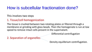


Albert Claude developed the technique of cell fractionation in 1930 which identified different organelles. Subcellular fractionation uses centrifugation to isolate organelles free from contamination so their structure and function can be precisely studied. It involves homogenizing tissues to break open cells and differential centrifugation to separate organelles by density. Specific marker enzymes are used to identify individual organelles and electron microscopy confirms purity. This technique provides information on potential cellular irregularities and correction methods.


