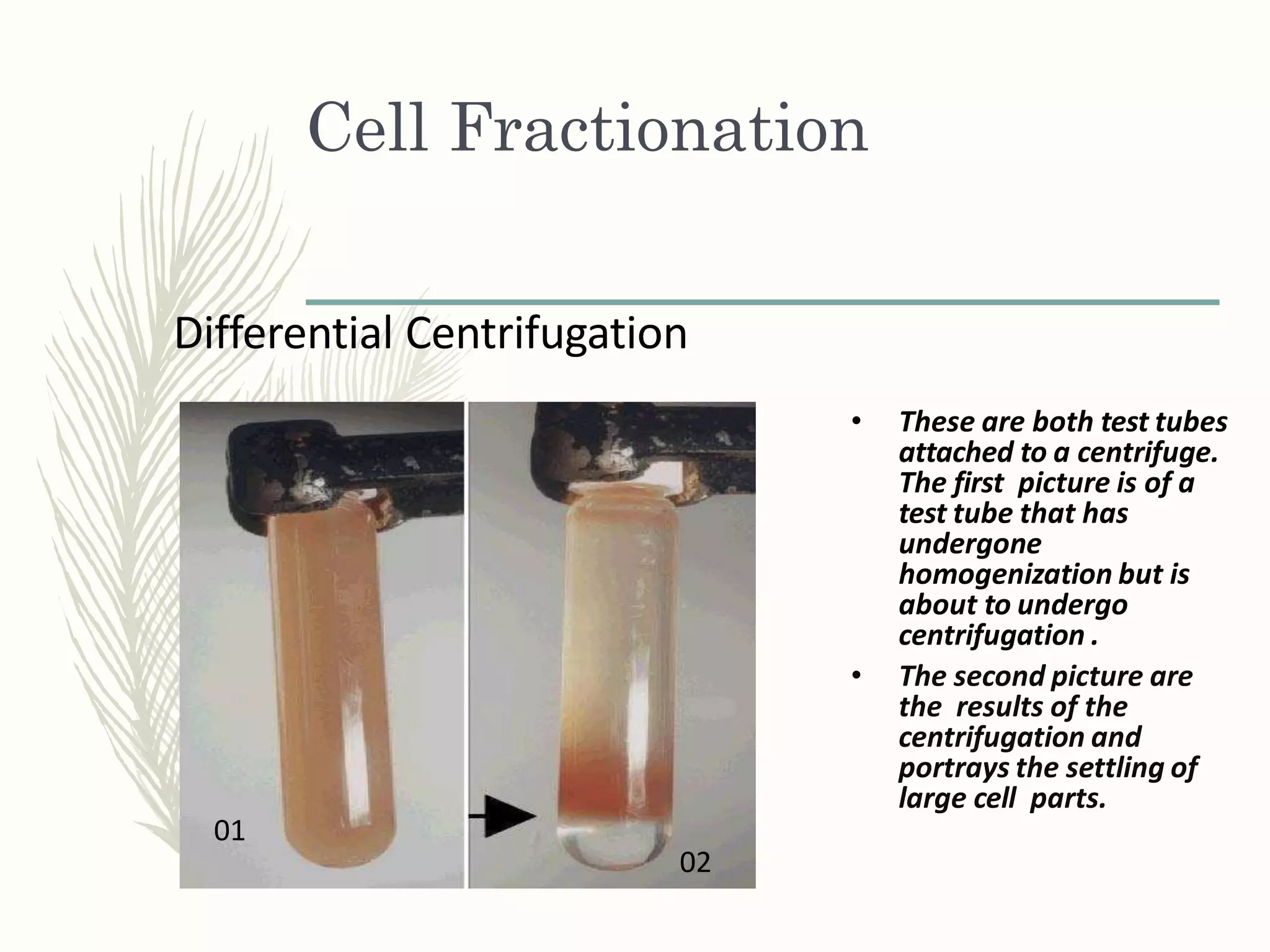
Cell
Fractionation
& Marker
Enzymes
Dr. Praveen Katiyar
Cell
Cell Fractionation
• Cell is the structural and functional unit of life.
• Cell contain organelles which perform a variety of specific functions.
• Electron micrographs explains only the structure but not functions
of the cell organelles.
• Toobtain precise information about the cell organelles, it is necessary to
isolate them free from contaminating organelles.
• Process of isolating cell organelles involves a variety of procedures. These
procedures are collectively called Cell fractionation.
• It can also be defined as : the separation of homogeneous sets , usually
organelles, from a larger population of cells.
• Individual organelle is identified using specific markers.
• This information is used to study potential cellular
irregularities and methods to correct them.
Cell Fractionation
Albert Claude in 1930 developed the technique of cell fractionation &
identified the different organelles using the technique of centrifugation. He
received Nobel Prize for the same in 1974.
Methods of cell fractionation-
These involve -
1-Homogenization-The tissue is crushed between two rotating plates or
filtered through a
membrane or grinding with glass beads. Then the homogenate is run at low
speed to remove intact cells present in the supernatant.
2-Seperation of Cell organelles-
A-Differential centrifugation
B-Density equilibrium centrifugation
Cell Fractionation
Cell Fractionation
Steps
1. Homogenization
2. Differential centrifugation
3. Further separation and purification by density gradient
centrifugation
4. Collection of fractions
5. Analysis of fractions
Cell Fractionation
Homogenization
• First the cells must be broken open.
• A variety of different methods are available; the method chosen
depends on the type of experiment and the type of sample
(bacterial culture or mammalian tissue sample, for example)
Detergents like SDS or Triton X disrupt the cell membrane so the
contents can flow out.
• Subjecting the cells to ultrasound waves or sonicating them will also
break them open, as will agitation in the presence of metal or glass
beads.
• Blenders may work with tissue samples but will not work with
bacteria or other microorganisms.
Cell Fractionation
Homogenization or Cell disruption
Chemical : alkali, organic solvents, detergents
Enzymatic : lysozyme , chitinase
Physical : osmotic shock, freeze/thaw
Mechanical : sonication, homogenization, French press
Chemical Disruption-
Detergents such as TritionX-100 or NP40 can
permeabilize cells by solubilizing membranes.
Detergents can be expensive, denature proteins,
andmust be removed after disruption.
Cell Fractionation
Homogenization or Cell disruption
Sonication-
A sonicator can be immersed directly into
a cell suspension.
The sonicator is vibrated and high frequency
sound waves disrupt cells.
Homogenization-
Cells are disrupted as they pass
between the plunger and
vessel wall.
Differential centrifugation of homogenate
Homogenize cells in 0.25 M sucrose,
1 mM EDTA, 10 mM Tris-HCl, pH 7.4 1000g/10 min
pellet
nuclei
supernatant
supernatant
15,000g/15 min
100,000g/60 min
Pellet (LMF)
mitochondria
lysosomes
peroxisomes
pellet
vesicles (microsomes)
supernatant cytosol
Cell Fractionation
Differential Centrifugation
• Differential centrifugation is the process where a homogenate (soup
of tissue and cells)undergoes repeat centrifugations and increasing
centrifugal force.
• Centrifugations is the use of increased gravity to quicken the
precipitation of substances to the bottom.
• The tool used here is the centrifuge, "merry-go- round for test
tubes" that spin at various speeds.
Cell Fractionation
Differential Centrifugation
• The centrifuge separates the cell's parts into pellet and supernatant.
• The pellet are the large cell structures that are settled at the
test tube's bottom.
• The supernatant are smaller parts of the cell suspending in liquid,
the supernatant is decanted and undergoes another
centrifugation.
• The process is repeated and increases speed with each trial to
collect successively smaller parts of a cell in pellets.
Cell Fractionation
Differential Centrifugation
Cell Fractionation
Differential Centrifugation
• These are both test tubes
attached to a centrifuge.
The first picture is of a
test tube that has
undergone
homogenization but is
about to undergo
centrifugation .
• The second picture are
the results of the
centrifugation and
portrays the settling of
large cell parts.
01
02
Cell Fractionation
Buoyant Density Centrifugation
• The buoyant density centrifugation involves viruses with densities of
1.1-1.2 g/cm and a sucrose gradient.
• The cell suspension is added to the top of the sucrose gradient.
• In this centrifugation the densest componentsmove
fastest down the tube and stops at the sucrose density equal to
its own.
• The sucrose gradient bands at the bottom contain
cell components with high buoyant densities and the components
at the top have low buoyant densities.
Cell Fractionation
An illustration of the sucrose gradient and the buoyant
density centrifugation.
Cell Fractionation
Collection of fractions
Collecting Fractions-keeping samples pure and intact
1. By hand: puncture sidewall of centrifuge tube with needle
and withdraw fractions through syringe
2. Machine: gradient uploader; introduces very dense, non-miscible
medium into bottom of tube, pushes fractions up to be collected
from top
3. If no pellet, can collect fractions through hole in bottom of tube
Cell Fractionation
Analysis of fractions
Analysis of fractions-need to identify and quantify the purified
fractions, so that they can be used successfully in downstream
applications.
Methods:
1-Light or electron microscopy
2-Biochemical-determine presence of marker enzymes
3.Assay for a protein marker with an antibody (western)
4.Determine the protein concentration by using a spectrophotometer,
e.g. Bradfordassay
5.Determine specific activity (the ratio of activity of the enzyme
of interest to the protein concentration
Cell Fractionation
How is cell fractionation used in cell biology?
• Differential Centrifugation allows us to look at each
organelle within the cell.
• We can look at the individual organelles and study them in
detail.
• This helps to determine each organelles function within the
cell.
Cell Fractionation
Investigating Cell function
• Differential Centrifugation allows us to look at each
organelle within the cell
• We can look at the individual organelles and study them in
detail
• This helps to determine each organelles function within the
cell
Cellular Markers or Enzyme
Markers
• These are enzymes specific to certain organelles.
• Following the activity of these enzymes, it is possible to locate the
organelles.
• By monitoring where each enzyme activity is found during a cell
fractionation protocol, one can monitor the fractionation of organelle
Protocol.
• Marker enzymes also provide information on the biochemical purity
of the fractionated organelles.
• The presence of unwanted marker enzyme activity in the preparation
indicates the level of contamination by other organelles.
• However electron microscopy is the final step to determine purity of
the preparation and the presence of organelle in the sample.
Common Enzyme markers for
Organelles
Organelle Marker enzyme
Nucleus Lamin A & C
Mitochondria –Inner Membrane ATP synthetase
Mitochondria- Outer Membrane Monoamine oxidase
Mitochondria - Matrix Citrate synthase
Lysosome Cathepsin
Golgi bodies Galactosyl transferase
Microsome Glucose -6-phosphatase
Cytoplasm Lactate dehydrogenase
Peroxisomes Catalase
Cell Fractionation 09 May 2020.pdf

Cell Fractionation 09 May 2020.pdf

  • 1.
  • 2.
  • 3.
    Cell Fractionation • Cellis the structural and functional unit of life. • Cell contain organelles which perform a variety of specific functions. • Electron micrographs explains only the structure but not functions of the cell organelles. • Toobtain precise information about the cell organelles, it is necessary to isolate them free from contaminating organelles. • Process of isolating cell organelles involves a variety of procedures. These procedures are collectively called Cell fractionation. • It can also be defined as : the separation of homogeneous sets , usually organelles, from a larger population of cells. • Individual organelle is identified using specific markers. • This information is used to study potential cellular irregularities and methods to correct them.
  • 4.
    Cell Fractionation Albert Claudein 1930 developed the technique of cell fractionation & identified the different organelles using the technique of centrifugation. He received Nobel Prize for the same in 1974. Methods of cell fractionation- These involve - 1-Homogenization-The tissue is crushed between two rotating plates or filtered through a membrane or grinding with glass beads. Then the homogenate is run at low speed to remove intact cells present in the supernatant. 2-Seperation of Cell organelles- A-Differential centrifugation B-Density equilibrium centrifugation
  • 5.
  • 6.
    Cell Fractionation Steps 1. Homogenization 2.Differential centrifugation 3. Further separation and purification by density gradient centrifugation 4. Collection of fractions 5. Analysis of fractions
  • 7.
    Cell Fractionation Homogenization • Firstthe cells must be broken open. • A variety of different methods are available; the method chosen depends on the type of experiment and the type of sample (bacterial culture or mammalian tissue sample, for example) Detergents like SDS or Triton X disrupt the cell membrane so the contents can flow out. • Subjecting the cells to ultrasound waves or sonicating them will also break them open, as will agitation in the presence of metal or glass beads. • Blenders may work with tissue samples but will not work with bacteria or other microorganisms.
  • 8.
    Cell Fractionation Homogenization orCell disruption Chemical : alkali, organic solvents, detergents Enzymatic : lysozyme , chitinase Physical : osmotic shock, freeze/thaw Mechanical : sonication, homogenization, French press Chemical Disruption- Detergents such as TritionX-100 or NP40 can permeabilize cells by solubilizing membranes. Detergents can be expensive, denature proteins, andmust be removed after disruption.
  • 9.
    Cell Fractionation Homogenization orCell disruption Sonication- A sonicator can be immersed directly into a cell suspension. The sonicator is vibrated and high frequency sound waves disrupt cells. Homogenization- Cells are disrupted as they pass between the plunger and vessel wall.
  • 10.
    Differential centrifugation ofhomogenate Homogenize cells in 0.25 M sucrose, 1 mM EDTA, 10 mM Tris-HCl, pH 7.4 1000g/10 min pellet nuclei supernatant supernatant 15,000g/15 min 100,000g/60 min Pellet (LMF) mitochondria lysosomes peroxisomes pellet vesicles (microsomes) supernatant cytosol
  • 11.
    Cell Fractionation Differential Centrifugation •Differential centrifugation is the process where a homogenate (soup of tissue and cells)undergoes repeat centrifugations and increasing centrifugal force. • Centrifugations is the use of increased gravity to quicken the precipitation of substances to the bottom. • The tool used here is the centrifuge, "merry-go- round for test tubes" that spin at various speeds.
  • 12.
    Cell Fractionation Differential Centrifugation •The centrifuge separates the cell's parts into pellet and supernatant. • The pellet are the large cell structures that are settled at the test tube's bottom. • The supernatant are smaller parts of the cell suspending in liquid, the supernatant is decanted and undergoes another centrifugation. • The process is repeated and increases speed with each trial to collect successively smaller parts of a cell in pellets.
  • 13.
  • 14.
    Cell Fractionation Differential Centrifugation •These are both test tubes attached to a centrifuge. The first picture is of a test tube that has undergone homogenization but is about to undergo centrifugation . • The second picture are the results of the centrifugation and portrays the settling of large cell parts. 01 02
  • 15.
    Cell Fractionation Buoyant DensityCentrifugation • The buoyant density centrifugation involves viruses with densities of 1.1-1.2 g/cm and a sucrose gradient. • The cell suspension is added to the top of the sucrose gradient. • In this centrifugation the densest componentsmove fastest down the tube and stops at the sucrose density equal to its own. • The sucrose gradient bands at the bottom contain cell components with high buoyant densities and the components at the top have low buoyant densities.
  • 16.
    Cell Fractionation An illustrationof the sucrose gradient and the buoyant density centrifugation.
  • 17.
    Cell Fractionation Collection offractions Collecting Fractions-keeping samples pure and intact 1. By hand: puncture sidewall of centrifuge tube with needle and withdraw fractions through syringe 2. Machine: gradient uploader; introduces very dense, non-miscible medium into bottom of tube, pushes fractions up to be collected from top 3. If no pellet, can collect fractions through hole in bottom of tube
  • 18.
    Cell Fractionation Analysis offractions Analysis of fractions-need to identify and quantify the purified fractions, so that they can be used successfully in downstream applications. Methods: 1-Light or electron microscopy 2-Biochemical-determine presence of marker enzymes 3.Assay for a protein marker with an antibody (western) 4.Determine the protein concentration by using a spectrophotometer, e.g. Bradfordassay 5.Determine specific activity (the ratio of activity of the enzyme of interest to the protein concentration
  • 19.
    Cell Fractionation How iscell fractionation used in cell biology? • Differential Centrifugation allows us to look at each organelle within the cell. • We can look at the individual organelles and study them in detail. • This helps to determine each organelles function within the cell.
  • 20.
    Cell Fractionation Investigating Cellfunction • Differential Centrifugation allows us to look at each organelle within the cell • We can look at the individual organelles and study them in detail • This helps to determine each organelles function within the cell
  • 21.
    Cellular Markers orEnzyme Markers • These are enzymes specific to certain organelles. • Following the activity of these enzymes, it is possible to locate the organelles. • By monitoring where each enzyme activity is found during a cell fractionation protocol, one can monitor the fractionation of organelle Protocol. • Marker enzymes also provide information on the biochemical purity of the fractionated organelles. • The presence of unwanted marker enzyme activity in the preparation indicates the level of contamination by other organelles. • However electron microscopy is the final step to determine purity of the preparation and the presence of organelle in the sample.
  • 22.
    Common Enzyme markersfor Organelles Organelle Marker enzyme Nucleus Lamin A & C Mitochondria –Inner Membrane ATP synthetase Mitochondria- Outer Membrane Monoamine oxidase Mitochondria - Matrix Citrate synthase Lysosome Cathepsin Golgi bodies Galactosyl transferase Microsome Glucose -6-phosphatase Cytoplasm Lactate dehydrogenase Peroxisomes Catalase