STERILITY TESTING OF PHARMACEUTICALS ppt by DR.C.P.PRINCE
Cell fractionationppt
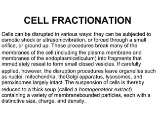
1. CELL FRACTIONATION
Cells can be disrupted in various ways: they can be subjected to
osmotic shock or ultrasonicvibration, or forced through a small
orifice, or ground up. These procedures break many of the
membranes of the cell (including the plasma membrane and
membranes of the endoplasmiceticulum) into fragments that
immediately reseal to form small closed vesicles. If carefully
applied, however, the disruption procedures leave organelles such
as nuclei, mitochondria, theGolgi apparatus, lysosomes, and
peroxisomes largely intact. The suspension of cells is thereby
reduced to a thick soup (called a homogenateor extract)
containing a variety of membranebounded particles, each with a
distinctive size, charge, and density.
2. • The various components of the homogenate are
seperated only after the commercial development in the
early 1940s of an instrument known as the preparative
ultracentrifuge, in which preparations of broken cells are
rotated at high speeds (Figure 4-34). This treatment
separates cell components on the basis of size and
density: in general, the largest units experience the
largest centrifugal force and move the most rapidly. At
relatively low speed,large components such as nuclei
and unbroken cells sediment to form a pellet at the
bottom of thecentrifuge tube; at slightly higher speed, a
pellet of mitochondria is deposited; and at even higher
• speeds and with longer periods of centrifugation,
3. • Centrifugation is the first step in most fractionations, but it
separates only components that differgreatly in size. A
finer degree of separation can be achieved by layering
the homogenate as anarrow band on top of a dilute salt
solution that fills a centrifuge tube. When centrifuged, the
• various components in the mixture move as a series of
distinct bands through the salt solution,each at a different
rate, in a process called velocity sedimentation (Figure 4-
36). For theprocedure to work effectively, the bands must
be protected from convective mixing, which
wouldnormally occur whenever a denser solution (for
example, one containing organelles) finds itself on top of
a lighter one (the salt solution) This is achieved by filling
the centrifuge tube with a shallow gradient of sucrose
prepared by a special mixing device; the resulting density
gradient.
4.
5. • The preparative ultracentrifuge. The sample is contained
in tubes that are inserted into a ring of cylindrical holes in
a metal rotor. Rapid rotation of the rotor generates
enormous centrifugal forces, which cause particles in the
sample to sediment. The vacuum reduces friction,
• preventing heating of the rotor and allowing the
refrigeration system to maintain the sample at 4 degree.
6. • with the dense end at the bottom of the tube, keeps each
region of the salt solution denser than any solution above
it and thereby prevents convective mixing from distorting
the separation. When sedimented through such dilute
sucrose gradients, different cell components separate
into distinct bands that can be collected individually. The
rate at which each component sediments depends
primarily on its size and shape and is normally described
in terms of its sedimentation
• coefficient, or s value.
7. • The ultracentrifuge is also used to separate cellular
components on the basis of their buoyant density,
independently of their size and shape. the sample is
usually sedimented through a steep density gradient that
contains a very high concentration of sucrose or cesium
• chloride. Each cellular component begins to move down
the gradient.