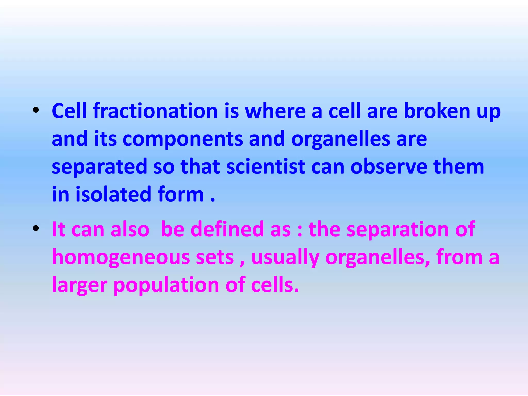
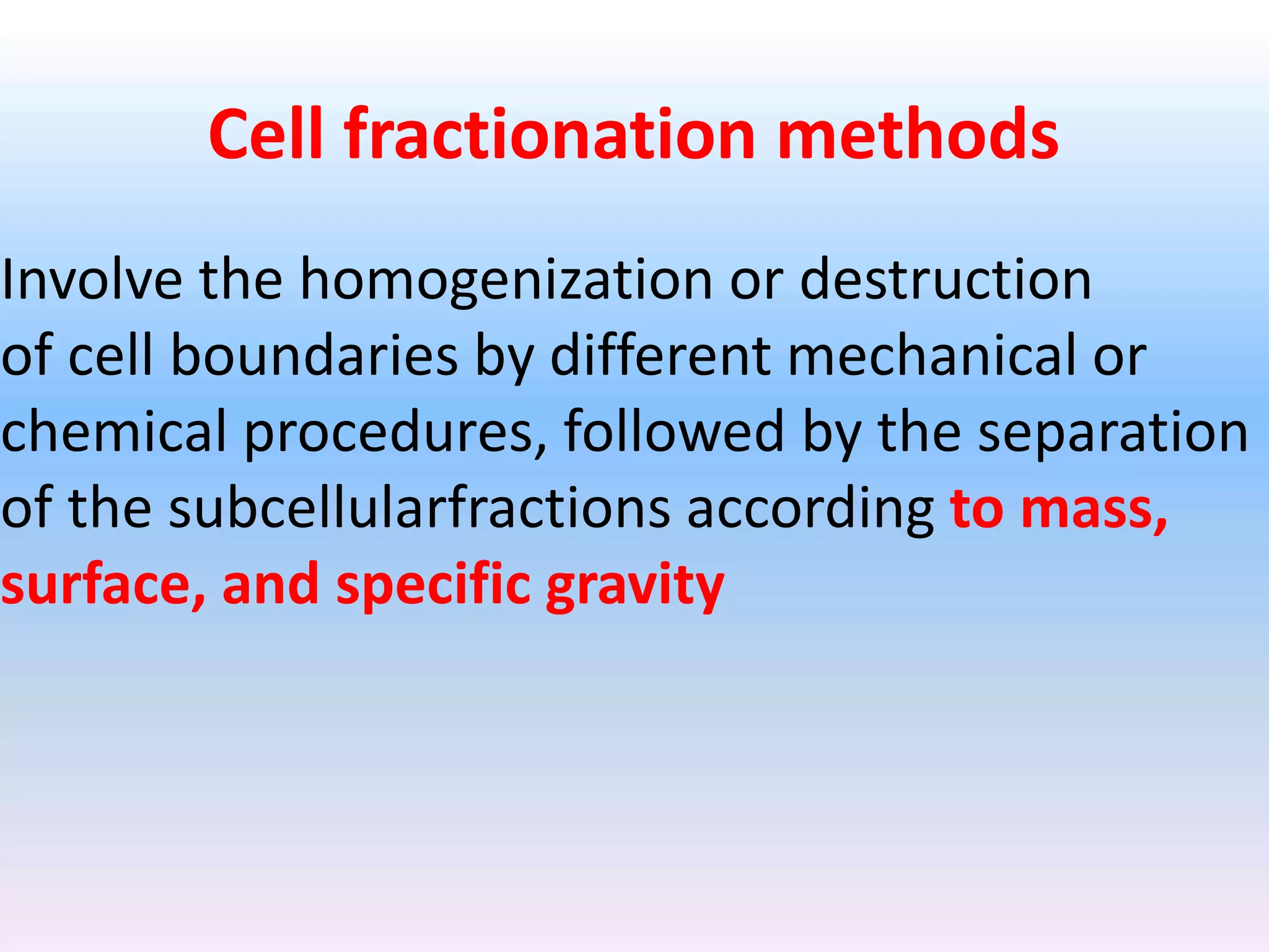
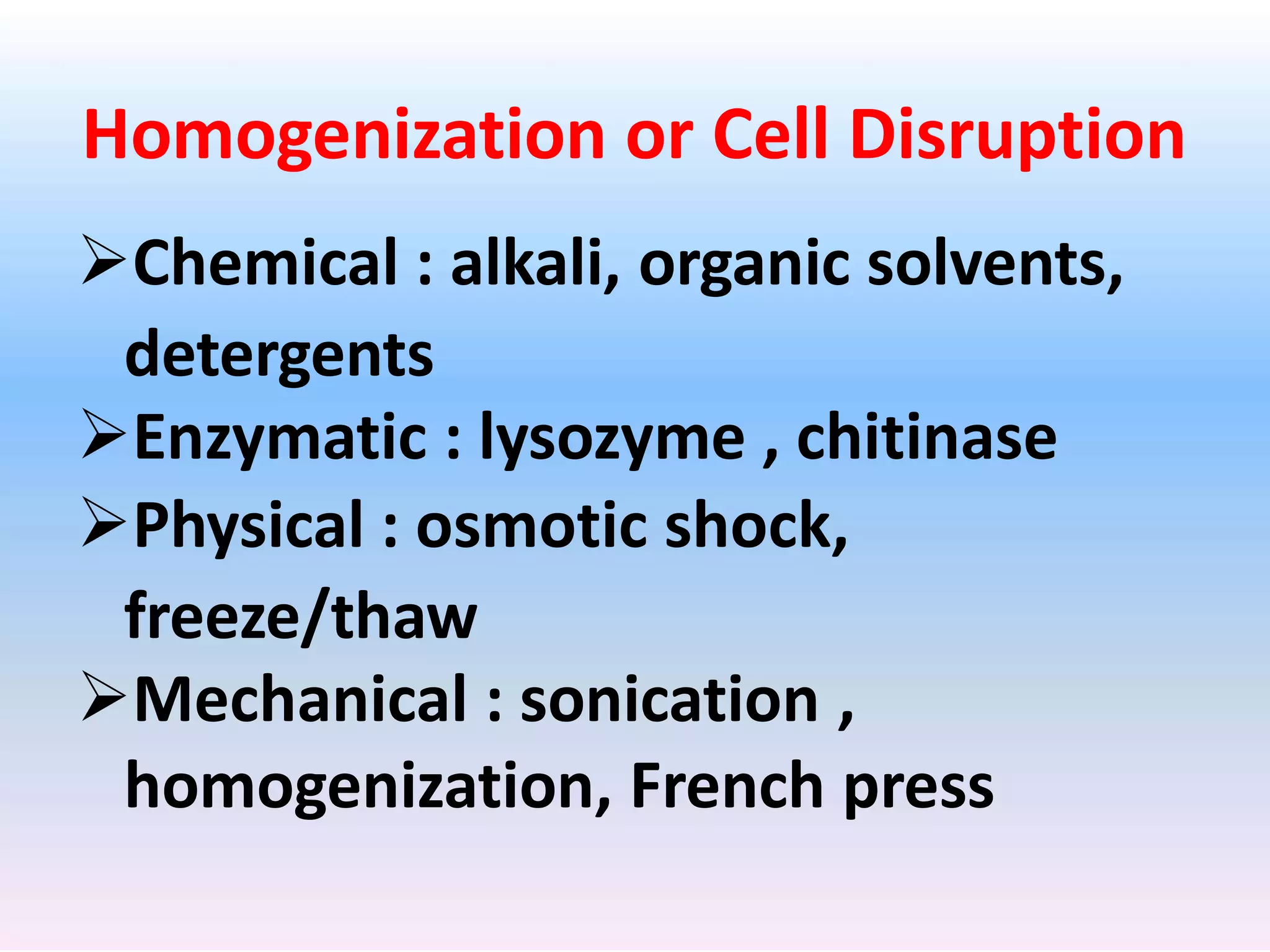
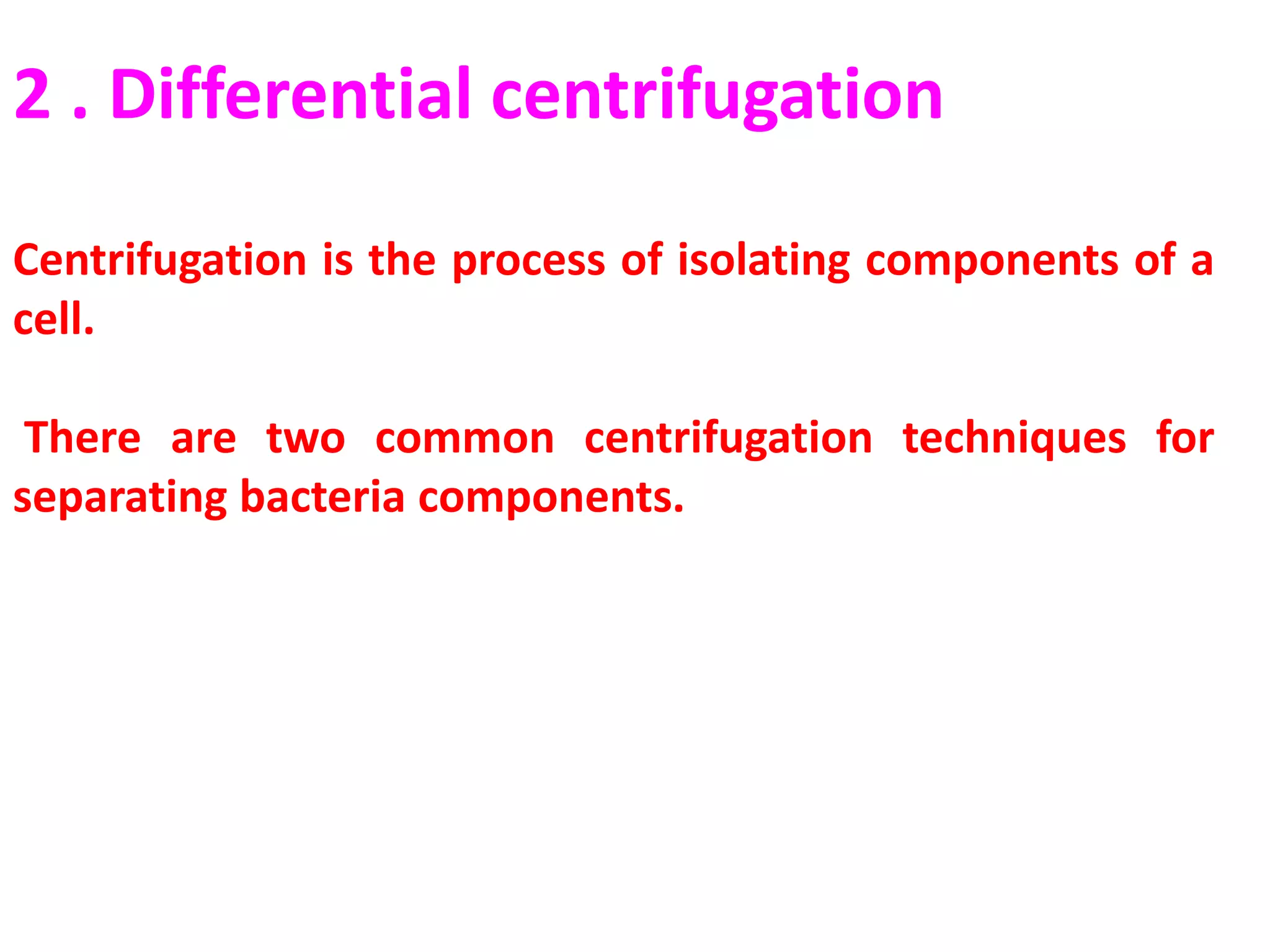
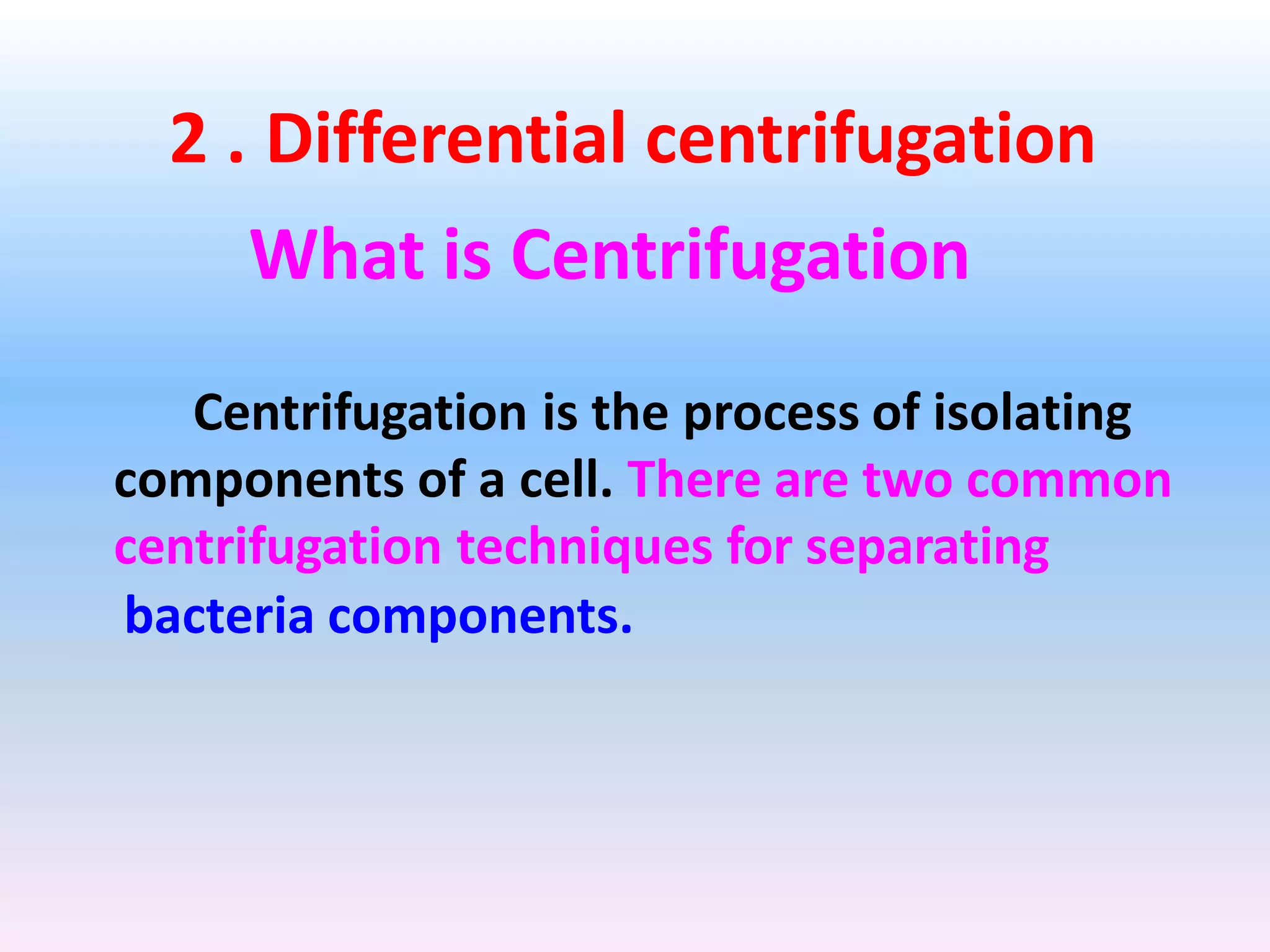
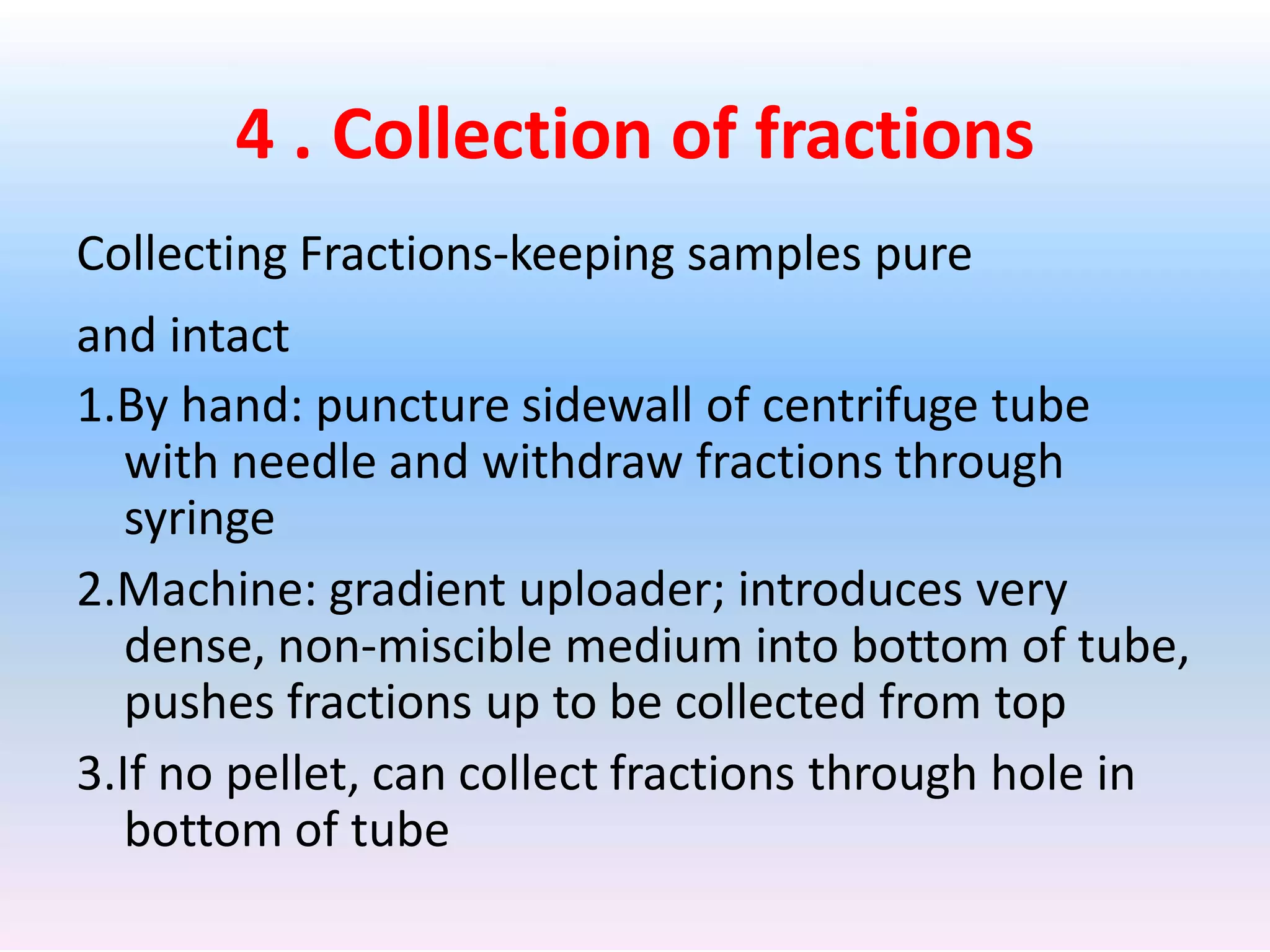
Historical Introduction
What is cell fractionation
• Biologists need to study certain organelles from a
cell(the mitochondria of a human cell or the
chloroplasts of a plant cell, for example Isolating
these organelles involves a variety of procedures
collectively called cell fractionation.
• As a method for studying processes within
organelles, cell fractionation has both advantages and
disadvantages.
• Cell fractionation is where a cell are broken up
and its components and organelles are
separated so that scientist can observe them
in isolated form .
• It can also be defined as : the separation of
homogeneous sets , usually organelles, from a
larger population of cells.
What we’ve learned so far using this
technique :
1.Mechanism of protein synthesis
2.DNA replication and transcription
3.RNA splicing
4.Muscle contraction
5.Microtubule assembly
6.Vesicular transport in the secretory pathway
7.Importance of mitachondria and chloroplasts
in energy interconversions .
Cell fractionation methods
Involve the homogenization or destruction
of cell boundaries by different mechanical or
chemical procedures, followed by the separation
of the subcellularfractions according to mass,
surface, and specific gravity
Steps of subcellular fractionation
1 . Homogenization
2 . Differential centrifugation
3 . Further separation and purification
by density gradient centrifugation
4 . Collection of fractions
5 . Analysis of fractions
1 . Homogenization
• First the cells must be broken open.
• A variety of different methods are available; the
method chosen depends on the type of experiment
and the type of sample (bacterial culture or
mammalian tissue sample, for example) Detergents
like SDS or Triton X disrupt the cell membrane so the
contents can flow out.
• Subjecting the cells to ultrasound waves or sonicating
them will also break them open, as will agitation in
the presence of metal or glass beads.
• Blenders may work with tissue samples but will not
work with bacteria or other microorganisms.
Homogenization or Cell Disruption
Chemical : alkali, organic solvents,
detergents
Enzymatic : lysozyme , chitinase
Physical : osmotic shock,
freeze/thaw
Mechanical : sonication ,
homogenization, French press
Chemical Disruption
• Detergents such as
TritionX-100 or NP40 can
permeabilize cells by
solubilizing membranes.
• Detergents can be
expensive, denature
proteins, and must be
removed after disruption
Sonication
• A sonicator can be immersed
directly into a cell suspension.
• The sonicator is vibrated and
high frequency sound waves
disrupt cells.
Homogenization
• Cells are placed in a closed
vessel (usually glass).
• A tight fitting plunger is inserted
and rotated with a downward
force.
• Cells are disrupted as they pass
between the plunger and vessel
wall.
Homogenize cells in 0.25 M sucrose,
1 mM EDTA, 10 mM Tris-HCl, pH 7.4 1000g/10 min
pellet
nuclei
supernatant
supernatant
15,000g/15 min
100,000g/60 min
Pellet (LMF)
mitochondria
lysosomes
peroxisomes
pellet
vesicles (microsomes)
supernatant cytosol
Differential centrifugation of homogenate
2 . Differential centrifugation
Centrifugation is the process of isolating components of a
cell.
There are two common centrifugation techniques for
separating bacteria components.
2 . Differential centrifugation
What is Centrifugation
Centrifugation is the process of isolating
components of a cell. There are two common
centrifugation techniques for separating
bacteria components.
2 . Differential centrifugation
What is Centrifugation
Centrifugation is the process of isolating
components of a cell. There are two common
centrifugation techniques for separating
bacteria components.
Differential Centrifugation
• Differential centrifugation is the process where a
homogenate (soup of tissue and cells)undergoes
repeat centrifugations and increasing centrifugal
force.
• Centrifugations is the use of increased gravity to
quicken the precipitation of substances tot the
bottom.
• The tool used here is the centrifuge, "merry-go-
round for test tubes" that spin at various speeds.
Differential Centrifugation
• The centrifuge separates the cell's parts into pellet
and supernatant.
• The pellet are the large cell structures that are
settled at the test tube's bottom.
• The supernatant are smaller parts of the cell
suspending in liquid, the supernatant is decanted
and undergoes another centrifugation.
• The process is repeated and increases speed with
each trial to collect successively smaller parts of a
cell in pellets.
• These are both test tubes attached to a centrifuge. The first
picture is of a test tube that has undergone homogenization but is
about to undergo centrifugation . The second picture are the
results of the centrifugation and portrays the settling of large cell
parts.
The preparative ultracentrifuge
• The preparative ultracentrifuge.
Sample is contained in tubes that are inserted into a
ring of cylindrical holes in a metal rotor.
Rapid rotation of the rotor generates enormous
centrifugal forces, which cause particles in the
sample to sediment.
The vacuum reduces friction, preventing heating of
the rotor and allowing the refrigeration system to
maintain the sample at 4°C.
When a centrifugal force is applied to an
aqueous mixture, components of larger size
and density will sediment faster
Low speed centrifugation is used to separate
intact cells from medium.
High speed centrifugation can be used to
separate subcellular components.
Tow types of centrifuge are there
Fixed-Angle Centrifugation
Swinging-Arm Centrifugation
Method of Differential
Centrifugation:
• 1. Cut tissue in an ice-
cold isotonic buffer.
It is cold to stop
enzyme reactions,
isotonic to stop
osmosis and a buffer
to stop pH changes.
• 2. Grind tissue in a
blender to break open
cells.
• 3. Filter to remove
insoluble tissue
• 4. Centrifuge
filtrate at low
speeds ( 1000 X g
for 10mins )
• This pellets the
nuclei as this is the
densest organelle
• 5. Centrifuge at
medium speeds ( 10
000 x g for 30
mins )
• This pellets
mitchondria which
are the second
densest organelle
• 6. Centrifuge at
high speeds ( 100
000 x g for 30
mins)
• This pellets ER,
golgi apparatus and
other membrane
fragments
• 7 Centrifuge at
very high speeds (
300 000 x g for
3hrs)
• This pellets
ribosomes
Buoyant density centrifugation
The buoyant density centrifugation involves viruses
with densities of 1.1-1.2 g/cm and a sucrose
gradient.
The cell suspension is added to the top of the
sucrose gradient.
In this centrifugation the densest components move
fastest down the tube and stops at the sucrose
density equal to its own.
The sucrose gradient bands at the bottom contain
cell components with high buoyant densities and
the components at the top have low buoyant
densities.
An illustration of the sucrose gradient and the
buoyant density centrifugation.
4 . Collection of fractions
Collecting Fractions-keeping samples pure
and intact
1.By hand: puncture sidewall of centrifuge tube
with needle and withdraw fractions through
syringe
2.Machine: gradient uploader; introduces very
dense, non-miscible medium into bottom of tube,
pushes fractions up to be collected from top
3.If no pellet, can collect fractions through hole in
bottom of tube
5 . Analysis of fractions
Analysis of fractions-need to identify and quantify the
purified fractions, so that they can be used successfully
in downstream applications
Methods:
1.Light or electron microscopy
2.Biochemical-determine presence of marker enzymes
3.Assay for a protein marker with an antibody (western)
4.Determine the protein concentration by using a
spectrophotometer, e.g. Bradford assay
5.Determine specific activity (the ratio of activity of
the enzyme of interest to the protein concentration
How is cell fractionation used in cell
biology?
• Scientists use this tool to increase their
knowledge of organelle functions. To be able to
do so they isolate organelles into pure groups,
such as isolating the mitochondria or the
nucleus.
For example, by centrifugation a specific cell
fraction was determined to have enzymes that
function in cellular respiration. This unknown cell
fraction was rich in mitochondria . Therefore
there researchers obtained evidence that helped
determine mitochondria were the site of cellular
respiration.
Investigating Cell Function
• Differential Centrifugation allows us to
look at each organelle within the cell
• We can look at the individual organelles
and study them in detail
• This helps to determine each organelles
function within the cell
Uses
• Separation of enzymes, hormones, RNA-DNA
hybrids, ribosomal subunits, subcellular
organelles, for the analysis of size distribution
of samples of polysomes and lipoprotein
fractions.
Isolation cell organelle by ankit

Isolation cell organelle by ankit

  • 2.
  • 4.
    What is cellfractionation • Biologists need to study certain organelles from a cell(the mitochondria of a human cell or the chloroplasts of a plant cell, for example Isolating these organelles involves a variety of procedures collectively called cell fractionation. • As a method for studying processes within organelles, cell fractionation has both advantages and disadvantages.
  • 5.
    • Cell fractionationis where a cell are broken up and its components and organelles are separated so that scientist can observe them in isolated form . • It can also be defined as : the separation of homogeneous sets , usually organelles, from a larger population of cells.
  • 7.
    What we’ve learnedso far using this technique : 1.Mechanism of protein synthesis 2.DNA replication and transcription 3.RNA splicing 4.Muscle contraction 5.Microtubule assembly 6.Vesicular transport in the secretory pathway 7.Importance of mitachondria and chloroplasts in energy interconversions .
  • 8.
    Cell fractionation methods Involvethe homogenization or destruction of cell boundaries by different mechanical or chemical procedures, followed by the separation of the subcellularfractions according to mass, surface, and specific gravity
  • 9.
    Steps of subcellularfractionation 1 . Homogenization 2 . Differential centrifugation 3 . Further separation and purification by density gradient centrifugation 4 . Collection of fractions 5 . Analysis of fractions
  • 10.
    1 . Homogenization •First the cells must be broken open. • A variety of different methods are available; the method chosen depends on the type of experiment and the type of sample (bacterial culture or mammalian tissue sample, for example) Detergents like SDS or Triton X disrupt the cell membrane so the contents can flow out. • Subjecting the cells to ultrasound waves or sonicating them will also break them open, as will agitation in the presence of metal or glass beads. • Blenders may work with tissue samples but will not work with bacteria or other microorganisms.
  • 11.
    Homogenization or CellDisruption Chemical : alkali, organic solvents, detergents Enzymatic : lysozyme , chitinase Physical : osmotic shock, freeze/thaw Mechanical : sonication , homogenization, French press
  • 12.
    Chemical Disruption • Detergentssuch as TritionX-100 or NP40 can permeabilize cells by solubilizing membranes. • Detergents can be expensive, denature proteins, and must be removed after disruption
  • 13.
    Sonication • A sonicatorcan be immersed directly into a cell suspension. • The sonicator is vibrated and high frequency sound waves disrupt cells.
  • 14.
    Homogenization • Cells areplaced in a closed vessel (usually glass). • A tight fitting plunger is inserted and rotated with a downward force. • Cells are disrupted as they pass between the plunger and vessel wall.
  • 15.
    Homogenize cells in0.25 M sucrose, 1 mM EDTA, 10 mM Tris-HCl, pH 7.4 1000g/10 min pellet nuclei supernatant supernatant 15,000g/15 min 100,000g/60 min Pellet (LMF) mitochondria lysosomes peroxisomes pellet vesicles (microsomes) supernatant cytosol Differential centrifugation of homogenate
  • 16.
    2 . Differentialcentrifugation Centrifugation is the process of isolating components of a cell. There are two common centrifugation techniques for separating bacteria components.
  • 17.
    2 . Differentialcentrifugation What is Centrifugation Centrifugation is the process of isolating components of a cell. There are two common centrifugation techniques for separating bacteria components.
  • 18.
    2 . Differentialcentrifugation What is Centrifugation Centrifugation is the process of isolating components of a cell. There are two common centrifugation techniques for separating bacteria components.
  • 20.
    Differential Centrifugation • Differentialcentrifugation is the process where a homogenate (soup of tissue and cells)undergoes repeat centrifugations and increasing centrifugal force. • Centrifugations is the use of increased gravity to quicken the precipitation of substances tot the bottom. • The tool used here is the centrifuge, "merry-go- round for test tubes" that spin at various speeds.
  • 21.
    Differential Centrifugation • Thecentrifuge separates the cell's parts into pellet and supernatant. • The pellet are the large cell structures that are settled at the test tube's bottom. • The supernatant are smaller parts of the cell suspending in liquid, the supernatant is decanted and undergoes another centrifugation. • The process is repeated and increases speed with each trial to collect successively smaller parts of a cell in pellets.
  • 22.
    • These areboth test tubes attached to a centrifuge. The first picture is of a test tube that has undergone homogenization but is about to undergo centrifugation . The second picture are the results of the centrifugation and portrays the settling of large cell parts.
  • 23.
  • 24.
    • The preparativeultracentrifuge. Sample is contained in tubes that are inserted into a ring of cylindrical holes in a metal rotor. Rapid rotation of the rotor generates enormous centrifugal forces, which cause particles in the sample to sediment. The vacuum reduces friction, preventing heating of the rotor and allowing the refrigeration system to maintain the sample at 4°C.
  • 25.
    When a centrifugalforce is applied to an aqueous mixture, components of larger size and density will sediment faster Low speed centrifugation is used to separate intact cells from medium. High speed centrifugation can be used to separate subcellular components.
  • 26.
    Tow types ofcentrifuge are there Fixed-Angle Centrifugation
  • 27.
  • 28.
    Method of Differential Centrifugation: •1. Cut tissue in an ice- cold isotonic buffer. It is cold to stop enzyme reactions, isotonic to stop osmosis and a buffer to stop pH changes. • 2. Grind tissue in a blender to break open cells. • 3. Filter to remove insoluble tissue
  • 29.
    • 4. Centrifuge filtrateat low speeds ( 1000 X g for 10mins ) • This pellets the nuclei as this is the densest organelle
  • 30.
    • 5. Centrifugeat medium speeds ( 10 000 x g for 30 mins ) • This pellets mitchondria which are the second densest organelle
  • 31.
    • 6. Centrifugeat high speeds ( 100 000 x g for 30 mins) • This pellets ER, golgi apparatus and other membrane fragments
  • 32.
    • 7 Centrifugeat very high speeds ( 300 000 x g for 3hrs) • This pellets ribosomes
  • 33.
    Buoyant density centrifugation Thebuoyant density centrifugation involves viruses with densities of 1.1-1.2 g/cm and a sucrose gradient. The cell suspension is added to the top of the sucrose gradient. In this centrifugation the densest components move fastest down the tube and stops at the sucrose density equal to its own. The sucrose gradient bands at the bottom contain cell components with high buoyant densities and the components at the top have low buoyant densities.
  • 34.
    An illustration ofthe sucrose gradient and the buoyant density centrifugation.
  • 35.
    4 . Collectionof fractions Collecting Fractions-keeping samples pure and intact 1.By hand: puncture sidewall of centrifuge tube with needle and withdraw fractions through syringe 2.Machine: gradient uploader; introduces very dense, non-miscible medium into bottom of tube, pushes fractions up to be collected from top 3.If no pellet, can collect fractions through hole in bottom of tube
  • 36.
    5 . Analysisof fractions Analysis of fractions-need to identify and quantify the purified fractions, so that they can be used successfully in downstream applications Methods: 1.Light or electron microscopy 2.Biochemical-determine presence of marker enzymes 3.Assay for a protein marker with an antibody (western) 4.Determine the protein concentration by using a spectrophotometer, e.g. Bradford assay 5.Determine specific activity (the ratio of activity of the enzyme of interest to the protein concentration
  • 37.
    How is cellfractionation used in cell biology? • Scientists use this tool to increase their knowledge of organelle functions. To be able to do so they isolate organelles into pure groups, such as isolating the mitochondria or the nucleus. For example, by centrifugation a specific cell fraction was determined to have enzymes that function in cellular respiration. This unknown cell fraction was rich in mitochondria . Therefore there researchers obtained evidence that helped determine mitochondria were the site of cellular respiration.
  • 38.
    Investigating Cell Function •Differential Centrifugation allows us to look at each organelle within the cell • We can look at the individual organelles and study them in detail • This helps to determine each organelles function within the cell
  • 40.
    Uses • Separation ofenzymes, hormones, RNA-DNA hybrids, ribosomal subunits, subcellular organelles, for the analysis of size distribution of samples of polysomes and lipoprotein fractions.