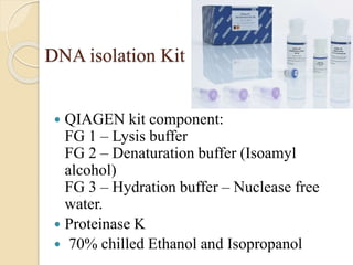
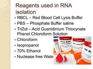
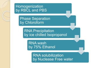
The technique of molecular biology like DNA isolation, RNA isolation, PCR, Western blot, RFLP, etc was developed with development in science. This presentation includes the method of DNA and RNA isolation and their Quantification techniques.
STS stands for sequence tagged site which is short DNA sequence, generally between 100 and 500 bp in length, that is easily recognizable and occurs only once in the chromosome or genome being studied.
This is technique used widely for protein separation from a mixture and is very easy and less costly method. Slides cover all essential points about EMSA and it is quite interesting to know that how it detect and separate different proteins and their mobility shift assay.
This presentation contains information about restriction enzymes, its nomenclature, restriction digestion, and its application. This also contains information about the chemicals used in restriction and also explains the general procedure of restriction digestion of DNA
Techniques of DNA Extraction, Purification and QuantificationBHUMI GAMETI
Introduction
The overall process…
Uses of isolated genomic DNA
Extraction of DNA from plant material
Components of DNA extraction solutions
Cell Lysis or Cell disruption :
Purification of DNA
CTAB Method
Phenol–chloroform extraction
PROTEINASE K
Salting out
Silica adsorption method
Magnetic beads
FTA Paper
Nucleic acid quantification
Agarose Gel Electrophoresis
UV spectroscopy
DNA quantification using NanoDrop
1. CENTRAL DOGMA OF MOLECULAR BIOLOGY
2. NUCLEIC ACID PREPARATION & APPLICATIONS
3. FUNDAMENTAL STEPS IN DNA PURIFICATION
4. ANALYSIS OF NUCLEIC ACIDS
5. STORAGE CONDITIONS
Streamline Your Research with Expert DNA Prep ServicesBlueheronbio
Elevate your molecular biology experiments with our precise and efficient DNA prep services. Trust our expertise to prepare high-quality DNA samples tailored to your specific research needs.
Some sample sources present special challenges in RNA isolation or contain substances that cause problems in RNA analysis. These guides to RNA isolation have tips for a whole range of sample types, including guidance on the best kits for each.
Simultaneous Isolation of RNA & DNA from one FFPE SampleQIAGEN
Worldwide, there are millions of tissue samples archived in tissue biobanks and biorepositories. These samples are extremely valuable for pharmacological and biomedical research and companion diagnostics, due to the linkage to patient history. The vast majority of archived tissue samples are formalin-fixed and paraffin-embedded (FFPE), since formalin is the standard fixative for tissue samples.
FFPE blocks serve as an excellent source for histomorphology studies, but their use in molecular studies is challenging, due to crosslinking and fragmentation caused by fixation, processing, embedding, and storage conditions. For reliable comparison of genomic and transcriptomic data from heterogeneous samples and to spare sample material, purification of DNA and RNA from
the same sample is essential. This is particularly important when working with tumorous tissues, which contain a heterogeneous distribution of healthy and malignant cells.
Richard's aventures in two entangled wonderlandsRichard Gill
Since the loophole-free Bell experiments of 2020 and the Nobel prizes in physics of 2022, critics of Bell's work have retreated to the fortress of super-determinism. Now, super-determinism is a derogatory word - it just means "determinism". Palmer, Hance and Hossenfelder argue that quantum mechanics and determinism are not incompatible, using a sophisticated mathematical construction based on a subtle thinning of allowed states and measurements in quantum mechanics, such that what is left appears to make Bell's argument fail, without altering the empirical predictions of quantum mechanics. I think however that it is a smoke screen, and the slogan "lost in math" comes to my mind. I will discuss some other recent disproofs of Bell's theorem using the language of causality based on causal graphs. Causal thinking is also central to law and justice. I will mention surprising connections to my work on serial killer nurse cases, in particular the Dutch case of Lucia de Berk and the current UK case of Lucy Letby.
This presentation explores a brief idea about the structural and functional attributes of nucleotides, the structure and function of genetic materials along with the impact of UV rays and pH upon them.
Introduction:
RNA interference (RNAi) or Post-Transcriptional Gene Silencing (PTGS) is an important biological process for modulating eukaryotic gene expression.
It is highly conserved process of posttranscriptional gene silencing by which double stranded RNA (dsRNA) causes sequence-specific degradation of mRNA sequences.
dsRNA-induced gene silencing (RNAi) is reported in a wide range of eukaryotes ranging from worms, insects, mammals and plants.
This process mediates resistance to both endogenous parasitic and exogenous pathogenic nucleic acids, and regulates the expression of protein-coding genes.
What are small ncRNAs?
micro RNA (miRNA)
short interfering RNA (siRNA)
Properties of small non-coding RNA:
Involved in silencing mRNA transcripts.
Called “small” because they are usually only about 21-24 nucleotides long.
Synthesized by first cutting up longer precursor sequences (like the 61nt one that Lee discovered).
Silence an mRNA by base pairing with some sequence on the mRNA.
Discovery of siRNA?
The first small RNA:
In 1993 Rosalind Lee (Victor Ambros lab) was studying a non- coding gene in C. elegans, lin-4, that was involved in silencing of another gene, lin-14, at the appropriate time in the
development of the worm C. elegans.
Two small transcripts of lin-4 (22nt and 61nt) were found to be complementary to a sequence in the 3' UTR of lin-14.
Because lin-4 encoded no protein, she deduced that it must be these transcripts that are causing the silencing by RNA-RNA interactions.
Types of RNAi ( non coding RNA)
MiRNA
Length (23-25 nt)
Trans acting
Binds with target MRNA in mismatch
Translation inhibition
Si RNA
Length 21 nt.
Cis acting
Bind with target Mrna in perfect complementary sequence
Piwi-RNA
Length ; 25 to 36 nt.
Expressed in Germ Cells
Regulates trnasposomes activity
MECHANISM OF RNAI:
First the double-stranded RNA teams up with a protein complex named Dicer, which cuts the long RNA into short pieces.
Then another protein complex called RISC (RNA-induced silencing complex) discards one of the two RNA strands.
The RISC-docked, single-stranded RNA then pairs with the homologous mRNA and destroys it.
THE RISC COMPLEX:
RISC is large(>500kD) RNA multi- protein Binding complex which triggers MRNA degradation in response to MRNA
Unwinding of double stranded Si RNA by ATP independent Helicase
Active component of RISC is Ago proteins( ENDONUCLEASE) which cleave target MRNA.
DICER: endonuclease (RNase Family III)
Argonaute: Central Component of the RNA-Induced Silencing Complex (RISC)
One strand of the dsRNA produced by Dicer is retained in the RISC complex in association with Argonaute
ARGONAUTE PROTEIN :
1.PAZ(PIWI/Argonaute/ Zwille)- Recognition of target MRNA
2.PIWI (p-element induced wimpy Testis)- breaks Phosphodiester bond of mRNA.)RNAse H activity.
MiRNA:
The Double-stranded RNAs are naturally produced in eukaryotic cells during development, and they have a key role in regulating gene expression .
Seminar of U.V. Spectroscopy by SAMIR PANDASAMIR PANDA
Spectroscopy is a branch of science dealing the study of interaction of electromagnetic radiation with matter.
Ultraviolet-visible spectroscopy refers to absorption spectroscopy or reflect spectroscopy in the UV-VIS spectral region.
Ultraviolet-visible spectroscopy is an analytical method that can measure the amount of light received by the analyte.
What is greenhouse gasses and how many gasses are there to affect the Earth.moosaasad1975
What are greenhouse gasses how they affect the earth and its environment what is the future of the environment and earth how the weather and the climate effects.
Earliest Galaxies in the JADES Origins Field: Luminosity Function and Cosmic ...Sérgio Sacani
We characterize the earliest galaxy population in the JADES Origins Field (JOF), the deepest
imaging field observed with JWST. We make use of the ancillary Hubble optical images (5 filters
spanning 0.4−0.9µm) and novel JWST images with 14 filters spanning 0.8−5µm, including 7 mediumband filters, and reaching total exposure times of up to 46 hours per filter. We combine all our data
at > 2.3µm to construct an ultradeep image, reaching as deep as ≈ 31.4 AB mag in the stack and
30.3-31.0 AB mag (5σ, r = 0.1” circular aperture) in individual filters. We measure photometric
redshifts and use robust selection criteria to identify a sample of eight galaxy candidates at redshifts
z = 11.5 − 15. These objects show compact half-light radii of R1/2 ∼ 50 − 200pc, stellar masses of
M⋆ ∼ 107−108M⊙, and star-formation rates of SFR ∼ 0.1−1 M⊙ yr−1
. Our search finds no candidates
at 15 < z < 20, placing upper limits at these redshifts. We develop a forward modeling approach to
infer the properties of the evolving luminosity function without binning in redshift or luminosity that
marginalizes over the photometric redshift uncertainty of our candidate galaxies and incorporates the
impact of non-detections. We find a z = 12 luminosity function in good agreement with prior results,
and that the luminosity function normalization and UV luminosity density decline by a factor of ∼ 2.5
from z = 12 to z = 14. We discuss the possible implications of our results in the context of theoretical
models for evolution of the dark matter halo mass function.
Observation of Io’s Resurfacing via Plume Deposition Using Ground-based Adapt...Sérgio Sacani
Since volcanic activity was first discovered on Io from Voyager images in 1979, changes
on Io’s surface have been monitored from both spacecraft and ground-based telescopes.
Here, we present the highest spatial resolution images of Io ever obtained from a groundbased telescope. These images, acquired by the SHARK-VIS instrument on the Large
Binocular Telescope, show evidence of a major resurfacing event on Io’s trailing hemisphere. When compared to the most recent spacecraft images, the SHARK-VIS images
show that a plume deposit from a powerful eruption at Pillan Patera has covered part
of the long-lived Pele plume deposit. Although this type of resurfacing event may be common on Io, few have been detected due to the rarity of spacecraft visits and the previously low spatial resolution available from Earth-based telescopes. The SHARK-VIS instrument ushers in a new era of high resolution imaging of Io’s surface using adaptive
optics at visible wavelengths.
(May 29th, 2024) Advancements in Intravital Microscopy- Insights for Preclini...Scintica Instrumentation
Intravital microscopy (IVM) is a powerful tool utilized to study cellular behavior over time and space in vivo. Much of our understanding of cell biology has been accomplished using various in vitro and ex vivo methods; however, these studies do not necessarily reflect the natural dynamics of biological processes. Unlike traditional cell culture or fixed tissue imaging, IVM allows for the ultra-fast high-resolution imaging of cellular processes over time and space and were studied in its natural environment. Real-time visualization of biological processes in the context of an intact organism helps maintain physiological relevance and provide insights into the progression of disease, response to treatments or developmental processes.
In this webinar we give an overview of advanced applications of the IVM system in preclinical research. IVIM technology is a provider of all-in-one intravital microscopy systems and solutions optimized for in vivo imaging of live animal models at sub-micron resolution. The system’s unique features and user-friendly software enables researchers to probe fast dynamic biological processes such as immune cell tracking, cell-cell interaction as well as vascularization and tumor metastasis with exceptional detail. This webinar will also give an overview of IVM being utilized in drug development, offering a view into the intricate interaction between drugs/nanoparticles and tissues in vivo and allows for the evaluation of therapeutic intervention in a variety of tissues and organs. This interdisciplinary collaboration continues to drive the advancements of novel therapeutic strategies.
Slide 1: Title Slide
Extrachromosomal Inheritance
Slide 2: Introduction to Extrachromosomal Inheritance
Definition: Extrachromosomal inheritance refers to the transmission of genetic material that is not found within the nucleus.
Key Components: Involves genes located in mitochondria, chloroplasts, and plasmids.
Slide 3: Mitochondrial Inheritance
Mitochondria: Organelles responsible for energy production.
Mitochondrial DNA (mtDNA): Circular DNA molecule found in mitochondria.
Inheritance Pattern: Maternally inherited, meaning it is passed from mothers to all their offspring.
Diseases: Examples include Leber’s hereditary optic neuropathy (LHON) and mitochondrial myopathy.
Slide 4: Chloroplast Inheritance
Chloroplasts: Organelles responsible for photosynthesis in plants.
Chloroplast DNA (cpDNA): Circular DNA molecule found in chloroplasts.
Inheritance Pattern: Often maternally inherited in most plants, but can vary in some species.
Examples: Variegation in plants, where leaf color patterns are determined by chloroplast DNA.
Slide 5: Plasmid Inheritance
Plasmids: Small, circular DNA molecules found in bacteria and some eukaryotes.
Features: Can carry antibiotic resistance genes and can be transferred between cells through processes like conjugation.
Significance: Important in biotechnology for gene cloning and genetic engineering.
Slide 6: Mechanisms of Extrachromosomal Inheritance
Non-Mendelian Patterns: Do not follow Mendel’s laws of inheritance.
Cytoplasmic Segregation: During cell division, organelles like mitochondria and chloroplasts are randomly distributed to daughter cells.
Heteroplasmy: Presence of more than one type of organellar genome within a cell, leading to variation in expression.
Slide 7: Examples of Extrachromosomal Inheritance
Four O’clock Plant (Mirabilis jalapa): Shows variegated leaves due to different cpDNA in leaf cells.
Petite Mutants in Yeast: Result from mutations in mitochondrial DNA affecting respiration.
Slide 8: Importance of Extrachromosomal Inheritance
Evolution: Provides insight into the evolution of eukaryotic cells.
Medicine: Understanding mitochondrial inheritance helps in diagnosing and treating mitochondrial diseases.
Agriculture: Chloroplast inheritance can be used in plant breeding and genetic modification.
Slide 9: Recent Research and Advances
Gene Editing: Techniques like CRISPR-Cas9 are being used to edit mitochondrial and chloroplast DNA.
Therapies: Development of mitochondrial replacement therapy (MRT) for preventing mitochondrial diseases.
Slide 10: Conclusion
Summary: Extrachromosomal inheritance involves the transmission of genetic material outside the nucleus and plays a crucial role in genetics, medicine, and biotechnology.
Future Directions: Continued research and technological advancements hold promise for new treatments and applications.
Slide 11: Questions and Discussion
Invite Audience: Open the floor for any questions or further discussion on the topic.
1. DNA & RNA
ISOLATION
Technique, Methodology and application
By Geetika Gupta
B.Sc (H) Medical Biotechnology
8th Semester
Amity Institute of Biotechnology.
2. Definition
DNA isolation is extraction/purification of
DNA from different sources through
physical and chemical reactions.
RNA isolation is extraction/purification of
RNA from various sources through
physical and chemical reactions.
3. DNA isolation
It was first performed by Friedrich Miescher in 1869.
It is used in various aspects of science e.g., Forensic sc.,
Genetic analysis, Molecular analysis, etc.
DNA isolation from different sources require different
methodology.
Isolation method depend on:
The source of DNA - blood, tissue, bacterial, viruses etc.
The final application – PCR, restriction, sequencing,
fingerprinting, library construction, etc
The type of DNA - genomic or plasmid.
5. Procedure of DNA isolation
Extraction of DNA involves 4 basic steps :
i) Preparation of cell extract from whole
blood
ii) Purification of DNA from cell extract.
iii) Concentration of DNA samples
iv) Measurement of purity of DNA
concentration
8. Quantification
Quantification is performed by
NanoDrop, a machine used to quantify
DNA, RNA, Protein amount in sample
and assess purity of the product.
Earlier spectrophotometer was used to
quantify isolated DNA.
10. DNA storage
DNA isolation kit Qiagen and Proteinase
K is stored in 4°C.
Whole blood (DNA sample) is stored at- -
20°C or -80°C.
70% Ethanol and isopropanol is also
stored at 4°C.
11. DNA Check Run
DNA check run is performed by in
0.8% of Agrose Gel Electrophoresis.
Components of Agrose:
0.8% Agrose (0.8g Agrose in 1ml TAE)
TAE buffer
13. Application of DNA isolation
Used to gather evidence in a crime
investigation ( Forensic science)
Used in genetic modification of plants
In fingerprinting/ genetic identification
Molecular biology investigations
Introduction of DNA into cells of
animals and plants
Diagnostic Purpose
14.
15. RNA isolation
Main purpose of RNA purification is to
isolate intact RNA for techniques like
cDNA library construction, RT-PCR, etc.
Isolation of RNA is very sensitive and
specific process that has to be performed
with utmost precautions and care.
16. Reagents used in RNA
isolation
RBCL – Red Blood Cell Lysis Buffer
PBS – Phosphate Buffer saline
TriZol – Acid Guanidinium Thiocynate
Phenol Chloroform Solution
Chloroform
Isopropanol
70% Ethanol
Nuclease free Water
18. Homogenization
by RBCL and PBS
Phase Separation
by Chloroform
RNA Precipitation
by ice chilled Isopropanol
RNA wash
by 75% Ethanol
RNA solubilization
by Nuclease Free water
21. RNA storage
RNA isolation reagents TriZol,
Chloroform, Isopropanol, 70%
Ethanol, Nuclease Free Water are
stored at 4°C
Total RNA extracted stored in -20°C
PBS and RBCL Buffer is stored at
room temperature.
22. Application of RNA isolation
Total RNA isolated can be used in:
Northern analysis
In-vitro translation
Poly-A selection
RNase protection assay
Molecular cloning
23. Reference
https://www.slideshare.net/AsmaAshraf7/dna-isolation-
73144905
DNA Extraction Created by George Rice, Montana State
University
Alaska BioPREP Virtual Textbook
http://www.nbpgr.ernet.in/Portals/6/DMX/GENOMIC_RESO
URCES/DNA%20extraction-
Comparison%20of%20methodologies.pdf
Rumaizah Muhamad, “RNA extraction” presentation
http://www.ukm.my/umbi/wp-
content/uploads/techrevision/RNA%20EXTRACTION.pdf
Chomczynski P, Sacchi N. Single-step method of RNA
isolation by acid guanidinium thiocyanate-phenol-chloroform
extraction. Anal Biochem. 1987 Apr;162(1):156-9.
Chomczynski, P. (1993) Biotechniques 15, 532.
Editor's Notes
2 - generating rna into protein in vitro
3 – to remove massive amt of rRNA from the sample
4 – detect and measure abundance of mRNA in total rna