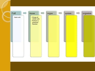
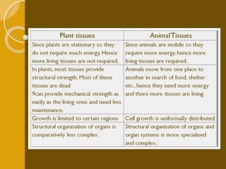
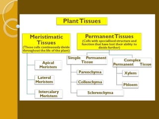
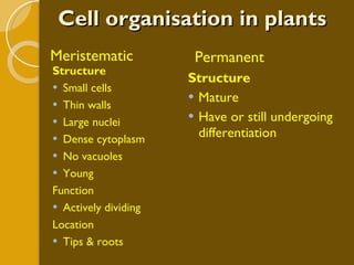
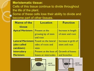
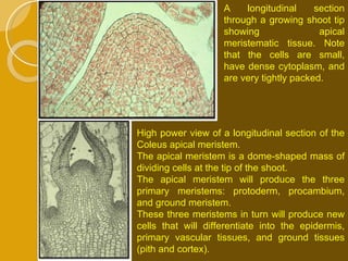
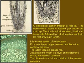
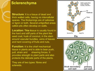
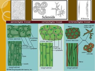
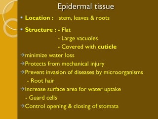
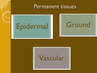
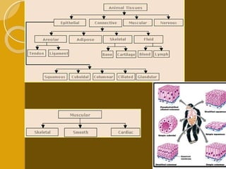
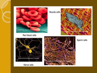
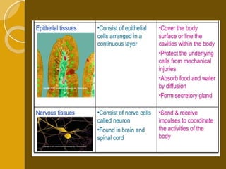
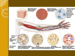
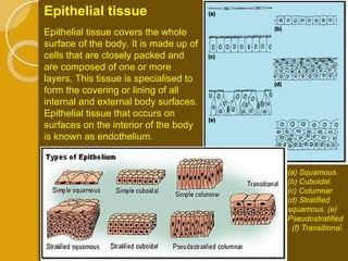
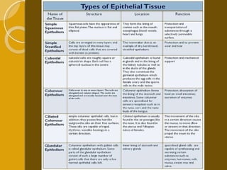
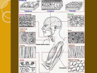
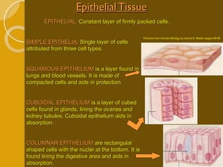
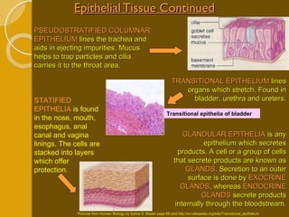
REKAYASA JARINGAN
Cell specialisation in multicellular organism Size of a cell cannot grow beyond a certain size So in order to grow    must increase number of cells! With increase in size, all living process will increase in terms of  size (more food, oxygen),  quality (efficient intake and outtake) and complexity (organelles    organs)
Cell specialisation in multicellular organism Specialisation  is modification in Structure Biochemistry Undergo process  differentiation
 
Organs Examples: heart, kidneys, lungs,  skin & stomach
 
T issue is a cellular organizational level intermediate between cells and a complete organism. Hence, a tissue is an ensemble of cells, not necessarily identical, but from the same origin, that together carry out a specific function.  T he study of tissue is known as  histology  or, in connection with disease,  histopathology.
 
Plant  tissue
 
Cell organisation in plants Meristematic Permanent Structure Small cells Thin walls Large nuclei Dense cytoplasm No vacuoles Young Function Actively dividing  Location Tips & roots Structure Mature Have or still undergoing differentiation
Meristematic tissue:   Cells of this tissue continue to divide throughout the life of the plant.  Some of these cells lose their ability to divide and become part of other tissues.
A longitudinal section through a growing shoot tip showing apical meristematic tissue. Note that the cells are small, have dense cytoplasm, and are very tightly packed. High power view of a longitudinal section of the Coleus apical meristem.  The apical meristem is a dome-shaped mass of dividing cells at the tip of the shoot.  The apical meristem will produce the three primary meristems: protoderm, procambium, and ground meristem.  These three meristems in turn will produce new cells that will differentiate into the epidermis, primary vascular tissues, and ground tissues (pith and cortex).
A longitudinal section through a root tip.  The meristematic tissue is located just above the root cap. This too is apical meristem; division of these cells followed by cell elongation results in the root growing in length. It is a cross section of a dicot stem.  Focus on the two large vascular bundles in the center of the slide.  The xylem tissue is stained red.  Just above the xylem is a layer of meristematic tissue, the vascular cambium. The phloem tissue is found outside of the vascular cambium.
This is a high-power view of a cross-section showing a lateral meristem, the vascular cambium, in the same plant shown in previous slide. Again, the xylem tissue is stained red, and the large cells on the top of the slide are phloem. The green brick-like cells between the xylem and phloem is the area in which the vascular cambium is located. The new cells produced by the cambium are initially like those of the cambium itself, but, as they grow and mature, their characteristics slowly change as they differentiate into other tissues. The vascular cambium is a single layer of cells within this brick like region; it is responsible for the growth in diameter of a stem. The tissues produced by the vascular cambium are secondary tissues.
Permanent tissue:  Cells of this tissue have lost their ability to divide and they have a specialized  structure to perform specific functions. Based on the type of cells present in the tissue, the Permanent tissue is divided into two categories: Simple Permanent Tissue  and  Complex Permanent Tissue. While the simple permanent tissue consist of only one type of cells (eg. Parenchyma),  the complex permanent tissue consists of more than one type of cells (eg. Xylem and phloem)
Types of parenchyma : i) Chlorenchyma :Certain parenchymatous tissue contain chloroplast and synthesize food by the process of photosynthesis. ii)  Aerenchyma: In aquatic plants parenchymatous cells have air cavities between them to store air, such a tissue is called Aerenchyma. It provides buoyancy to the aquatic plants so that they can float in water. Simple Permanent Tissues Parenchyma Structure :It is the fundamental tissue composed of thin walled, living cells whose cell wall is composed of cellulose. Small intercellular spaces are present between the cells. Location and function : It occurs in all soft parts of plants and is meant for storage of food and to provide turgidity to softer parts of plants. Parenchyma
Collenchyma in Transverse Section Showing Wall Thickenings 1. Cell Wall 2. Wall Thickenings 3. Protoplasm 4. Vacuole  Collenchyma Structure : This tissue is composed of somewhat elongated cells with cell walls that are irregularly thickened at corners due to deposition of cellulose or pectin. They may be oval, circular or polygonal. Very little intercellular spaces are present. Location : It occurs below the epidermis of stem and petiole (stalk of the leaf) and around veins.  Function : This tissue provides mechanical support and flexibility and in some cases it may possess chloroplasts to perform Photosynthesis. The stem and leaves are able to bend easily and then come back to their original position due to the presence of collenchyma.
Sclerenchyma Structure : It is a tissue of dead and thick walled cells, having no intercellular spaces. The thickenings are of cellulose or lignin or both. Several unlignified areas called pits often develop on walls. Location : This t issue is usually found in the hard and stiff parts of the plant like seed coat, husk of coconut, in the stem around vascular bundles, veins of leaves and hard covering of fruits and nuts. Function :  It is the chief mechanical tissue in plants and is able to bear push, pull, strain and  shearing forces. It provides strength to plant parts and also protects the delicate parts of the plants. They are of two types: fibres and sclereids.
 
Epidermal tissue Location :  stem, leaves & roots Structure : -  Flat  - Large vacuoles - Covered with  cuticle  minimize water loss Protects from mechanical injury Prevent invasion of diseases by microorganisms - Root hair  Increase surface area for water uptake - Guard cells Control opening & closing of stomata
Permanent tissues
Epidermis and Bark The protective tissues The  epidermis  usually consists of a single-layered group of cells that covers plants leaves, flowers, roots and stems. It forms a boundary between the plant and the external world. Bark  is formed from the meristem that appears later in the life cycle of a plant. Woody stems and some other stem structures produce a secondary covering called the secondary meristem or  periderm  or cork cambium that replaces the epidermis as the protective covering. The periderm replaces the epidermis, and acts as a protective covering like the epidermis.  Cells produced on the outside by periderm form the cork. Cells of have suberin in their walls to protect the stem from drying and pathogen attack. Older cork cells are dead, as is the case with woody stems. As the stem grows, the cork cambium produces new layers of cork which are impermeable to gases and water.
 
Periderm is found on the surface of woody plants; it includes the cork cells on the surface of older woody stems. The periderm replaces the epidermis in plants that have secondary growth. The cork cells are dead; it is their waterproofed cell walls that function as the protective outer covering of plants. Meristematic cells within the periderm (cork cambium, the other lateral meristem) produce the cork cells. type of surface tissue, the outer bark or periderm (stained red in this slide).
Complex Permanent Tissues Xylem and Phloem
They are:  Xylem tracheids Xylem tracheae or vessels Xylem fibers and Xylem parenchyma Xylem It is a complex permanent tissue, which is specialized for the conduction of water and mineral substances in the plant body. Xylem is a heterogenous tissue made up of four different types of cellular elements.
Phloem is also a heterogenous tissue, made up of four different types of cellular elements, namely,  Sieve tubes Companion cells Phloem parenchyma and Phloem fibres Phloem is a complex permanent tissue, which is specialized for the conduction of food and other organic substances.  Phloem
animal /human tissue
Multicellular (large) organisms function more efficiently if cells become specialized for specific functions. There are types of tissues found :  Connective Tissue Muscular Tissue Nervous Tissue Epithelial Tissue and Cell Junctions While also the system in Organism : Integumentary System Organ System Homeostasis *Sponges do not have tissues.


Connective Tissue It is an  tissue that is characterized by the abundance of extracellular components (such as fibers and intercellular substances).  The tissue derives its name from its function in connecting, supporting, surrounding  or binding cells and tissues.  Connective tissue is composed of:  cells extracellular matrix Extracellular matrix is a special feature that distinguishes connective tissue from the other tissues of the body. This matrix may be jelly-like, fluid, dense or rigid. The nature of matrix differs according to the function of that particular connective tissue.
Connective Tissue Connective tissue helps attach materials together through fibrous, supportive, bone and fluid connective tissues. All connective tissues have three common properties: specialized cells, ground substance, and protein fibers.  The  GROUND SUBSTANCE  is chemical substances that saturates space in between cells and fibers. There are three different types of fibers:  COLLAGEN FIBERS  are constructed of a protein (collagen) which aids in flexibility and durability. Similar to collagen fibers, the  RETICULAR FIBERS  branch outward to form thin support systems. Lastly, a protein known as elastin makes up  ELASTIC FIBERS  which offer flexibility. FIBROUS CONNECTIVE TISSUE Loose fibrous – Fibroblast cells; forms protective layer over organs and aids epithelium. Dense fibrous – Fibroblast cells; created from closely bundled collagen fibers. Often found in tendons/ligaments connections to bones/joints. Adipose tissue – Loose connective tissue where cells swell to store fat. Tissue protects organs, insulates organs, and is used for energy. Picture from http://en.wikipedia.org/wiki/Fibrous_connective_tissue


Connective Tissue: - Cells - Extracellular Matrix Characteristics Of Connective Tissue:  Cells -  widely spaced in the extracellular matrix Components of extracellular matrix:  -  Water  -  Ground substance:  semisolid gel   containing  Tissue fluids, salts & Glycoconjugates  two types Proteoglycans – core protein – GAG (Glycoproteins) is attached  five – Chondrotin sulphate, dermatan sulphate, keratan sulphate & heparan sulphate.  Glycoproteins – shorter branched oligosaccharides - Fibronectin & laminin - Fibres Collagen fibres (white fibres)  widely distributed Elastic fibres (yellow fibres) found  in the lungs, blood vessels  Reticular fibres are fine collagen  fibres found in spleen and liver
Functions – based on mechanical properties  Binding together Compartmentalization Support Physical  Immunologic protection & Storage
CELLS Younger or Undifferentiated cell  Fibrocytes- most numerous, synthesis and turnover of both fibres and non fibrillar ground substance Macrophages- and other phagocytic cells  Mast cells- secrete histamine & heparin which mediate inflammatory process  Plasma cells- secrete antibodies  Adipocytes-stores fat Eosinophils,neutrophils, etc
Fibers:  Reticular fibres Delicate  Form fine networks – scaffolding  for support resident cells Found mostly in the Lymphoid organs  Consist of collagen  - main type – tropocollagen type III  Stroma = supporting network of an organ/gland Parenchyma = specific cells that characterize the gland/organ E.g in Lymph node, stroma = reticular tissue, principal parenchymal cells – lymphocytes
Fibers:  Elastic fibres Embedded in an amorphous matrix.  - matrix is composed of the protein elastin.  Microfibrils- individual Fibres are branched - branches end in hook like fashion Normally - Thinner than the collagen fibres
Fibers: Collagen Dominant  Unbranched Function  - strength to the connective tissue. Thickness of the fibres varies Each of these fibrils is composed of microfibrils Types: different tropocollagen types (named type I to XX) Types I, II and III are the major fibre-forming tropocollagens. Tropocollagen type IV - component of the basal lamina.
Connective tissue
Connective Tissue Continued SUPPORTIVE TISSUE  aids in the formation of bone and cartilage. Due to its solid matrix, this tissues cells usually occupy small cavities known as  LACUNAE . There are three different types of cartilage:  HYALINE CARTILAGE  is constructed of a whitish matrix with thin collagen fibers. This type of cartilage is found at the end of bones, in the trachea, and in the nose. Similar to Hyaline,  ELASTIC CARTILAGE  contains more elastic fibers which yields more flexibility. Elastic cartilage is found in the outer ear area, the voice box (larynx), and the epiglottis. Lastly, the  FIBROCARTILAGE  consists of very durable collagen fibers which can endure pressure/weight and absorb shock. Fibrocartilage is found in the joint of the pubic bones, spinal disks, and in knee cartilage. Picture from http://en.wikipedia.org/wiki/Fibrocartilage Supportive Connective Tissue Fibrocartilage Hyaline Cartilage Picture from http://www.victoriacollege.edu/dept/bio/Belltutorials/Histology%20Tutorial/Basic%20Tissues/Connective%20Tissue.html#Elastic%20CT
Connective Tissue Continued BONES  are the firmest connective tissue which is constructed of a hard matrix and collagen fibers. Bones get some of their structure from  OSSEOUS TISSUE , which lends to the cylindrical shape of them  (OSTEONS) . Long and dense  COMPACT BONES  have rings of hard matrix in the osteons. At the ends of long bones is a lighter structure known as  SPONGY BONE . This material has a separated formation permitting space for marrow and blood vessels. Bone Pictures from  http://en.wikipedia.org/wiki/Bone#Compact_bone  and Human Biology by Sylvia S. Mader Page 63 Spongy Bone Compact Bone
Connective Tissue Continued Fluid BLOOD:  Plasma liquid with formed elements (red and white blood cells, and platelets). Bloods duties include carrying oxygen and nutrients throughout the body to other tissues fluid and circulates heat.  RED BLOOD CELLS  transport oxygen throughout the body by loosely binding the oxygen with the cells  HEMOGLOBIN ,   an iron containing structure. Red blood cells do not contain a nucleus. On the other hand,  WHITE BLOOD CELLS  contain a nucleus and are larger in size. Also, they have a more translucent appearance. White blood cells help to fight infections by either consuming the pathogens through phagocytosis or creating antibodies to fight infections.  PLATELETS  are present in bone marrow where they aid in the reconstruction of broken blood vessels. Platelets are pieces of cells. LYMPH:  Yellowish fluid containing white blood cells. Lymph originates from tissue fluid and is cleansed in the  LYMPH NODES  which is lymphatic tissue on a lymphatic vessel. Picture of elements found in blood Picture from Human Biology by Sylvia S. Mader page 64
CONNECTIVE TISSUE PROPER: TYPES
Loose Areolar Connective Tissue - numerous cells and blood vessels - abundant ground substance, flexible, not resistant to stress - Found in -  under epithelial surfaces, around blood vessels & glands
The 5 primary components of the superficial fascia (loose irregular areolar connective tissue):  1. Fibroblasts 2. Collagen Fibers 3. Elastic Fibers 4. Tissue Fluid 5. Fat  In contrast to the overlying dermis and the underlying deep fascia, the superficial fascia may be distinguished by the presence of fat
Dense irregular connective tissue Dense irregular connective tissue forms the dermis of the skin.  Are very extensive.  High density of collagen fibres and their irregular distribution. Few cells. Dark spots scattered between the collagen fibres represent the nuclei of the cells  Dense irregular
Dense Regular Connective Tissue Coarse collagen fibres are aligned with each other with only very narrow opens spaces between them.  Like in most other connective tissues, there will be only a few cells between the fibres.  Their cytoplasm is difficult to identify but the nuclei can be seen scattered among the collagen fibres.  Nuclei are often elongated,  and their long axis runs parallel to the course of the collagen  fibres found in areas of tension,  tendons, ligaments &  aponeuroses, stroma of spleen & lymph nodes. Dense regular
Adipose tissue (white) An aggregation of fat cells (adipocytes) Contains a large droplet of fat that fills it. Nucleus is pushed against the plasma membrane & is flattened. Cells are supported by reticular fibres
Brown adipose tissue Here Adipocytes have nucleus centrally placed Cytoplasm has a frothy appearance Cell borders are not clear Capillaries are very frequent
Epithelial tissue covers the whole surface of the body. It is made up of cells that are closely packed and are composed of one or more layers. This tissue is specialised to form the covering or lining of all internal and external body surfaces. Epithelial tissue that occurs on surfaces on the interior of the body is known as endothelium.  Epithelial tissue  (a) Squamous. (b) Cuboidal.  (c) Columnar. (d) Stratified squamous. (e) Pseudostratified. (f) Transitional.
 
 
Epithelial Tissue EPITHELIAL:  Constant layer of firmly packed cells. SIMPLE EPITHELIA:  Single layer of cells attributed from three cell types. SQUAMOUS EPITHELIUM  is a layer found in lungs and blood vessels. It is made of compacted cells and aids in protection. CUBOIDAL EPITHELIUM  is a layer of cubed cells found in glands, lining the ovaries and kidney tubules. Cuboidal epithelium aids in absorption. COLUMNAR EPITHELIUM  are rectangular shaped cells with the nuclei at the bottom. It is found lining the digestive area and aids in absorption. Pictures from Human Biology by Sylvia S. Mader pages 68-69
Epithelial Tissue Continued PSEUDOSTRATIFIED COLUMNAR EPITHELIUM  lines the trachea and aids in ejecting impurities. Mucus helps to trap particles and cilia carries it to the throat area. TRANSITIONAL EPITHELIUM  lines organs which stretch. Found in bladder, urethra and ureters. STATIFIED EPITHELIA  is found in the nose, mouth, esophagus, anal canal and vagina linings. The cells are stacked into layers which offer protection. GLANDULAR EPITHELIA  is any epithelium which secretes products. A cell or a group of cells that secrete products are known as  GLANDS . Secretion to an outer surface is done by  EXOCRINE GLANDS , whereas  ENDOCRINE GLANDS  secrete products internally through the bloodstream. Pictures from Human Biology by Sylvia S. Mader page 69 and http://en.wikipedia.org/wiki/Transitional_epithelium Transitional epithelia of bladder
Muscular tissue  Muscles of the body are made up of elongated muscle cells also known as muscle fibre. The movement of the body is brought about by the contraction and relaxation of contractile protein present in muscle cells. These contractile proteins are actin and myosin.
Muscular Tissue SKELETON MUSCLE:  Found where muscles attach to bone and aid in movement. The fibers are long cylinder shapes that are formed by the combining of cells, resulting in multiple nuclei. SMOOTH MUSCLE:  Found in the walls of blood vessels and some internal organs. Aids in the transfer of substances. CARDIAC MUSLE:  Found in heart walls. Its function is to pump blood. Pictures from Human Biology by Sylvia S. Mader page 65
 
 
All living cells have the ability to react to stimuli.  Nervous tissue  is  specialised  to  react to stimuli  and to  conduct impulses  to various organs in the body which bring about a  response to the stimulus .  Nerve tissue  (as in the brain, spinal cord and peripheral nerves that branch throughout the body) are all made up of specialised nerve cells called  neurons . Nervous Tissue
Neurons have many different shapes and sizes. However, a typical neuron in a human consists of four major regions: a cell body, dendrites, an  axon , and synaptic terminals. Like all cells, the entire neuron is surrounded by a cell membrane.  The cell body is the enlarged portion of a neuron that most closely resembles other cells. It contains the  nucleus  and other  organelles  (for example, the  mitochondria  and  endoplasmic reticulum ).  The dendrites and axon are thin  cytoplasmic  extensions of the neuron. The dendrites, which branch out in treelike fashion from the cell body, are specialized to receive signals and transmit them toward the cell body. The single long axon carries signals away from the cell body. In humans, a single axon may be as long as 1 meter (about 3 feet). Some neurons that have cell bodies in the spinal cord have axons that extend all the way down to the toes.
A  nerve  is an enclosed, cable-like bundle of axons (the long, slender projections of neurons). A nerve provides a common pathway for the electrochemical nerve impulses that are transmitted along each of the axons.
Nervous Tissue Nerve tissue consists of Neurons and Neuroglia. NEURONS:  A cell consisting of dendrites, a body and axon.  DENDRITES  are branches off the cell body that receive signals. The  CELL BODY  contains a nucleus and   cytoplasm. An  AXON  carries out nerve impulses from the body. NEUROGLIA:  Cells found in nervous tissue. Neuroglia support neurons through nourishment. Picture from Human Biology by Sylvia S. Mader Page 66 Axon Dendrites
Cell Junctions Cell junctions aid tissues in their functions by joining cells together either by tight junctions, adhesion junctions or gap junctions. A. TIGHT JUNCTIONS:  Cell layers become resilient by creating a tough barrier by the joining of plasma membrane proteins. B. ADHESION JUNCTIONS:  Cells cytoskeleton fibers are attached to one another. Found in tissues which stretch, like skin. C. GAP JUNCTIONS:  A junction formed by two neighboring plasma membranes, allowing molecules/ions to circulate through channels. Pictures from Human Biology by Sylvia S. Mader Page 70
Integumentary System :  Skin and other organs SKIN:  Covers the body, protects tissues, prevents H 2 O loss, regulates temperature, and protects against diseases from entering the body. The skin contains two sections, the epidermis and the dermis. Certain cells can produce Vitamin D with the help of UV radiation. EPIDERMIS:  Stratified squamous epithelium. Stem cells get new epidermal cells for skin renewal. Picture from Human Biology by Sylvia S. Mader Page 71 Epidermis Dermis Subcutaneous layer SUBCUTANEOUS LAYER :  Constructed of adipose and loose connective tissue. Offers protective layer against external abuse. DERMIS:  Thick fibrous (collagen and elastic) tissue under the epidermis. Allows movement and flexibility without tearing. Blood vessels deliver nutrients to the skin while regulating body temperature. Contains sensory receptors. Nails, hair follicles, and sweat glands are accessory organs of skin.
 
The Integumentary System Integumentary System (inte = whole; -gument = body covering ) Two major components:  Cutaneous membrane / skin Accessory structure: hair, nails, & multicellular exocrine glands
General Function of the integumentary system Protection Excretion Maintenance of normal body temperature Thermoreceptors Alterations in cutaneous blood flow conserve or release heat Neural control (primary determining factor)  Local control Synthesis of vitamin D 3 Storage of lipids Detection of touch, pressure, pain and temperature stimuli
Synthesis of vitamin D 3
The Integumentary System The integument does not function in isolation. There are also:  extensive network of blood vessels sensory receptors (touch, pressure, temperature & pain) hypodermis / subcutaneous layer  : separates the integument from the deep fascia around other organs Accessible Varied in function Underappreciated
Nerve endings in skin and subcutaneous tissue provide input to the brain for touch, pressure, thermal and pain sensations Control blood flow and sweat gland activity for thermoregulation NERVOUS SYSTEM The integumentary system affects other systems in the body The integumentary system is affected by other systems in the body RESPIRATORY SYSTEM Hairs in nose filter dust particles from inhaled air Stimulation of pain nerve endings in skin may alter breathing rate Provide oxygen to skin cells and eliminates carbon dioxide via gas exchange with blood
Keratinocytes in skin help activate 7 dehydrocholesterol to cholecalciferol(vit D3), later on vit D3 will be changed into calcitriol in kidney, a hormone that aids absorption of dietary calcium and phosphorus Sex hormones  Stimulate sebaceous gland activity Influence growth, distribution of subcutaneous fat, and apocrine sweat gland activity Adrenal hormone Alter dermal blood flow and help mobilize lipids from adipocytes ENDOCRINE SYSTEM DIGESTIVE SYSTEM Helps activate vitamin D  to the hormone calcitriol, which promotes absorption of dietary calcium and phosphorus in the small intestine Provides nutrients for all cells and lipids for storage by adipocytes
CARDIOVASCULAR SYSTEM Local chemical changes in dermis cause widening and narrowing of skin blood vessels, which help adjust blood flow to the skin Prevents fluid loss of the body Serves as blood reservoir Provides oxygen and nutrients for the skin; delivers hormones and cells of immune system to the skin; provide substances needed by skin glands to make their secretions Carries away carbon dioxide, waste products and toxins from the skin Provides heat to maintain normal skin temperature URINARY SYSTEM Assists in excretion of water and solutes in sweat Keratinized epidermis limits fluid loss through skin Excretes waste products Maintains normal pH and ion composition of body fluids
LYMPHATIC SYSTEM & IMMUNITY Discourage penetration and growth of microbes: Provide mechanical barriers Langerhans cells in epidermis : recognizing & processing foreign antigens Macrophages in the dermis : phagocytize microbes that penetrate the skin surface Mast cells trigger inflammation and initiate the immune response Defending the integument by providing additional macrophages and mobilizing lymphocytes REPRODUCTIVE SYSTEM Nerve endings in skin and subcutaneous tissue respond to erotic stimuli    contributing to sexual pleasure Mammary glands (modified sweat glands) produce milk  Suckling of a baby stimulates nerve endings in skin    leading to milk ejection Skin stretches during pregnancy as fetus enlarges Sex hormones affect hair distribution, adipose tissue distribution in subcutaneous layer, and mammary gland development
SKIN Largest organ of the body, both surface area & weight In adults, covers an area of about 2m 2  and weighs 4.5 – 5 kg (16% of total body weight) Has 3 components: Epidermis (5 layers) Dermis (2 Layers) Papillary dermis Reticular dermis Subcutaneous (Hypodermis/Fatty layer) )
Eight Functions of Human Skin Protect underlying tissues from injury: mechanical, heat, cold, biological. Prevent excess water loss. Act as a temperature regulator. Serve as a reservoir for food and water: adipose tissue Assist in the process of excretion: H 2 0, Salt, Urea, Lactic Acid. Serve as a sense organ for cutaneous senses: pain, heat, cold, pressure, touch. Prevent entrance of foreign bodies: microorganisms. Serve as a seat of origin for Vitamin D.
The Anatomy of Human Skin
The  epidermis   is the ectodermally derived outer layer composed of  keratinized stratified squamous epithelium.  Keratinocytes provide protective properties. Melanocytes provide pigmentation. Langerhans’ cells help immune system. Merkel cells provide sensory receptors. The epidermis is the most superficial layer - provides first barrier of protection from invasion of foreign substances . The dermis functions in thermoregulation and supports the vascular network to supply the avascular epidermis
 
5 layers Stratum basale (germinativum) Stratum spinosum Stratum granulosum Stratum lucidum Stratum corneum
The stratum Germinatum ( SG ) provides the germinal cells necessary for the regeneration of the layers of the epidermis. These germinal cells are separated from the dermis by a thin layer of basement membrane.  Continuous mitotic activity Consisting of a wide layer of cuboidal or columnar  cells in contact with the dermis 1 layer
Stratum spinosum  The cells that divide in the statum germinativum soon begin to accumulate many desmosomes on their outer surface. These provide the characteristic “prickles” of the stratum spinosum ( SS ), which is called the prickle-cell layer.  Consisting of several  rows of polyhedral cells  with round nuclei
Stratum Granulosum  Consisting of 3-5 layers of polygonal cells that gradually become flattened The cytoplasm contains KERATOHYALINE granules The progressive maturation of a keratinocyte is charcterized by the accumulation of keratin, called keratinization.  The cells of the stratum granulosum ( SGR )  accumulate dense basophilic  Keratohyalin granules These granules contain  lipids, which along with  the desmosomal  connections, help to form  a waterproof barrier
Stratum lucidum  (eosinophilic) Composed of a thin layer of transparent flattened cells More evident in thick skin The stratum lucidum is normally only well seen in thick epidermis and represents a transition from stratum granulosum to stratum corneum Epidermis varies in thickness depending mainly on frictional forces and is thickest on the palms and soles.
Stratum corneum  (horny layer) Cells in this layer are dead having lost their nuclei and other organelles Squamous and extremelly flattened and scale like Cells contain protein keratin  Consists of many layers of flattened non-nucleated keratinized cells In SC a keratinocyte gradually migrates to the surface and is sloughed off in a process called desquamation.  S C
 
The Dermis Papillary layer Superficial thin layer of loose connective tissue beneath the epithelium  Reticular layer Deeper and thicker layer which contains skin appendages, blood vessels, and nerves The  dermis  is the mesodermally derived layer of dense irregular collagenous connective tissue that underlies and interdigitates with the epidermis.  Collagen, glycoaminoglycans, elastine, ect. Fibroblasts are principal cellular constituent. Vascular structures, nerves, skin appendages.
The dermis is the mid layer of skin, thick inner layer of skin, which comprises  blood vessels ,  connective tissue ,  nerves ,  lymph vessels ,  sweat glands  and hair shafts. It has two main layers: The upper layer for touch,  pain  and heat, which communicate with the central nervous system and is responsible for the folds of the  fingerprints . The lower layer made of dense elastic fibers that house the  hair follicles , nerves,  gland , and gives the skin most of its stretchiness and strength. The Dermis
The Dermis contains mostly fibroblasts secrete collagen, elastin and ground substance support and elasticity of skin  Also present are immune cells that are involved in defense against foreign invaders passing through the epidermis.  is typically subdivided into two zones, a  papillary dermis  and a  reticular layer .
The papillary dermis (PD) contains vascular networks that have two functions: support the avascular epidermis with nutrients  provide a network for thermoregulation.  The vasculature by increasing or decreasing blood flow, can conserve or dissipate heat.  The vasculature interdigitates in areas = dermal papillae ( DP ).  PD also contains free sensory nerve endings and Meissner’s corpuscles
Melanocytes are melanin-pigment forming cells derived from neural crest widely distributed throughout the body. present in the epidermis and its appendages
Sebaceous glands
Merckels cells  In the basal layer of the epidermis, and intimately associated with a nerve terminal.  Also present in outer root sheath of large hair follicles They are mechanoreceptors Clusters of Merkel cell–neurite complexes in glabrous skin = touch corpuscles (Tastscheiben), and in hairy skin =tactile hair discs. Detect vertical, shearing, or other directional deformations (continuous touch), and direction of hair movement
changes in epidermal cell shape during keratinisation  and three specialised cells within the epidermis:  m = melanocyte; L = Langerhans cell; M = Merkel cell (associated with nerve ending).
Meissner's corpuscles are located in the dermal papillae of skin and are in contact with basal cells.  They are most numerous in the finger tips, palm and sole,  lips, and nipple.   They are rapidly adapting mechanoreceptors that subserve discriminative touch sensations   Elongated ovoid bodies found in the dermal papillae (papillary) Base of epidermis Meissner's Corpuscle   (tactile)
Sensory receptors  Free nerve endings are used to sense both temperature and pain
The Reticular Layer (RD) consists of dense irregular connective tissue, It differs from papillary layer ( PD ), which is made up of loose connective tissue (less number of cells)  This layer of the dermis gives the skin’s overall strength and elasticity It houses other important epithelial derived structures such as glands and hair follicles.
Hypodermis: composed of loose connective tissue with large numbers of adipose cells.  provides insulation, shock absorption, energy storage, and the ability of skin to slide over joints.  contains the major blood vessels of the skin. Adipose tissue plus connective tissue. Anchors skin to underlying tissues. Shook absorber and insulator.
Pacinian Corpuscle These are located deep in the hypodermis.  Their function is for deep pressure reception. Detection of vibration and mediate deep pressure sensation Sliced onion-like lamellar structure located in the deep dermis and subcutaneous tissue
Skin appendages Hair or Hair shaft Elongated keratinized structure derived from invagination of epidermal epithelium Hair follicle A tubular invagination of the epidermis which extends down into the dermis Sweat gland Appears as group of ducts bound by cuboidal cells Sebaceous gland Connected to the hair follicle, compound of polygonal cells with clear cytoplasm and centrally located nucleus  which are all embryologically epidermal in origin.  The skin contains a variety of appendages:
Sebaceous Glands Simple alveolar glands found everywhere except palms of the hands and soles of the feet. Do the ducts of these glands branch? Are these glands exo- or endocrine? Secrete an oily, lipid-rich secretion called  sebum . Lanolin is actually sheep sebum Sebum is typically secreted into a hair follicle or occasionally onto the body surface. Sebum softens and lubricates the skin.  It also decreases the skin’s permeability to water and is quite bactericidal. arrector pili muscle
The sebaceous gland is indicated by the arrow.  Note how its duct is unbranched and how it empties into a hair follicle.
Sudoriferous/Sweat Glands Sweat glands. Distributed over the entire body except the nipples and portions of the external genitalia. Over 2.5 million per person. 2 types: Merocrine sweat glands Apocrine sweat glands
Merocrine Sweat Glands More numerous than apocrine sweat glands. Especially prominent on the palms, soles, and forehead. Simple, coiled, tubular glands. Duct empties into a funnel-shaped pore at the skin surface. Major function of merocrine sweating is to cool the body –  thermoregulation .
Merocrine Sweat Glands Merocrine sweat is a dilute watery solution of some salts (including NaCl), vitamin C, antibodies, small amounts of nitrogenous wastes (urea, uric acid, and ammonia), and lactic acid. pH of sweat is 4-6 creating a film on the body known as the  acid mantle .  Such an acidic environment is  bacteriostatic  – prevents bacterial reproduction and growth.
Hair and Hair Follicles The hair follicle surrounds much of the hair root. It contains an outer connective tissue sheath and an inner epithelial root sheath. At the base of the hair follicle is a single layer of mitotic cells derived from the stratum basale.  This is the  hair matrix . All the cells of the hair are derived from the hair matrix.  Just beneath the hair matrix is an obvious dermal papilla called the  hair papilla .  It contains the blood vessels that nourish the matrix and the cells of the hair follicle.
Notice the hair shaft, hair follicle, papilla, and the multiple sebaceous glands.
Hair and Hair Follicles Wrapped around the bulb of the follicle is a network of sensory nerve endings known as the  hair root plexus . Allow the hairs to serve a sensory function. Attached to each hair is a bundle of smooth muscle known as an  arrector pili muscle .  In times of fright or cold, these muscles contract and cause the hair to stand on end – and produces goose bumps. Increases airflow in mammals with significant hair (i.e., not humans) and increases the apparent size of an animal with significant hair.  Vestigial in humans. arrector pili muscle.
The arrow indicates an arrector pili muscle.  In this picture, you should also try to identify the shaft, root, follicle, hair papilla, and sebaceous gland.
Hair follicles with well developed sebaceous glands and their ducts.
SKIN, vertical section. Epidermis with keratinized cells, hair in hair follicles, sebaceous glands. Thick dermis.
Nail Located on dorsal distal phalanx of each finger & toe Nail plate composed of hard keratin Stratum corneum of the epidermis that overlies the nail root forms the eponychium(cuticle) Hyponychium or nail plate consisits of the stratum corneum of the underlying nail bed and so is a keratinised epithelial layer Nail bed epidermis Has only stratum basale & spinosum Growth due to cells in nail matrix & nail root
Organ System Overview INTEGUMENTARY:  Skin, hair, hair muscles, nails, blood vessels, glands, and nerves protect body, regulates body temperature, and creates vitamin D from UV radiation. CARDIOVASCULAR:  Heart muscle pumps blood into vessels where nutrients/oxygen are delivered to cells. Blood removes carbon dioxide and cell waste while circulating heat. Red blood cells transport oxygen while white blood cells fight infections. Platelets aid in vessel repair. Controls Fluid and pH. LYMPHATIC:  System collects tissue fluids, absorbs fat, and stores white blood cells. Helps regulate fluid balance. IMMUNE:  Includes all cells which help to protect the organism from disease. DIGESTIVE:  Takes food and breaks it down into nutrient molecules for cells. Eliminates waste. RESPIRATORY:  Sustains breathing by taking oxygen in and removing carbon dioxide. Also helps manage normal pH. URINARY:  Helps regulate fluid balance and manage normal pH through excretion of waste products. SKELETAL:  Gives body shape while protecting organs. Skeletal muscles aid in movement. Blood cells are created from marrow and minerals are stored in the system. MUSCULAR:  Muscle contraction aids in movement and posture. Smooth muscle helps organs contract which releases heat and warms the body. NERVOUS:  Brain, spinal cord and nerves receive sensory data which is stored. Nerve impulses are sent to muscles to aid in movement. ENDOCRINE:  Glands produce hormones into blood. Aids in control of fluids, pH balance, and metabolism. Helps maintain reproductive organs. REPRODUCTIVE:  Creates and moves gametes and hormones; births offspring.
Organ System Overview Continued DORSAL CAVITY:  Contains the Cranial Cavity and the Vertebral Cavity. CRANIAL:  Brain VERTEBRAL:  Spinal Cord VENTRAL CAVITY:   Contains the Thoracic Cavity, Abdominal Cavity, and Pelvic Cavity. THORACIC:  Heart, lungs, and esophagus ABDOMINAL:  Stomach, Liver, Spleen, Pancreas, Gallbladder, and Intestines PELVIC:  Reproductive organs A muscle known as  DIAPHRAM  separates the thoracic and abdominal cavities.  Four Types of Body Membranes MUCOUS:  Loose fibrous epithelial tissue which lines the interior respiratory, digestive, urinary, and reproductive systems. Goblet cells secrete mucus to from bacterial or virus penetration. SEROUS:  Thoracic cavity and lungs are covered by pleurae, heart is covered by pericardial sac, and the abdominal cavity is covered by the peritoneum. The abdominal organs are attached to the wall by mesentery (double layer peritoneum). Membranes remain lubricated by watery secretion. SYNOVIAL:  Loose connective tissue lines cavities of joints and secrete lubricative solution to keep bones moving freely. MENINGES:  Protective tissue which covers the brain and spinal cord.
Organ System Overview Continued Thoracic Cavity Abdominal Cavity Ventral Cavity Dorsal Cavity Picture from Human Biology by Sylvia S. Mader Page 77
Homeostasis:   The body’s capacity to physically regulate its internal environment is known as  HOMEOSTASIS . All systems work together to help maintain homeostasis. The normal conditions upheld in a cell or organism is known as Homeostasis. For example, the organ systems of humans all work together to perform certain functions such as absorbing nutrients and oxygen,  and excreting waste.  Picture from Human Biology by Sylvia S. Mader Page 79 They also adjust their processes to maintain regularity such as sweating when the body temperature starts to rise.
Homeostasis Continued NEGATIVE FEEDBACK:  The internal environment stays fairly stable due to negative feedback mechanisms through sensors and the control center. When a change occurs, a sensor will notify the control center which release an effect to overturn the change. POSITIVE FEEDBACK:  When the internal environment senses stimulation from nerve impulses, the brain sends positive signals to not only keep the stimulation going, but to make it stronger. Example of Negative Feedback Cycle
REKAYASA JARINGAN
Tissue Engineering – a definition Creation of a functional biological substitute using living cells and a matrix to maintain,  improve or restore damage to tissues and organs  (Atala, A. Engineering tissues, organs and cells. 2007 J Tissue Eng Regen Med 1: 83-96) Bringing together the fields of medicine, biology, engineering and biotechnology http://www.henryfordhealth.org/ http://www.ipeinc.com/l http://rgcb.res.in/
Tissue engineering Tissue engineering is the process of creating living, physiological 3D tissues and organs. The process starts with a source of cells derived from a patient or from a donor. The cells may be immature cells, in the stem cell stage, or cells that are already capable of carrying out tissue functions; often, a mixture of different cell types (e.g., liver cells and blood vessel cells) and cell maturity levels is needed. Many therapeutic applications of tissue engineering involve disease processes that might be prevented or treated if better drugs were available or if the processes could be better understood .
Tissue Engineering
Important Variables
In general, there are three main approaches to tissue engineering: (1) To use isolated cells or cell substitutes as cellular replacement parts;  (2) To use acellular materials capable of inducing tissue regeneration; and  (3) To use a combination of cells and materials (typically in the form of scaffolds and this approach be categorized into two categories: Open  and  closed  systems. These systems are distinguished based on the exposure of the cells to the immune system upon implantation
The materials used for tissue engineering are either synthetic biodegradable materials  such as polylactic acid (PLA), polyglycolic acid (PGA),  poly lactic-glycolic acid (PLGA), polypropylene fumarate,  poly ethylene glycol (PEG) and polyarylates) or natural materials such as collagen, hydroxyapatite, calcium carbonate, and alginate.  Natural materials are typically more favorable to cell adherence, whereas the properties of synthetic materials such as degradation rate, mechanical properties, structure, and porosity can be better controlled
Current approaches for tissue engineering using tissue (postnatal) stem cells: (A) Expansion of a population ex vivo prior to transplantation into the host,  (B) Ex vivo recreation of a tissue or organ for transplantation, and (C) Design of substances and/or devices for in vivo activation of stem cells, either local or distant, to induce appropriate tissue repair
Delivery Methods Injectable stem cells Cells or  cell-polymer mix Less invasive Adopt shape of environment Controlled growth factor release Solid scaffold manufacturing Computer-aided design Match defect shape
The need for TE Failing tissues and organs Shortfalls of current options Autologous tissues Allogeneic tissues Xenogeneic tissues Synthetic materials http://www.myskin-info.com http://www.stanford.edu/
SKIN GRAFFTING Definitions Graft A skin graft is a tissue of epidermis and varying amounts of dermis that is detached from its own blood supply and placed in a new area with a new blood supply. Flap Any tissue used for reconstruction or wound closure that retains all or part of its original blood supply after the tissue has been moved to the recipient location.
Classification of Grafts Autografts  – A tissue transferred from one part of the body to another. Homografts/Allograft   – tissue transferred from a genetically different individual of the same species. Xenografts  – a graft transferred from an individual of one species to an individual of another species.
Graft vs. Flap Graft Does not maintain original blood supply. Flap Maintains original blood supply.
Types of Grafts Grafts are typically described in terms of thickness or depth. Split Thickness :  Contains 100% of the epidermis and a portion of the dermis. Split thickness grafts are further classified as  thin  or  thick . Full Thickness :  Contains 100% of the epidermis and dermis. The amount of primary contraction is directly related to the thickness of dermis in the graft.
The Process of Take Phase 1 (0-48h)  – Plasmatic Imbibition Diffusion of nutrition from the recipient bed .  Phase 2 –  Inosculation Vessels in graft connect with those in recipient bed. Phase 3 (day 3-5)  – Neovascular Ingrowth Graft revascularized by ingrowth of new vessels into bed.
Requirements for Survival Bed must be well vascularized. The contact between graft and recipient must be fully immobile. Low bacterial count at the site.
Split Thickness Used when cosmetic appearance is not a primary issue or when the size of the wound is too large to use a full thickness graft. Chronic Ulcers Temporary coverage Correction of pigmentation disorders Burns
Full Thickness Indications for full thickness skin grafts include: If adjacent tissue has premalignant or malignant lesions and precludes the use of a flap.  Specific locations that lend themselves well to FTSGs include the nasal tip, helical rim, forehead, eyelids, medial canthus, concha, and digits.
 
Tissue Transplantation Definition: to transfer a graft (an organ or tissue) from  one part or individual to another May take place between: Different parts of same organism(autografting) Different  organisms of the same specie (allografting) Different species(xenografting)
Autologous tissues Tissue from the same patient The ideal option Biocompatible, no immune response Natural Decreasing availability of healthy tissue from patients with disease Atherosclerosis http://commons.wikimedia.org/
Autografting The transfer of self tissue from one body site to another in the same individual Due to the genetic homology of the tissue, the immune system does not respond to it Use: synthetic implantation skin grafts bone marrow transplantation
Allogeneic tissues Tissue from another person More practical than autologous harvest Severe shortage of donors Increased immune response to foreign material  (Santos, T. et al. TERMIS, NA 2007) Immunosuppressant drugs required Unpleasant side effects Expensive
Allografting Definition: The transfer of organs or tissue from human to human or from cadaver to human As there are more and more people every year waiting for donor organs and tissues, allografting transplantation has become quite common. Allografting transplantation has many applications.
Application of allografting transplantation
Xenogeneic tissues Tissue harvested from animals Potentially readily available Immune response from host to foreign material  (Santos, T. et al. TERMIS, NA 2007) Risk of disease transmission from animal to human e.g. prions and PERVs Significant ethical considerations www.wallpaperbase.com  http://www.sheppardsoftware.com/
Xenografting Definition: Xenotransplantation – the  transfer of tissue from one species to another Usually refers to the implantation of animal tissue in humans provides a new source of organs for humans many different types of tissue can be transplanted: e.g. heart, kidney, liver or lung
From which animals are we able to transplant organs 1. The Chimpanzee: Its DNA sequence differs from ours by only 2% 2. The Baboon: Its organs are too small for a large adult human 3. The Pig: Surprisingly similar too our anatomy and physiology
TE – in comparison Biocompatible  made with the patient’s own cells Should not elicit immune or inflammatory response Engineered to fill the exact role required Degradation rate  Composition Size  Mechanical properties Off-the-shelf availability Durability Functional Adequate mechanical and hemodynamic function, Mature ECM, Living Growth and remodeling capabilities of the construct should mimic the native structure
Models for Tissue Engineering In vitro  differentiation Construct tissues outside body before transplantation Ultimate goal Most economical Least waiting time In situ  methodology Host remodeling of environment Ex vivo  approach Excision and remodeling in culture Combine physical and chemical factors Optimize stem cell differentiation and organization
Applications for TE In surgery Transplantation of failing tissues/organs Aiding tissues in the healing process In the laboratory Observing immunological, pathological and healing changes in human tissue without harming patients Drug therapies: efficacy and side effects of drugs
Current TE models Skin – collagen matrix seeded with fibroblasts or keratinocytes http://www.cbte.group.shef.ac.uk/research/ http://www.myskin-info.com/index.php Myskin™
Current TE models Vascular – small diameter vessels  Decellularised Porcine Ureter Mr Chris Derham
Current TE models Heart valves - decellularised http://www.biomed.metu.edu.tr/courses/ www.chir.unizh.ch/
Current TE models Urethra Atala et al. A novel inert collagen matrix for hypospadias repair. (1999) J Urol. 162(3 pt 2):1148-1151
Current TE models Bladder Construction of engineered bladder Scaffold seeded with cells (A) and engineered bladder anastamosed to native bladder with running 4–0 polyglycolic sutures (B).  Implant covered with fibrin glue and omentum (C).  Atala et al. Tissue-engineered autologous bladders for patients needing cystoplasty. (2006) Lancet. 367(9518):1241-6
Current TE models Kidney Lanza et al. Generation of histocompatible tissues using nuclear transplantation. (2002) Nat Biotechnol. 20(7):689-696.
Current TE models Cartilage news.bbc.co.uk  Foetal lamb tracheal defects Fuchs et al. Fetal tracheal augmentation with cartilage engineered from bone marrow–derived mesenchymal progenitor cells. (2003) J Pediatr Surg 38: 984–987
Example: Vascular TE Large diameter grafts ( > 6mm lumen) There is already a viable synthetic alternative Small diameter ( < 6mm) PTFE/ePTFE used in large diameter vessels is unsuitable here Low velocity blood flow in small diameter vessels  -> thrombus formation Need confluent endothelium Current TE models Availability Autologous cells needs previous banking Not useful in emergencies How do we predict the onset of disease? Correct choice of animal model to simulate the human body Benefits over conventional means must outweigh costs
Tissue Engineered Heart Valves (TEHV) Heart valve disease occurs when one or more of the four heart valves cease to adequately perform their function, thereby failing to maintain unidirectional blood flow through the heart Surgical procedures or total valve replacement are necessary  Adapted from http://z.about.com/d/p/440/e/f/19011.jpg
Tissue Engineered Blood Vessels (TEBV) From  An Introduction to Biomaterials.  Ch 24.  Fig.4  Ramaswami, P and Wagner, WR. 2005.  Atherosclerosis, in the form of coronary artery disease results in over coronary artery bypass graft procedures. Many patients do not have suitable vessels due to age, disease, or previous use. Synthetic coronary bypass vessels have not performed adequately to be employed to any significant degree
Problems of Transplantation There are not enough organs patients in industrially developed countries badly need donor organs and tissues people die because of the lack of available organs for transplant each day.   Rejection: When the immune system of the host detects foreign graft tissue, it launches an attack, resulting in tissue rejection
Immune system rejection Often a transplanted organ is not identified by the immune system as the tissue of the organism    It can be attacked and destroyed. Against this effect the patient has to swallow Immunesuppressiva which cause symptoms like suffering from AIDS. In 15-20 minutes the organ dies, unable to withstand the immune system attack. Rejection  of a heart
The Future of TE Some tissues already in clinical use Improvements needed to increase availability and safety For widespread use, reduced cost is essential Further work should focus on: vascularisation of new tissue; maintaining nutrient supply to cells in matrix with increasing size Achieving full potential of stem cells to differentiate into desired cell types
Challenges Unforeseen hurdles in the creation of multicellular constructs In the laboratory Supply of nutrients  Removal of waste  Size  Mechanical stability In the patient Availability Evidence of efficacy Safety; graft rejection Cost
Creation of collagen matrices Containing fibroblasts Seeded with endothelial cells Incorporation of fibrin Novel imaging techniques of tissue-engineered constructs Challenges occur at the most basic level Challenges
Making a TE model It will need: A scaffold  to allow growth and differentiation of cells Cells  of the relevant type and number Optimum conditions for cell growth Growth factors Physical environment (bioreactors) www.bioexpress.com
Tissue engineering requires three things: Cells Signals Scaffold The scaffold refers to the tissue model construct The signals refer to molecular signaling molecules, also known as growth factors
Scaffold Structural integrity to support cell attachment, growth and differentiation Correct pore size Mechanical strength to withstand  in vivo  compression Can be natural, synthetic or combined A sheet of small intestinal submucosal scaffold http://www.rcsed.ac.uk/journal/
 
Basic scaffold criteria: Portions must be biodegradable Usually designed in the shape of the tissue product the researcher is working on www.eng.nus.edu.sg/ EResnews/0210/rd/rd_10.html
Biomimetic Scaffold Fabrication                                                                                                                                            bms.dent.umich.edu/research/malab.html
www.millenium-biologix.com/Html/00_ScientificInformationCartiGraft.htm Autologous de novo cartilage formed on Skelite™ tissue engineering scaffold (grown  in vitro ), illustrating the configuration of the implant that provides functional cartilage tissue at the articular surface. The presence of functional cartilage tissue represents a major advance over current cell therapy techniques. Cell therapy involves the implantation of cells that still have to make new cartilage  in vivo  at the defect site under very challenging conditions. The histology image  on the right shows that cells are healthy and growing, while attaching themselves to the Skelite™ and beginning to differentiate into mature cartilage.
Cells Able to cause change and affect structure and function of a graft Examples: Skin model :  fibroblasts and keratinocytes  Vascular construct: smooth muscle and endothelial cells Cartilage: chondrocytes Stem cells – much potential but difficult to direct differentiation and achieve sufficient cell numbers Skin: Ikuta et al Mouse epidermal keratinocytes in three-dimensional organotypic coculture with dermal fibroblasts form a stratified sheet resembling skin. (2006) Bioscience, Biotechnology & Biochemistry. 70(11):2669-75  Vascular: L’heureux et al. A completely biological tissue-engineered human blood vessel. (1998) FASEB J., 12: 47 – 56. Cartilage: Schaefer et al. Tissue-engineered composites for the repair of large osteochondral defects. (2002) Arthrit Rheum 46(9): 2524-2534
Cell sourcing Ideally autologous Difficulties in isolating cells  from diseased tissue Stem cells Adult Embryonic ‘ Immunoprivileged’ Potentially could differentiate into a wide variety of cell types  ( Ungrin, M. and Zandstra, P. TERMIS, NA 2007 ) Potentially teratogenic/carcinogenic Ethically, legally and financially questionable www.udel.edu
Environment Growth factors Directing cellular activity Bioreactors Designed to expose cells to  physical stimuli and/or maintain desired conditions Examples Bladder urothelium: cyclic strain Vascular graft: pulsatile flow ITEMS Bioreactor:        Six Station Vascular Bioreactor   www.tissuegrowth.com Bladder: Kerr et al. The bladder as a bioreactor: urothelium production and secretion of growth hormone into urine.(1998) Nature Biotechnology 16(1):75-9  Vascular: Engbers-Buijtenhuijs et al. Biological characterisation of vascular grafts cultured in a bioreactor. (2006) Biomaterials. 27(11):2390-7
References Biomedical Engineering Handbook - J.D.Bronzino Journal of US-China Medical Science ,  Jul. 2007, Volume 4, No.7 (Serial No.32) Successful Use of a Physiologically Acceptable Artificial Skin in the Treatment of Extensive Burn Injury – John F Burke , MD Tissue Engineering – Concepts and Strategies , Amulya K Saxena  Internet
Works Cited http://www.nigms.nih.gov/Publications/Factsheet_ArtificialSkin.htm   http://www.seattlepi.com/local/burn231.shtml   http://www.discoveriesinmedicine.com/images/mdis_0000_0001_0_img0032.jpg   http://www.scielo.org.za/img/revistas/sajs/v104n11-12/a30fig02.gif   http://pubs.acs.org/subscribe/archive/mdd/v07/i09/html/904feature_willis1.html   http://www.integra-ls.com/products/default.aspx?product=46#Product%20Description   http://www.nlm.nih.gov/medlineplus/ency/article/002363.htm   http://www.webmd.com/skin-problems-and-treatments/guide/skin-biopsies   http://www.burnsurvivor.com/skin_substitutes.html
Works Cited Amarin. Lipophilic Platform – Plasma Membrane. Accessed 4 Feb 2008. < http://www.amarincorp.com/science_and_technology/lipophilic_platform.452.399.html >. &quot;Bone.&quot;  Wikipedia, The Free Encyclopedia . 6 Feb 2008, 05:32 UTC. Wikimedia Foundation, Inc. 2 Feb 2008  < http://en.wikipedia.org/w/index.php?title=Bone&oldid=189439576 >.  &quot;Cell.&quot;  Wikipedia, The Free Encyclopedia . 1 Feb 2008, 15:34 UTC. Wikimedia Foundation, Inc. 2 Feb 2008 < http://en.wikipedia.org/w/index.php?title=Cell_%28biology%29&oldid=188815215 >. &quot;Cell nucleus.&quot;  Wikipedia, The Free Encyclopedia . 1 Feb 2008, 01:41 UTC. Wikimedia Foundation, Inc. 2 Feb 2008  < http://en.wikipedia.org/w/index.php?title=Cell_nucleus&oldid=188933496 >. &quot;Eukaryote.&quot;  Wikipedia, The Free Encyclopedia . 1 Feb 2008, 21:56 UTC. Wikimedia Foundation, Inc. 2 Feb 2008  < http://en.wikipedia.org/w/index.php?title=Eukaryote&oldid=189118974 >. &quot;Fibrocartilage.&quot;  Wikipedia, The Free Encyclopedia . 27 Oct 2007, 13:26 UTC. Wikimedia Foundation, Inc. 2 Feb 2008 < http://en.wikipedia.org/w/index.php?title=Fibrocartilage&oldid=167424710 >. &quot;Fibrous connective tissue.&quot;  Wikipedia, The Free Encyclopedia . 7 Dec 2007, 11:12 UTC. Wikimedia Foundation, Inc. 2 Feb 2008  < http://en.wikipedia.org/w/index.php?title=Fibrous_connective_tissue&oldid=176345853 >.  Mader, Sylvia S.  Human Biology . New York: The McGraw-Hill Companies, Inc, 2008. Pages 41-82. Menlo School. Cytoskeleton. Accessed 5 Feb 2008. < http://sun.menloschool.org/~birchler/cells/animals/cytoskeleton/ >. &quot;Mitochondrion.&quot;  Wikipedia, The Free Encyclopedia . 22 Jan 2008, 18:59 UTC. Wikimedia Foundation, Inc. 6 Feb 2008 < http://en.wikipedia.org/w/index.php?title=Mitochondrion&oldid=186166731 >.  &quot;Osmosis.&quot;  Wikipedia, The Free Encyclopedia . 4 Feb 2008, 21:04 UTC. Wikimedia Foundation, Inc. 5 Feb 2008  < http://en.wikipedia.org/w/index.php?title=Osmosis&oldid=189106016 >. &quot;Transitional epithelium.&quot;  Wikipedia, The Free Encyclopedia . 23 Jan 2008, 07:06 UTC. Wikimedia Foundation, Inc. 3 Feb 2008  < http://en.wikipedia.org/w/index.php?title=Transitional_epithelium&oldid=186300028 >. Victoria College. Biology Department - Connective Tissues. Accessed 5 Feb 2008. < http://www.victoriacollege.edu/dept/bio/Belltutorials/Histology%20Tutorial/Basic%20Tissues/Connective%20Tissue.h tml>.

Rekayasa jaringan kulit

  • 1.
  • 2.
    Cell specialisation inmulticellular organism Size of a cell cannot grow beyond a certain size So in order to grow  must increase number of cells! With increase in size, all living process will increase in terms of size (more food, oxygen), quality (efficient intake and outtake) and complexity (organelles  organs)
  • 3.
    Cell specialisation inmulticellular organism Specialisation is modification in Structure Biochemistry Undergo process differentiation
  • 4.
  • 5.
    Organs Examples: heart,kidneys, lungs, skin & stomach
  • 6.
  • 7.
    T issue isa cellular organizational level intermediate between cells and a complete organism. Hence, a tissue is an ensemble of cells, not necessarily identical, but from the same origin, that together carry out a specific function. T he study of tissue is known as histology or, in connection with disease, histopathology.
  • 8.
  • 9.
  • 10.
  • 11.
    Cell organisation inplants Meristematic Permanent Structure Small cells Thin walls Large nuclei Dense cytoplasm No vacuoles Young Function Actively dividing Location Tips & roots Structure Mature Have or still undergoing differentiation
  • 12.
    Meristematic tissue: Cells of this tissue continue to divide throughout the life of the plant. Some of these cells lose their ability to divide and become part of other tissues.
  • 13.
    A longitudinal sectionthrough a growing shoot tip showing apical meristematic tissue. Note that the cells are small, have dense cytoplasm, and are very tightly packed. High power view of a longitudinal section of the Coleus apical meristem. The apical meristem is a dome-shaped mass of dividing cells at the tip of the shoot. The apical meristem will produce the three primary meristems: protoderm, procambium, and ground meristem. These three meristems in turn will produce new cells that will differentiate into the epidermis, primary vascular tissues, and ground tissues (pith and cortex).
  • 14.
    A longitudinal sectionthrough a root tip. The meristematic tissue is located just above the root cap. This too is apical meristem; division of these cells followed by cell elongation results in the root growing in length. It is a cross section of a dicot stem. Focus on the two large vascular bundles in the center of the slide. The xylem tissue is stained red. Just above the xylem is a layer of meristematic tissue, the vascular cambium. The phloem tissue is found outside of the vascular cambium.
  • 15.
    This is ahigh-power view of a cross-section showing a lateral meristem, the vascular cambium, in the same plant shown in previous slide. Again, the xylem tissue is stained red, and the large cells on the top of the slide are phloem. The green brick-like cells between the xylem and phloem is the area in which the vascular cambium is located. The new cells produced by the cambium are initially like those of the cambium itself, but, as they grow and mature, their characteristics slowly change as they differentiate into other tissues. The vascular cambium is a single layer of cells within this brick like region; it is responsible for the growth in diameter of a stem. The tissues produced by the vascular cambium are secondary tissues.
  • 16.
    Permanent tissue: Cells of this tissue have lost their ability to divide and they have a specialized structure to perform specific functions. Based on the type of cells present in the tissue, the Permanent tissue is divided into two categories: Simple Permanent Tissue and Complex Permanent Tissue. While the simple permanent tissue consist of only one type of cells (eg. Parenchyma), the complex permanent tissue consists of more than one type of cells (eg. Xylem and phloem)
  • 17.
    Types of parenchyma: i) Chlorenchyma :Certain parenchymatous tissue contain chloroplast and synthesize food by the process of photosynthesis. ii) Aerenchyma: In aquatic plants parenchymatous cells have air cavities between them to store air, such a tissue is called Aerenchyma. It provides buoyancy to the aquatic plants so that they can float in water. Simple Permanent Tissues Parenchyma Structure :It is the fundamental tissue composed of thin walled, living cells whose cell wall is composed of cellulose. Small intercellular spaces are present between the cells. Location and function : It occurs in all soft parts of plants and is meant for storage of food and to provide turgidity to softer parts of plants. Parenchyma
  • 18.
    Collenchyma in TransverseSection Showing Wall Thickenings 1. Cell Wall 2. Wall Thickenings 3. Protoplasm 4. Vacuole Collenchyma Structure : This tissue is composed of somewhat elongated cells with cell walls that are irregularly thickened at corners due to deposition of cellulose or pectin. They may be oval, circular or polygonal. Very little intercellular spaces are present. Location : It occurs below the epidermis of stem and petiole (stalk of the leaf) and around veins. Function : This tissue provides mechanical support and flexibility and in some cases it may possess chloroplasts to perform Photosynthesis. The stem and leaves are able to bend easily and then come back to their original position due to the presence of collenchyma.
  • 19.
    Sclerenchyma Structure :It is a tissue of dead and thick walled cells, having no intercellular spaces. The thickenings are of cellulose or lignin or both. Several unlignified areas called pits often develop on walls. Location : This t issue is usually found in the hard and stiff parts of the plant like seed coat, husk of coconut, in the stem around vascular bundles, veins of leaves and hard covering of fruits and nuts. Function : It is the chief mechanical tissue in plants and is able to bear push, pull, strain and shearing forces. It provides strength to plant parts and also protects the delicate parts of the plants. They are of two types: fibres and sclereids.
  • 20.
  • 21.
    Epidermal tissue Location: stem, leaves & roots Structure : - Flat - Large vacuoles - Covered with cuticle minimize water loss Protects from mechanical injury Prevent invasion of diseases by microorganisms - Root hair Increase surface area for water uptake - Guard cells Control opening & closing of stomata
  • 22.
  • 23.
    Epidermis and BarkThe protective tissues The epidermis usually consists of a single-layered group of cells that covers plants leaves, flowers, roots and stems. It forms a boundary between the plant and the external world. Bark is formed from the meristem that appears later in the life cycle of a plant. Woody stems and some other stem structures produce a secondary covering called the secondary meristem or periderm or cork cambium that replaces the epidermis as the protective covering. The periderm replaces the epidermis, and acts as a protective covering like the epidermis. Cells produced on the outside by periderm form the cork. Cells of have suberin in their walls to protect the stem from drying and pathogen attack. Older cork cells are dead, as is the case with woody stems. As the stem grows, the cork cambium produces new layers of cork which are impermeable to gases and water.
  • 24.
  • 25.
    Periderm is foundon the surface of woody plants; it includes the cork cells on the surface of older woody stems. The periderm replaces the epidermis in plants that have secondary growth. The cork cells are dead; it is their waterproofed cell walls that function as the protective outer covering of plants. Meristematic cells within the periderm (cork cambium, the other lateral meristem) produce the cork cells. type of surface tissue, the outer bark or periderm (stained red in this slide).
  • 26.
    Complex Permanent TissuesXylem and Phloem
  • 27.
    They are: Xylem tracheids Xylem tracheae or vessels Xylem fibers and Xylem parenchyma Xylem It is a complex permanent tissue, which is specialized for the conduction of water and mineral substances in the plant body. Xylem is a heterogenous tissue made up of four different types of cellular elements.
  • 28.
    Phloem is alsoa heterogenous tissue, made up of four different types of cellular elements, namely, Sieve tubes Companion cells Phloem parenchyma and Phloem fibres Phloem is a complex permanent tissue, which is specialized for the conduction of food and other organic substances. Phloem
  • 29.
  • 30.
    Multicellular (large) organismsfunction more efficiently if cells become specialized for specific functions. There are types of tissues found : Connective Tissue Muscular Tissue Nervous Tissue Epithelial Tissue and Cell Junctions While also the system in Organism : Integumentary System Organ System Homeostasis *Sponges do not have tissues.
  • 31.
  • 32.
  • 33.
  • 34.
  • 35.
    Connective Tissue Itis an tissue that is characterized by the abundance of extracellular components (such as fibers and intercellular substances). The tissue derives its name from its function in connecting, supporting, surrounding or binding cells and tissues. Connective tissue is composed of: cells extracellular matrix Extracellular matrix is a special feature that distinguishes connective tissue from the other tissues of the body. This matrix may be jelly-like, fluid, dense or rigid. The nature of matrix differs according to the function of that particular connective tissue.
  • 36.
    Connective Tissue Connectivetissue helps attach materials together through fibrous, supportive, bone and fluid connective tissues. All connective tissues have three common properties: specialized cells, ground substance, and protein fibers. The GROUND SUBSTANCE is chemical substances that saturates space in between cells and fibers. There are three different types of fibers: COLLAGEN FIBERS are constructed of a protein (collagen) which aids in flexibility and durability. Similar to collagen fibers, the RETICULAR FIBERS branch outward to form thin support systems. Lastly, a protein known as elastin makes up ELASTIC FIBERS which offer flexibility. FIBROUS CONNECTIVE TISSUE Loose fibrous – Fibroblast cells; forms protective layer over organs and aids epithelium. Dense fibrous – Fibroblast cells; created from closely bundled collagen fibers. Often found in tendons/ligaments connections to bones/joints. Adipose tissue – Loose connective tissue where cells swell to store fat. Tissue protects organs, insulates organs, and is used for energy. Picture from http://en.wikipedia.org/wiki/Fibrous_connective_tissue
  • 37.
  • 38.
  • 39.
  • 40.
  • 41.
    Connective Tissue: -Cells - Extracellular Matrix Characteristics Of Connective Tissue: Cells - widely spaced in the extracellular matrix Components of extracellular matrix: - Water - Ground substance: semisolid gel containing Tissue fluids, salts & Glycoconjugates two types Proteoglycans – core protein – GAG (Glycoproteins) is attached five – Chondrotin sulphate, dermatan sulphate, keratan sulphate & heparan sulphate. Glycoproteins – shorter branched oligosaccharides - Fibronectin & laminin - Fibres Collagen fibres (white fibres) widely distributed Elastic fibres (yellow fibres) found in the lungs, blood vessels Reticular fibres are fine collagen fibres found in spleen and liver
  • 42.
    Functions – basedon mechanical properties Binding together Compartmentalization Support Physical Immunologic protection & Storage
  • 43.
    CELLS Younger orUndifferentiated cell Fibrocytes- most numerous, synthesis and turnover of both fibres and non fibrillar ground substance Macrophages- and other phagocytic cells Mast cells- secrete histamine & heparin which mediate inflammatory process Plasma cells- secrete antibodies Adipocytes-stores fat Eosinophils,neutrophils, etc
  • 44.
    Fibers: Reticularfibres Delicate Form fine networks – scaffolding for support resident cells Found mostly in the Lymphoid organs Consist of collagen - main type – tropocollagen type III Stroma = supporting network of an organ/gland Parenchyma = specific cells that characterize the gland/organ E.g in Lymph node, stroma = reticular tissue, principal parenchymal cells – lymphocytes
  • 45.
    Fibers: Elasticfibres Embedded in an amorphous matrix. - matrix is composed of the protein elastin. Microfibrils- individual Fibres are branched - branches end in hook like fashion Normally - Thinner than the collagen fibres
  • 46.
    Fibers: Collagen Dominant Unbranched Function - strength to the connective tissue. Thickness of the fibres varies Each of these fibrils is composed of microfibrils Types: different tropocollagen types (named type I to XX) Types I, II and III are the major fibre-forming tropocollagens. Tropocollagen type IV - component of the basal lamina.
  • 47.
  • 48.
    Connective Tissue ContinuedSUPPORTIVE TISSUE aids in the formation of bone and cartilage. Due to its solid matrix, this tissues cells usually occupy small cavities known as LACUNAE . There are three different types of cartilage: HYALINE CARTILAGE is constructed of a whitish matrix with thin collagen fibers. This type of cartilage is found at the end of bones, in the trachea, and in the nose. Similar to Hyaline, ELASTIC CARTILAGE contains more elastic fibers which yields more flexibility. Elastic cartilage is found in the outer ear area, the voice box (larynx), and the epiglottis. Lastly, the FIBROCARTILAGE consists of very durable collagen fibers which can endure pressure/weight and absorb shock. Fibrocartilage is found in the joint of the pubic bones, spinal disks, and in knee cartilage. Picture from http://en.wikipedia.org/wiki/Fibrocartilage Supportive Connective Tissue Fibrocartilage Hyaline Cartilage Picture from http://www.victoriacollege.edu/dept/bio/Belltutorials/Histology%20Tutorial/Basic%20Tissues/Connective%20Tissue.html#Elastic%20CT
  • 49.
    Connective Tissue ContinuedBONES are the firmest connective tissue which is constructed of a hard matrix and collagen fibers. Bones get some of their structure from OSSEOUS TISSUE , which lends to the cylindrical shape of them (OSTEONS) . Long and dense COMPACT BONES have rings of hard matrix in the osteons. At the ends of long bones is a lighter structure known as SPONGY BONE . This material has a separated formation permitting space for marrow and blood vessels. Bone Pictures from http://en.wikipedia.org/wiki/Bone#Compact_bone and Human Biology by Sylvia S. Mader Page 63 Spongy Bone Compact Bone
  • 50.
    Connective Tissue ContinuedFluid BLOOD: Plasma liquid with formed elements (red and white blood cells, and platelets). Bloods duties include carrying oxygen and nutrients throughout the body to other tissues fluid and circulates heat. RED BLOOD CELLS transport oxygen throughout the body by loosely binding the oxygen with the cells HEMOGLOBIN , an iron containing structure. Red blood cells do not contain a nucleus. On the other hand, WHITE BLOOD CELLS contain a nucleus and are larger in size. Also, they have a more translucent appearance. White blood cells help to fight infections by either consuming the pathogens through phagocytosis or creating antibodies to fight infections. PLATELETS are present in bone marrow where they aid in the reconstruction of broken blood vessels. Platelets are pieces of cells. LYMPH: Yellowish fluid containing white blood cells. Lymph originates from tissue fluid and is cleansed in the LYMPH NODES which is lymphatic tissue on a lymphatic vessel. Picture of elements found in blood Picture from Human Biology by Sylvia S. Mader page 64
  • 51.
  • 52.
    Loose Areolar ConnectiveTissue - numerous cells and blood vessels - abundant ground substance, flexible, not resistant to stress - Found in - under epithelial surfaces, around blood vessels & glands
  • 53.
    The 5 primarycomponents of the superficial fascia (loose irregular areolar connective tissue): 1. Fibroblasts 2. Collagen Fibers 3. Elastic Fibers 4. Tissue Fluid 5. Fat In contrast to the overlying dermis and the underlying deep fascia, the superficial fascia may be distinguished by the presence of fat
  • 54.
    Dense irregular connectivetissue Dense irregular connective tissue forms the dermis of the skin. Are very extensive. High density of collagen fibres and their irregular distribution. Few cells. Dark spots scattered between the collagen fibres represent the nuclei of the cells Dense irregular
  • 55.
    Dense Regular ConnectiveTissue Coarse collagen fibres are aligned with each other with only very narrow opens spaces between them. Like in most other connective tissues, there will be only a few cells between the fibres. Their cytoplasm is difficult to identify but the nuclei can be seen scattered among the collagen fibres. Nuclei are often elongated, and their long axis runs parallel to the course of the collagen fibres found in areas of tension, tendons, ligaments & aponeuroses, stroma of spleen & lymph nodes. Dense regular
  • 56.
    Adipose tissue (white)An aggregation of fat cells (adipocytes) Contains a large droplet of fat that fills it. Nucleus is pushed against the plasma membrane & is flattened. Cells are supported by reticular fibres
  • 57.
    Brown adipose tissueHere Adipocytes have nucleus centrally placed Cytoplasm has a frothy appearance Cell borders are not clear Capillaries are very frequent
  • 58.
    Epithelial tissue coversthe whole surface of the body. It is made up of cells that are closely packed and are composed of one or more layers. This tissue is specialised to form the covering or lining of all internal and external body surfaces. Epithelial tissue that occurs on surfaces on the interior of the body is known as endothelium. Epithelial tissue (a) Squamous. (b) Cuboidal. (c) Columnar. (d) Stratified squamous. (e) Pseudostratified. (f) Transitional.
  • 59.
  • 60.
  • 61.
    Epithelial Tissue EPITHELIAL: Constant layer of firmly packed cells. SIMPLE EPITHELIA: Single layer of cells attributed from three cell types. SQUAMOUS EPITHELIUM is a layer found in lungs and blood vessels. It is made of compacted cells and aids in protection. CUBOIDAL EPITHELIUM is a layer of cubed cells found in glands, lining the ovaries and kidney tubules. Cuboidal epithelium aids in absorption. COLUMNAR EPITHELIUM are rectangular shaped cells with the nuclei at the bottom. It is found lining the digestive area and aids in absorption. Pictures from Human Biology by Sylvia S. Mader pages 68-69
  • 62.
    Epithelial Tissue ContinuedPSEUDOSTRATIFIED COLUMNAR EPITHELIUM lines the trachea and aids in ejecting impurities. Mucus helps to trap particles and cilia carries it to the throat area. TRANSITIONAL EPITHELIUM lines organs which stretch. Found in bladder, urethra and ureters. STATIFIED EPITHELIA is found in the nose, mouth, esophagus, anal canal and vagina linings. The cells are stacked into layers which offer protection. GLANDULAR EPITHELIA is any epithelium which secretes products. A cell or a group of cells that secrete products are known as GLANDS . Secretion to an outer surface is done by EXOCRINE GLANDS , whereas ENDOCRINE GLANDS secrete products internally through the bloodstream. Pictures from Human Biology by Sylvia S. Mader page 69 and http://en.wikipedia.org/wiki/Transitional_epithelium Transitional epithelia of bladder
  • 63.
    Muscular tissue Muscles of the body are made up of elongated muscle cells also known as muscle fibre. The movement of the body is brought about by the contraction and relaxation of contractile protein present in muscle cells. These contractile proteins are actin and myosin.
  • 64.
    Muscular Tissue SKELETONMUSCLE: Found where muscles attach to bone and aid in movement. The fibers are long cylinder shapes that are formed by the combining of cells, resulting in multiple nuclei. SMOOTH MUSCLE: Found in the walls of blood vessels and some internal organs. Aids in the transfer of substances. CARDIAC MUSLE: Found in heart walls. Its function is to pump blood. Pictures from Human Biology by Sylvia S. Mader page 65
  • 65.
  • 66.
  • 67.
    All living cellshave the ability to react to stimuli. Nervous tissue is specialised to react to stimuli and to conduct impulses to various organs in the body which bring about a response to the stimulus . Nerve tissue (as in the brain, spinal cord and peripheral nerves that branch throughout the body) are all made up of specialised nerve cells called neurons . Nervous Tissue
  • 68.
    Neurons have manydifferent shapes and sizes. However, a typical neuron in a human consists of four major regions: a cell body, dendrites, an axon , and synaptic terminals. Like all cells, the entire neuron is surrounded by a cell membrane. The cell body is the enlarged portion of a neuron that most closely resembles other cells. It contains the nucleus and other organelles (for example, the mitochondria and endoplasmic reticulum ). The dendrites and axon are thin cytoplasmic extensions of the neuron. The dendrites, which branch out in treelike fashion from the cell body, are specialized to receive signals and transmit them toward the cell body. The single long axon carries signals away from the cell body. In humans, a single axon may be as long as 1 meter (about 3 feet). Some neurons that have cell bodies in the spinal cord have axons that extend all the way down to the toes.
  • 69.
    A nerve is an enclosed, cable-like bundle of axons (the long, slender projections of neurons). A nerve provides a common pathway for the electrochemical nerve impulses that are transmitted along each of the axons.
  • 70.
    Nervous Tissue Nervetissue consists of Neurons and Neuroglia. NEURONS: A cell consisting of dendrites, a body and axon. DENDRITES are branches off the cell body that receive signals. The CELL BODY contains a nucleus and cytoplasm. An AXON carries out nerve impulses from the body. NEUROGLIA: Cells found in nervous tissue. Neuroglia support neurons through nourishment. Picture from Human Biology by Sylvia S. Mader Page 66 Axon Dendrites
  • 71.
    Cell Junctions Celljunctions aid tissues in their functions by joining cells together either by tight junctions, adhesion junctions or gap junctions. A. TIGHT JUNCTIONS: Cell layers become resilient by creating a tough barrier by the joining of plasma membrane proteins. B. ADHESION JUNCTIONS: Cells cytoskeleton fibers are attached to one another. Found in tissues which stretch, like skin. C. GAP JUNCTIONS: A junction formed by two neighboring plasma membranes, allowing molecules/ions to circulate through channels. Pictures from Human Biology by Sylvia S. Mader Page 70
  • 72.
    Integumentary System : Skin and other organs SKIN: Covers the body, protects tissues, prevents H 2 O loss, regulates temperature, and protects against diseases from entering the body. The skin contains two sections, the epidermis and the dermis. Certain cells can produce Vitamin D with the help of UV radiation. EPIDERMIS: Stratified squamous epithelium. Stem cells get new epidermal cells for skin renewal. Picture from Human Biology by Sylvia S. Mader Page 71 Epidermis Dermis Subcutaneous layer SUBCUTANEOUS LAYER : Constructed of adipose and loose connective tissue. Offers protective layer against external abuse. DERMIS: Thick fibrous (collagen and elastic) tissue under the epidermis. Allows movement and flexibility without tearing. Blood vessels deliver nutrients to the skin while regulating body temperature. Contains sensory receptors. Nails, hair follicles, and sweat glands are accessory organs of skin.
  • 73.
  • 74.
    The Integumentary SystemIntegumentary System (inte = whole; -gument = body covering ) Two major components: Cutaneous membrane / skin Accessory structure: hair, nails, & multicellular exocrine glands
  • 75.
    General Function ofthe integumentary system Protection Excretion Maintenance of normal body temperature Thermoreceptors Alterations in cutaneous blood flow conserve or release heat Neural control (primary determining factor) Local control Synthesis of vitamin D 3 Storage of lipids Detection of touch, pressure, pain and temperature stimuli
  • 76.
  • 77.
    The Integumentary SystemThe integument does not function in isolation. There are also: extensive network of blood vessels sensory receptors (touch, pressure, temperature & pain) hypodermis / subcutaneous layer : separates the integument from the deep fascia around other organs Accessible Varied in function Underappreciated
  • 78.
    Nerve endings inskin and subcutaneous tissue provide input to the brain for touch, pressure, thermal and pain sensations Control blood flow and sweat gland activity for thermoregulation NERVOUS SYSTEM The integumentary system affects other systems in the body The integumentary system is affected by other systems in the body RESPIRATORY SYSTEM Hairs in nose filter dust particles from inhaled air Stimulation of pain nerve endings in skin may alter breathing rate Provide oxygen to skin cells and eliminates carbon dioxide via gas exchange with blood
  • 79.
    Keratinocytes in skinhelp activate 7 dehydrocholesterol to cholecalciferol(vit D3), later on vit D3 will be changed into calcitriol in kidney, a hormone that aids absorption of dietary calcium and phosphorus Sex hormones Stimulate sebaceous gland activity Influence growth, distribution of subcutaneous fat, and apocrine sweat gland activity Adrenal hormone Alter dermal blood flow and help mobilize lipids from adipocytes ENDOCRINE SYSTEM DIGESTIVE SYSTEM Helps activate vitamin D to the hormone calcitriol, which promotes absorption of dietary calcium and phosphorus in the small intestine Provides nutrients for all cells and lipids for storage by adipocytes
  • 80.
    CARDIOVASCULAR SYSTEM Localchemical changes in dermis cause widening and narrowing of skin blood vessels, which help adjust blood flow to the skin Prevents fluid loss of the body Serves as blood reservoir Provides oxygen and nutrients for the skin; delivers hormones and cells of immune system to the skin; provide substances needed by skin glands to make their secretions Carries away carbon dioxide, waste products and toxins from the skin Provides heat to maintain normal skin temperature URINARY SYSTEM Assists in excretion of water and solutes in sweat Keratinized epidermis limits fluid loss through skin Excretes waste products Maintains normal pH and ion composition of body fluids
  • 81.
    LYMPHATIC SYSTEM &IMMUNITY Discourage penetration and growth of microbes: Provide mechanical barriers Langerhans cells in epidermis : recognizing & processing foreign antigens Macrophages in the dermis : phagocytize microbes that penetrate the skin surface Mast cells trigger inflammation and initiate the immune response Defending the integument by providing additional macrophages and mobilizing lymphocytes REPRODUCTIVE SYSTEM Nerve endings in skin and subcutaneous tissue respond to erotic stimuli  contributing to sexual pleasure Mammary glands (modified sweat glands) produce milk Suckling of a baby stimulates nerve endings in skin  leading to milk ejection Skin stretches during pregnancy as fetus enlarges Sex hormones affect hair distribution, adipose tissue distribution in subcutaneous layer, and mammary gland development
  • 82.
    SKIN Largest organof the body, both surface area & weight In adults, covers an area of about 2m 2 and weighs 4.5 – 5 kg (16% of total body weight) Has 3 components: Epidermis (5 layers) Dermis (2 Layers) Papillary dermis Reticular dermis Subcutaneous (Hypodermis/Fatty layer) )
  • 83.
    Eight Functions ofHuman Skin Protect underlying tissues from injury: mechanical, heat, cold, biological. Prevent excess water loss. Act as a temperature regulator. Serve as a reservoir for food and water: adipose tissue Assist in the process of excretion: H 2 0, Salt, Urea, Lactic Acid. Serve as a sense organ for cutaneous senses: pain, heat, cold, pressure, touch. Prevent entrance of foreign bodies: microorganisms. Serve as a seat of origin for Vitamin D.
  • 84.
    The Anatomy ofHuman Skin
  • 85.
    The epidermis is the ectodermally derived outer layer composed of keratinized stratified squamous epithelium. Keratinocytes provide protective properties. Melanocytes provide pigmentation. Langerhans’ cells help immune system. Merkel cells provide sensory receptors. The epidermis is the most superficial layer - provides first barrier of protection from invasion of foreign substances . The dermis functions in thermoregulation and supports the vascular network to supply the avascular epidermis
  • 86.
  • 87.
    5 layers Stratumbasale (germinativum) Stratum spinosum Stratum granulosum Stratum lucidum Stratum corneum
  • 88.
    The stratum Germinatum( SG ) provides the germinal cells necessary for the regeneration of the layers of the epidermis. These germinal cells are separated from the dermis by a thin layer of basement membrane. Continuous mitotic activity Consisting of a wide layer of cuboidal or columnar cells in contact with the dermis 1 layer
  • 89.
    Stratum spinosum The cells that divide in the statum germinativum soon begin to accumulate many desmosomes on their outer surface. These provide the characteristic “prickles” of the stratum spinosum ( SS ), which is called the prickle-cell layer. Consisting of several rows of polyhedral cells with round nuclei
  • 90.
    Stratum Granulosum Consisting of 3-5 layers of polygonal cells that gradually become flattened The cytoplasm contains KERATOHYALINE granules The progressive maturation of a keratinocyte is charcterized by the accumulation of keratin, called keratinization. The cells of the stratum granulosum ( SGR ) accumulate dense basophilic Keratohyalin granules These granules contain lipids, which along with the desmosomal connections, help to form a waterproof barrier
  • 91.
    Stratum lucidum (eosinophilic) Composed of a thin layer of transparent flattened cells More evident in thick skin The stratum lucidum is normally only well seen in thick epidermis and represents a transition from stratum granulosum to stratum corneum Epidermis varies in thickness depending mainly on frictional forces and is thickest on the palms and soles.
  • 92.
    Stratum corneum (horny layer) Cells in this layer are dead having lost their nuclei and other organelles Squamous and extremelly flattened and scale like Cells contain protein keratin Consists of many layers of flattened non-nucleated keratinized cells In SC a keratinocyte gradually migrates to the surface and is sloughed off in a process called desquamation. S C
  • 93.
  • 94.
    The Dermis Papillarylayer Superficial thin layer of loose connective tissue beneath the epithelium Reticular layer Deeper and thicker layer which contains skin appendages, blood vessels, and nerves The dermis is the mesodermally derived layer of dense irregular collagenous connective tissue that underlies and interdigitates with the epidermis. Collagen, glycoaminoglycans, elastine, ect. Fibroblasts are principal cellular constituent. Vascular structures, nerves, skin appendages.
  • 95.
    The dermis isthe mid layer of skin, thick inner layer of skin, which comprises blood vessels , connective tissue , nerves , lymph vessels , sweat glands and hair shafts. It has two main layers: The upper layer for touch, pain and heat, which communicate with the central nervous system and is responsible for the folds of the fingerprints . The lower layer made of dense elastic fibers that house the hair follicles , nerves, gland , and gives the skin most of its stretchiness and strength. The Dermis
  • 96.
    The Dermis containsmostly fibroblasts secrete collagen, elastin and ground substance support and elasticity of skin Also present are immune cells that are involved in defense against foreign invaders passing through the epidermis. is typically subdivided into two zones, a papillary dermis and a reticular layer .
  • 97.
    The papillary dermis(PD) contains vascular networks that have two functions: support the avascular epidermis with nutrients provide a network for thermoregulation. The vasculature by increasing or decreasing blood flow, can conserve or dissipate heat. The vasculature interdigitates in areas = dermal papillae ( DP ). PD also contains free sensory nerve endings and Meissner’s corpuscles
  • 98.
    Melanocytes are melanin-pigmentforming cells derived from neural crest widely distributed throughout the body. present in the epidermis and its appendages
  • 99.
  • 100.
    Merckels cells In the basal layer of the epidermis, and intimately associated with a nerve terminal. Also present in outer root sheath of large hair follicles They are mechanoreceptors Clusters of Merkel cell–neurite complexes in glabrous skin = touch corpuscles (Tastscheiben), and in hairy skin =tactile hair discs. Detect vertical, shearing, or other directional deformations (continuous touch), and direction of hair movement
  • 101.
    changes in epidermalcell shape during keratinisation and three specialised cells within the epidermis: m = melanocyte; L = Langerhans cell; M = Merkel cell (associated with nerve ending).
  • 102.
    Meissner's corpuscles arelocated in the dermal papillae of skin and are in contact with basal cells. They are most numerous in the finger tips, palm and sole, lips, and nipple. They are rapidly adapting mechanoreceptors that subserve discriminative touch sensations Elongated ovoid bodies found in the dermal papillae (papillary) Base of epidermis Meissner's Corpuscle (tactile)
  • 103.
    Sensory receptors Free nerve endings are used to sense both temperature and pain
  • 104.
    The Reticular Layer(RD) consists of dense irregular connective tissue, It differs from papillary layer ( PD ), which is made up of loose connective tissue (less number of cells) This layer of the dermis gives the skin’s overall strength and elasticity It houses other important epithelial derived structures such as glands and hair follicles.
  • 105.
    Hypodermis: composed ofloose connective tissue with large numbers of adipose cells. provides insulation, shock absorption, energy storage, and the ability of skin to slide over joints. contains the major blood vessels of the skin. Adipose tissue plus connective tissue. Anchors skin to underlying tissues. Shook absorber and insulator.
  • 106.
    Pacinian Corpuscle Theseare located deep in the hypodermis. Their function is for deep pressure reception. Detection of vibration and mediate deep pressure sensation Sliced onion-like lamellar structure located in the deep dermis and subcutaneous tissue
  • 107.
    Skin appendages Hairor Hair shaft Elongated keratinized structure derived from invagination of epidermal epithelium Hair follicle A tubular invagination of the epidermis which extends down into the dermis Sweat gland Appears as group of ducts bound by cuboidal cells Sebaceous gland Connected to the hair follicle, compound of polygonal cells with clear cytoplasm and centrally located nucleus which are all embryologically epidermal in origin. The skin contains a variety of appendages:
  • 108.
    Sebaceous Glands Simplealveolar glands found everywhere except palms of the hands and soles of the feet. Do the ducts of these glands branch? Are these glands exo- or endocrine? Secrete an oily, lipid-rich secretion called sebum . Lanolin is actually sheep sebum Sebum is typically secreted into a hair follicle or occasionally onto the body surface. Sebum softens and lubricates the skin. It also decreases the skin’s permeability to water and is quite bactericidal. arrector pili muscle
  • 109.
    The sebaceous glandis indicated by the arrow. Note how its duct is unbranched and how it empties into a hair follicle.
  • 110.
    Sudoriferous/Sweat Glands Sweatglands. Distributed over the entire body except the nipples and portions of the external genitalia. Over 2.5 million per person. 2 types: Merocrine sweat glands Apocrine sweat glands
  • 111.
    Merocrine Sweat GlandsMore numerous than apocrine sweat glands. Especially prominent on the palms, soles, and forehead. Simple, coiled, tubular glands. Duct empties into a funnel-shaped pore at the skin surface. Major function of merocrine sweating is to cool the body – thermoregulation .
  • 112.
    Merocrine Sweat GlandsMerocrine sweat is a dilute watery solution of some salts (including NaCl), vitamin C, antibodies, small amounts of nitrogenous wastes (urea, uric acid, and ammonia), and lactic acid. pH of sweat is 4-6 creating a film on the body known as the acid mantle . Such an acidic environment is bacteriostatic – prevents bacterial reproduction and growth.
  • 113.
    Hair and HairFollicles The hair follicle surrounds much of the hair root. It contains an outer connective tissue sheath and an inner epithelial root sheath. At the base of the hair follicle is a single layer of mitotic cells derived from the stratum basale. This is the hair matrix . All the cells of the hair are derived from the hair matrix. Just beneath the hair matrix is an obvious dermal papilla called the hair papilla . It contains the blood vessels that nourish the matrix and the cells of the hair follicle.
  • 114.
    Notice the hairshaft, hair follicle, papilla, and the multiple sebaceous glands.
  • 115.
    Hair and HairFollicles Wrapped around the bulb of the follicle is a network of sensory nerve endings known as the hair root plexus . Allow the hairs to serve a sensory function. Attached to each hair is a bundle of smooth muscle known as an arrector pili muscle . In times of fright or cold, these muscles contract and cause the hair to stand on end – and produces goose bumps. Increases airflow in mammals with significant hair (i.e., not humans) and increases the apparent size of an animal with significant hair. Vestigial in humans. arrector pili muscle.
  • 116.
    The arrow indicatesan arrector pili muscle. In this picture, you should also try to identify the shaft, root, follicle, hair papilla, and sebaceous gland.
  • 117.
    Hair follicles withwell developed sebaceous glands and their ducts.
  • 118.
    SKIN, vertical section.Epidermis with keratinized cells, hair in hair follicles, sebaceous glands. Thick dermis.
  • 119.
    Nail Located ondorsal distal phalanx of each finger & toe Nail plate composed of hard keratin Stratum corneum of the epidermis that overlies the nail root forms the eponychium(cuticle) Hyponychium or nail plate consisits of the stratum corneum of the underlying nail bed and so is a keratinised epithelial layer Nail bed epidermis Has only stratum basale & spinosum Growth due to cells in nail matrix & nail root
  • 120.
    Organ System OverviewINTEGUMENTARY: Skin, hair, hair muscles, nails, blood vessels, glands, and nerves protect body, regulates body temperature, and creates vitamin D from UV radiation. CARDIOVASCULAR: Heart muscle pumps blood into vessels where nutrients/oxygen are delivered to cells. Blood removes carbon dioxide and cell waste while circulating heat. Red blood cells transport oxygen while white blood cells fight infections. Platelets aid in vessel repair. Controls Fluid and pH. LYMPHATIC: System collects tissue fluids, absorbs fat, and stores white blood cells. Helps regulate fluid balance. IMMUNE: Includes all cells which help to protect the organism from disease. DIGESTIVE: Takes food and breaks it down into nutrient molecules for cells. Eliminates waste. RESPIRATORY: Sustains breathing by taking oxygen in and removing carbon dioxide. Also helps manage normal pH. URINARY: Helps regulate fluid balance and manage normal pH through excretion of waste products. SKELETAL: Gives body shape while protecting organs. Skeletal muscles aid in movement. Blood cells are created from marrow and minerals are stored in the system. MUSCULAR: Muscle contraction aids in movement and posture. Smooth muscle helps organs contract which releases heat and warms the body. NERVOUS: Brain, spinal cord and nerves receive sensory data which is stored. Nerve impulses are sent to muscles to aid in movement. ENDOCRINE: Glands produce hormones into blood. Aids in control of fluids, pH balance, and metabolism. Helps maintain reproductive organs. REPRODUCTIVE: Creates and moves gametes and hormones; births offspring.
  • 121.
    Organ System OverviewContinued DORSAL CAVITY: Contains the Cranial Cavity and the Vertebral Cavity. CRANIAL: Brain VERTEBRAL: Spinal Cord VENTRAL CAVITY: Contains the Thoracic Cavity, Abdominal Cavity, and Pelvic Cavity. THORACIC: Heart, lungs, and esophagus ABDOMINAL: Stomach, Liver, Spleen, Pancreas, Gallbladder, and Intestines PELVIC: Reproductive organs A muscle known as DIAPHRAM separates the thoracic and abdominal cavities. Four Types of Body Membranes MUCOUS: Loose fibrous epithelial tissue which lines the interior respiratory, digestive, urinary, and reproductive systems. Goblet cells secrete mucus to from bacterial or virus penetration. SEROUS: Thoracic cavity and lungs are covered by pleurae, heart is covered by pericardial sac, and the abdominal cavity is covered by the peritoneum. The abdominal organs are attached to the wall by mesentery (double layer peritoneum). Membranes remain lubricated by watery secretion. SYNOVIAL: Loose connective tissue lines cavities of joints and secrete lubricative solution to keep bones moving freely. MENINGES: Protective tissue which covers the brain and spinal cord.
  • 122.
    Organ System OverviewContinued Thoracic Cavity Abdominal Cavity Ventral Cavity Dorsal Cavity Picture from Human Biology by Sylvia S. Mader Page 77
  • 123.
    Homeostasis: The body’s capacity to physically regulate its internal environment is known as HOMEOSTASIS . All systems work together to help maintain homeostasis. The normal conditions upheld in a cell or organism is known as Homeostasis. For example, the organ systems of humans all work together to perform certain functions such as absorbing nutrients and oxygen, and excreting waste. Picture from Human Biology by Sylvia S. Mader Page 79 They also adjust their processes to maintain regularity such as sweating when the body temperature starts to rise.
  • 124.
    Homeostasis Continued NEGATIVEFEEDBACK: The internal environment stays fairly stable due to negative feedback mechanisms through sensors and the control center. When a change occurs, a sensor will notify the control center which release an effect to overturn the change. POSITIVE FEEDBACK: When the internal environment senses stimulation from nerve impulses, the brain sends positive signals to not only keep the stimulation going, but to make it stronger. Example of Negative Feedback Cycle
  • 125.
  • 126.
    Tissue Engineering –a definition Creation of a functional biological substitute using living cells and a matrix to maintain, improve or restore damage to tissues and organs (Atala, A. Engineering tissues, organs and cells. 2007 J Tissue Eng Regen Med 1: 83-96) Bringing together the fields of medicine, biology, engineering and biotechnology http://www.henryfordhealth.org/ http://www.ipeinc.com/l http://rgcb.res.in/
  • 127.
    Tissue engineering Tissueengineering is the process of creating living, physiological 3D tissues and organs. The process starts with a source of cells derived from a patient or from a donor. The cells may be immature cells, in the stem cell stage, or cells that are already capable of carrying out tissue functions; often, a mixture of different cell types (e.g., liver cells and blood vessel cells) and cell maturity levels is needed. Many therapeutic applications of tissue engineering involve disease processes that might be prevented or treated if better drugs were available or if the processes could be better understood .
  • 128.
  • 129.
  • 130.
    In general, thereare three main approaches to tissue engineering: (1) To use isolated cells or cell substitutes as cellular replacement parts; (2) To use acellular materials capable of inducing tissue regeneration; and (3) To use a combination of cells and materials (typically in the form of scaffolds and this approach be categorized into two categories: Open and closed systems. These systems are distinguished based on the exposure of the cells to the immune system upon implantation
  • 131.
    The materials usedfor tissue engineering are either synthetic biodegradable materials such as polylactic acid (PLA), polyglycolic acid (PGA), poly lactic-glycolic acid (PLGA), polypropylene fumarate, poly ethylene glycol (PEG) and polyarylates) or natural materials such as collagen, hydroxyapatite, calcium carbonate, and alginate. Natural materials are typically more favorable to cell adherence, whereas the properties of synthetic materials such as degradation rate, mechanical properties, structure, and porosity can be better controlled
  • 132.
    Current approaches fortissue engineering using tissue (postnatal) stem cells: (A) Expansion of a population ex vivo prior to transplantation into the host, (B) Ex vivo recreation of a tissue or organ for transplantation, and (C) Design of substances and/or devices for in vivo activation of stem cells, either local or distant, to induce appropriate tissue repair
  • 133.
    Delivery Methods Injectablestem cells Cells or cell-polymer mix Less invasive Adopt shape of environment Controlled growth factor release Solid scaffold manufacturing Computer-aided design Match defect shape
  • 134.
    The need forTE Failing tissues and organs Shortfalls of current options Autologous tissues Allogeneic tissues Xenogeneic tissues Synthetic materials http://www.myskin-info.com http://www.stanford.edu/
  • 135.
    SKIN GRAFFTING DefinitionsGraft A skin graft is a tissue of epidermis and varying amounts of dermis that is detached from its own blood supply and placed in a new area with a new blood supply. Flap Any tissue used for reconstruction or wound closure that retains all or part of its original blood supply after the tissue has been moved to the recipient location.
  • 136.
    Classification of GraftsAutografts – A tissue transferred from one part of the body to another. Homografts/Allograft – tissue transferred from a genetically different individual of the same species. Xenografts – a graft transferred from an individual of one species to an individual of another species.
  • 137.
    Graft vs. FlapGraft Does not maintain original blood supply. Flap Maintains original blood supply.
  • 138.
    Types of GraftsGrafts are typically described in terms of thickness or depth. Split Thickness : Contains 100% of the epidermis and a portion of the dermis. Split thickness grafts are further classified as thin or thick . Full Thickness : Contains 100% of the epidermis and dermis. The amount of primary contraction is directly related to the thickness of dermis in the graft.
  • 139.
    The Process ofTake Phase 1 (0-48h) – Plasmatic Imbibition Diffusion of nutrition from the recipient bed . Phase 2 – Inosculation Vessels in graft connect with those in recipient bed. Phase 3 (day 3-5) – Neovascular Ingrowth Graft revascularized by ingrowth of new vessels into bed.
  • 140.
    Requirements for SurvivalBed must be well vascularized. The contact between graft and recipient must be fully immobile. Low bacterial count at the site.
  • 141.
    Split Thickness Usedwhen cosmetic appearance is not a primary issue or when the size of the wound is too large to use a full thickness graft. Chronic Ulcers Temporary coverage Correction of pigmentation disorders Burns
  • 142.
    Full Thickness Indicationsfor full thickness skin grafts include: If adjacent tissue has premalignant or malignant lesions and precludes the use of a flap. Specific locations that lend themselves well to FTSGs include the nasal tip, helical rim, forehead, eyelids, medial canthus, concha, and digits.
  • 143.
  • 144.
    Tissue Transplantation Definition:to transfer a graft (an organ or tissue) from one part or individual to another May take place between: Different parts of same organism(autografting) Different organisms of the same specie (allografting) Different species(xenografting)
  • 145.
    Autologous tissues Tissuefrom the same patient The ideal option Biocompatible, no immune response Natural Decreasing availability of healthy tissue from patients with disease Atherosclerosis http://commons.wikimedia.org/
  • 146.
    Autografting The transferof self tissue from one body site to another in the same individual Due to the genetic homology of the tissue, the immune system does not respond to it Use: synthetic implantation skin grafts bone marrow transplantation
  • 147.
    Allogeneic tissues Tissuefrom another person More practical than autologous harvest Severe shortage of donors Increased immune response to foreign material (Santos, T. et al. TERMIS, NA 2007) Immunosuppressant drugs required Unpleasant side effects Expensive
  • 148.
    Allografting Definition: Thetransfer of organs or tissue from human to human or from cadaver to human As there are more and more people every year waiting for donor organs and tissues, allografting transplantation has become quite common. Allografting transplantation has many applications.
  • 149.
  • 150.
    Xenogeneic tissues Tissueharvested from animals Potentially readily available Immune response from host to foreign material (Santos, T. et al. TERMIS, NA 2007) Risk of disease transmission from animal to human e.g. prions and PERVs Significant ethical considerations www.wallpaperbase.com http://www.sheppardsoftware.com/
  • 151.
    Xenografting Definition: Xenotransplantation– the transfer of tissue from one species to another Usually refers to the implantation of animal tissue in humans provides a new source of organs for humans many different types of tissue can be transplanted: e.g. heart, kidney, liver or lung
  • 152.
    From which animalsare we able to transplant organs 1. The Chimpanzee: Its DNA sequence differs from ours by only 2% 2. The Baboon: Its organs are too small for a large adult human 3. The Pig: Surprisingly similar too our anatomy and physiology
  • 153.
    TE – incomparison Biocompatible made with the patient’s own cells Should not elicit immune or inflammatory response Engineered to fill the exact role required Degradation rate Composition Size Mechanical properties Off-the-shelf availability Durability Functional Adequate mechanical and hemodynamic function, Mature ECM, Living Growth and remodeling capabilities of the construct should mimic the native structure
  • 154.
    Models for TissueEngineering In vitro differentiation Construct tissues outside body before transplantation Ultimate goal Most economical Least waiting time In situ methodology Host remodeling of environment Ex vivo approach Excision and remodeling in culture Combine physical and chemical factors Optimize stem cell differentiation and organization
  • 155.
    Applications for TEIn surgery Transplantation of failing tissues/organs Aiding tissues in the healing process In the laboratory Observing immunological, pathological and healing changes in human tissue without harming patients Drug therapies: efficacy and side effects of drugs
  • 156.
    Current TE modelsSkin – collagen matrix seeded with fibroblasts or keratinocytes http://www.cbte.group.shef.ac.uk/research/ http://www.myskin-info.com/index.php Myskin™
  • 157.
    Current TE modelsVascular – small diameter vessels Decellularised Porcine Ureter Mr Chris Derham
  • 158.
    Current TE modelsHeart valves - decellularised http://www.biomed.metu.edu.tr/courses/ www.chir.unizh.ch/
  • 159.
    Current TE modelsUrethra Atala et al. A novel inert collagen matrix for hypospadias repair. (1999) J Urol. 162(3 pt 2):1148-1151
  • 160.
    Current TE modelsBladder Construction of engineered bladder Scaffold seeded with cells (A) and engineered bladder anastamosed to native bladder with running 4–0 polyglycolic sutures (B). Implant covered with fibrin glue and omentum (C). Atala et al. Tissue-engineered autologous bladders for patients needing cystoplasty. (2006) Lancet. 367(9518):1241-6
  • 161.
    Current TE modelsKidney Lanza et al. Generation of histocompatible tissues using nuclear transplantation. (2002) Nat Biotechnol. 20(7):689-696.
  • 162.
    Current TE modelsCartilage news.bbc.co.uk Foetal lamb tracheal defects Fuchs et al. Fetal tracheal augmentation with cartilage engineered from bone marrow–derived mesenchymal progenitor cells. (2003) J Pediatr Surg 38: 984–987
  • 163.
    Example: Vascular TELarge diameter grafts ( > 6mm lumen) There is already a viable synthetic alternative Small diameter ( < 6mm) PTFE/ePTFE used in large diameter vessels is unsuitable here Low velocity blood flow in small diameter vessels -> thrombus formation Need confluent endothelium Current TE models Availability Autologous cells needs previous banking Not useful in emergencies How do we predict the onset of disease? Correct choice of animal model to simulate the human body Benefits over conventional means must outweigh costs
  • 164.
    Tissue Engineered HeartValves (TEHV) Heart valve disease occurs when one or more of the four heart valves cease to adequately perform their function, thereby failing to maintain unidirectional blood flow through the heart Surgical procedures or total valve replacement are necessary Adapted from http://z.about.com/d/p/440/e/f/19011.jpg
  • 165.
    Tissue Engineered BloodVessels (TEBV) From An Introduction to Biomaterials. Ch 24. Fig.4 Ramaswami, P and Wagner, WR. 2005. Atherosclerosis, in the form of coronary artery disease results in over coronary artery bypass graft procedures. Many patients do not have suitable vessels due to age, disease, or previous use. Synthetic coronary bypass vessels have not performed adequately to be employed to any significant degree
  • 166.
    Problems of TransplantationThere are not enough organs patients in industrially developed countries badly need donor organs and tissues people die because of the lack of available organs for transplant each day. Rejection: When the immune system of the host detects foreign graft tissue, it launches an attack, resulting in tissue rejection
  • 167.
    Immune system rejectionOften a transplanted organ is not identified by the immune system as the tissue of the organism  It can be attacked and destroyed. Against this effect the patient has to swallow Immunesuppressiva which cause symptoms like suffering from AIDS. In 15-20 minutes the organ dies, unable to withstand the immune system attack. Rejection of a heart
  • 168.
    The Future ofTE Some tissues already in clinical use Improvements needed to increase availability and safety For widespread use, reduced cost is essential Further work should focus on: vascularisation of new tissue; maintaining nutrient supply to cells in matrix with increasing size Achieving full potential of stem cells to differentiate into desired cell types
  • 169.
    Challenges Unforeseen hurdlesin the creation of multicellular constructs In the laboratory Supply of nutrients Removal of waste Size Mechanical stability In the patient Availability Evidence of efficacy Safety; graft rejection Cost
  • 170.
    Creation of collagenmatrices Containing fibroblasts Seeded with endothelial cells Incorporation of fibrin Novel imaging techniques of tissue-engineered constructs Challenges occur at the most basic level Challenges
  • 171.
    Making a TEmodel It will need: A scaffold to allow growth and differentiation of cells Cells of the relevant type and number Optimum conditions for cell growth Growth factors Physical environment (bioreactors) www.bioexpress.com
  • 172.
    Tissue engineering requiresthree things: Cells Signals Scaffold The scaffold refers to the tissue model construct The signals refer to molecular signaling molecules, also known as growth factors
  • 173.
    Scaffold Structural integrityto support cell attachment, growth and differentiation Correct pore size Mechanical strength to withstand in vivo compression Can be natural, synthetic or combined A sheet of small intestinal submucosal scaffold http://www.rcsed.ac.uk/journal/
  • 174.
  • 175.
    Basic scaffold criteria:Portions must be biodegradable Usually designed in the shape of the tissue product the researcher is working on www.eng.nus.edu.sg/ EResnews/0210/rd/rd_10.html
  • 176.
    Biomimetic Scaffold Fabrication                                                                                                                                          bms.dent.umich.edu/research/malab.html
  • 177.
    www.millenium-biologix.com/Html/00_ScientificInformationCartiGraft.htm Autologous denovo cartilage formed on Skelite™ tissue engineering scaffold (grown in vitro ), illustrating the configuration of the implant that provides functional cartilage tissue at the articular surface. The presence of functional cartilage tissue represents a major advance over current cell therapy techniques. Cell therapy involves the implantation of cells that still have to make new cartilage in vivo at the defect site under very challenging conditions. The histology image on the right shows that cells are healthy and growing, while attaching themselves to the Skelite™ and beginning to differentiate into mature cartilage.
  • 178.
    Cells Able tocause change and affect structure and function of a graft Examples: Skin model : fibroblasts and keratinocytes Vascular construct: smooth muscle and endothelial cells Cartilage: chondrocytes Stem cells – much potential but difficult to direct differentiation and achieve sufficient cell numbers Skin: Ikuta et al Mouse epidermal keratinocytes in three-dimensional organotypic coculture with dermal fibroblasts form a stratified sheet resembling skin. (2006) Bioscience, Biotechnology & Biochemistry. 70(11):2669-75 Vascular: L’heureux et al. A completely biological tissue-engineered human blood vessel. (1998) FASEB J., 12: 47 – 56. Cartilage: Schaefer et al. Tissue-engineered composites for the repair of large osteochondral defects. (2002) Arthrit Rheum 46(9): 2524-2534
  • 179.
    Cell sourcing Ideallyautologous Difficulties in isolating cells from diseased tissue Stem cells Adult Embryonic ‘ Immunoprivileged’ Potentially could differentiate into a wide variety of cell types ( Ungrin, M. and Zandstra, P. TERMIS, NA 2007 ) Potentially teratogenic/carcinogenic Ethically, legally and financially questionable www.udel.edu
  • 180.
    Environment Growth factorsDirecting cellular activity Bioreactors Designed to expose cells to physical stimuli and/or maintain desired conditions Examples Bladder urothelium: cyclic strain Vascular graft: pulsatile flow ITEMS Bioreactor:        Six Station Vascular Bioreactor www.tissuegrowth.com Bladder: Kerr et al. The bladder as a bioreactor: urothelium production and secretion of growth hormone into urine.(1998) Nature Biotechnology 16(1):75-9 Vascular: Engbers-Buijtenhuijs et al. Biological characterisation of vascular grafts cultured in a bioreactor. (2006) Biomaterials. 27(11):2390-7
  • 181.
    References Biomedical EngineeringHandbook - J.D.Bronzino Journal of US-China Medical Science , Jul. 2007, Volume 4, No.7 (Serial No.32) Successful Use of a Physiologically Acceptable Artificial Skin in the Treatment of Extensive Burn Injury – John F Burke , MD Tissue Engineering – Concepts and Strategies , Amulya K Saxena Internet
  • 182.
    Works Cited http://www.nigms.nih.gov/Publications/Factsheet_ArtificialSkin.htm http://www.seattlepi.com/local/burn231.shtml http://www.discoveriesinmedicine.com/images/mdis_0000_0001_0_img0032.jpg http://www.scielo.org.za/img/revistas/sajs/v104n11-12/a30fig02.gif http://pubs.acs.org/subscribe/archive/mdd/v07/i09/html/904feature_willis1.html http://www.integra-ls.com/products/default.aspx?product=46#Product%20Description http://www.nlm.nih.gov/medlineplus/ency/article/002363.htm http://www.webmd.com/skin-problems-and-treatments/guide/skin-biopsies http://www.burnsurvivor.com/skin_substitutes.html
  • 183.
    Works Cited Amarin.Lipophilic Platform – Plasma Membrane. Accessed 4 Feb 2008. < http://www.amarincorp.com/science_and_technology/lipophilic_platform.452.399.html >. &quot;Bone.&quot; Wikipedia, The Free Encyclopedia . 6 Feb 2008, 05:32 UTC. Wikimedia Foundation, Inc. 2 Feb 2008 < http://en.wikipedia.org/w/index.php?title=Bone&oldid=189439576 >. &quot;Cell.&quot; Wikipedia, The Free Encyclopedia . 1 Feb 2008, 15:34 UTC. Wikimedia Foundation, Inc. 2 Feb 2008 < http://en.wikipedia.org/w/index.php?title=Cell_%28biology%29&oldid=188815215 >. &quot;Cell nucleus.&quot; Wikipedia, The Free Encyclopedia . 1 Feb 2008, 01:41 UTC. Wikimedia Foundation, Inc. 2 Feb 2008 < http://en.wikipedia.org/w/index.php?title=Cell_nucleus&oldid=188933496 >. &quot;Eukaryote.&quot; Wikipedia, The Free Encyclopedia . 1 Feb 2008, 21:56 UTC. Wikimedia Foundation, Inc. 2 Feb 2008 < http://en.wikipedia.org/w/index.php?title=Eukaryote&oldid=189118974 >. &quot;Fibrocartilage.&quot; Wikipedia, The Free Encyclopedia . 27 Oct 2007, 13:26 UTC. Wikimedia Foundation, Inc. 2 Feb 2008 < http://en.wikipedia.org/w/index.php?title=Fibrocartilage&oldid=167424710 >. &quot;Fibrous connective tissue.&quot; Wikipedia, The Free Encyclopedia . 7 Dec 2007, 11:12 UTC. Wikimedia Foundation, Inc. 2 Feb 2008 < http://en.wikipedia.org/w/index.php?title=Fibrous_connective_tissue&oldid=176345853 >. Mader, Sylvia S. Human Biology . New York: The McGraw-Hill Companies, Inc, 2008. Pages 41-82. Menlo School. Cytoskeleton. Accessed 5 Feb 2008. < http://sun.menloschool.org/~birchler/cells/animals/cytoskeleton/ >. &quot;Mitochondrion.&quot; Wikipedia, The Free Encyclopedia . 22 Jan 2008, 18:59 UTC. Wikimedia Foundation, Inc. 6 Feb 2008 < http://en.wikipedia.org/w/index.php?title=Mitochondrion&oldid=186166731 >. &quot;Osmosis.&quot; Wikipedia, The Free Encyclopedia . 4 Feb 2008, 21:04 UTC. Wikimedia Foundation, Inc. 5 Feb 2008 < http://en.wikipedia.org/w/index.php?title=Osmosis&oldid=189106016 >. &quot;Transitional epithelium.&quot; Wikipedia, The Free Encyclopedia . 23 Jan 2008, 07:06 UTC. Wikimedia Foundation, Inc. 3 Feb 2008 < http://en.wikipedia.org/w/index.php?title=Transitional_epithelium&oldid=186300028 >. Victoria College. Biology Department - Connective Tissues. Accessed 5 Feb 2008. < http://www.victoriacollege.edu/dept/bio/Belltutorials/Histology%20Tutorial/Basic%20Tissues/Connective%20Tissue.h tml>.

Editor's Notes

  • #127 Biological substitutes that mimic tissues
  • #141 No shearing. Fully Immobile.
  • #146 E.g. patient needing a CABG has atherosclerosis and has associated disease which may be varicose veins which cannot be used for grafting. Burns patient needs skin graft but not enough available from own body
  • #148 Immune response in graft rejection can be hyperacute (mins/hours) polymorphonuclear invasion after cytotoxic antibodies encounter foreign antigens. Causes vascular deterioration. acute (days/months) mononuclear exudate of inflammation that is reversible if treated quickly. chronic (months/years after good function); slow deterioration. Can be confused with recurrence of previous disease. ABO and rhesus crossmatching required, then HLA tissue typing before transplant.
  • #151 Can hyperacute or acute. Again, vascular system affected because antibodies attack endothelial cells and trigger complement causing micro thrombi so rejection. More genetic differences between species so greater immune response. Diseases: prions such as bovine BSE to human CJD. PERV porcine endogenous retrovirus, porcine tissues decell in use e.g. heart valves but infection risk unknown. Ethics and religion: rearing animals for sacrifice; animal parts inside body.
  • #154 Most important is off the shelf availability
  • #157 Myskin in sheffiled. Dermagraft. Clinically: Used to induce wound healing in diabetic foot ulcers, burns. In the lab: studying disease processes e.g. psoriasis, melanoma.
  • #158 Porcine ureter – advantages: natural, long without branches Many other models, including wrapping cell sheets around a mandrel
  • #159 Developed by synergraft. Trials were stopped due to early deaths occuring in children. Leaflets did not re-endothelialize in vivo like they had in specifically controlled conditions in vitro. Collagen matrix which was not supposed to be antigenic caused inflammatory response which led to fibrosis around the valve. Synergraft have also developed a bovine ureter-based arteriovenous shunt to be used in patients on renal dialysis. Reduces the risks of infection associated with multiple needle puncture sites.
  • #160 Collagen matrix decellularised and implanted in sections of urethra in patients with strictures. Stayed patent and functional for up to 7 years. Ideal off-the-shelf availability with autologous cell seeding.
  • #161 Bladder dysfunction causes not only urinary problems but can also affect renal function. PGA scaffold with autologous urothelial cells seeded, anastamosed to native bladder and covered with omentum. Improved symptoms.
  • #162 Bovine. Renal cells cloned, seeded onto a membranous scaffold. Urine collected in sub cut. Bags and analysed. Cells shown to have the capacity to filter, reabsorb and secrete. There was no immune response that was expected in using mitochondrial DNA of oocytes in cloning renal cells.
  • #163 Chondrocytes seeded into matrices made e.g. from PGA or fibrin glue. Useful in patients with cartilage defects or impingent in the knee joint. Picture: lamb model: biodegradable tube seeded with mesenchymal stem cells collected from amnion to fix congenital tracheal defect in lamb. Convenient because trachea not needed before birth so implantation carried out in utero.
  • #170 TE came to the forefront of research when it emerged as a positive alternative to current surgical and scientific methods. When hurdles became more obvious, interest dwindled. Now scientists are finally getting over some of these problems with pioneering new ideas. Safety – e.g. how do you monitor the patency of a graft after implantation into a patient?
  • #174 Typical pore size 100 micrometers. Has to allow movement of cells through it so cells don’t aggregate and die. Natural: collagen, fibrin which can be extracted and purified from animals/humans. Can be supplemented with GAGs like hyaluronic acid. Natural materials and no cells so no immune response and less risk of infection Synthetic: PLA, PGA. Foreign so risk of infection but allow precise engineering of properties like mechanical strength, pore size, rate of degradation Hybrids: natural with natural, synthetic and natural, synthetic and synthetic. Can have best of both, improved properties. E.g. getting same burst pressure as native arteries. Niklason et al used smooth muscle cells and PGA, very high burst pressure but not natural. Compromises.
  • #179 Commercial eg Skin: Myskin in sheffield.
  • #180 Adult stem cells – could become any cell type within the organ of origin. Embryonic: before differentiation into 3 embryonic tissues so could become any tissue. Blessing because can make them become any tissue. But disadvantage is that their differentiation in vivo is difficult to control. Teratogenic Eg. Stem cells used for a corneal transplant might form wrong cell type eg hair, teeth. Ethics: creating embryos for sacrifice.
  • #181 E.g. VEGF and FBF to make endothelial cells grow.