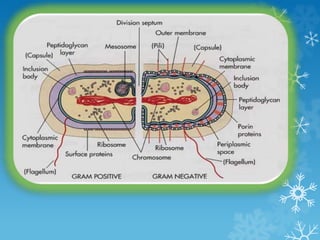
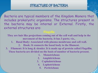
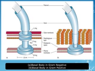
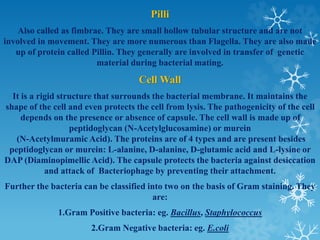
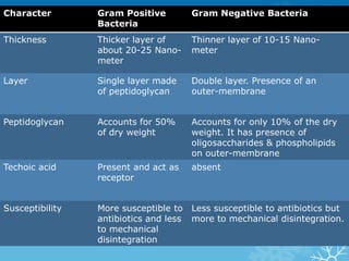
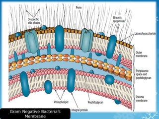
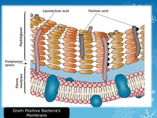
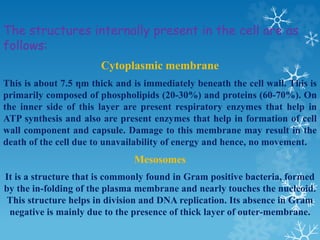
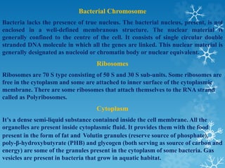
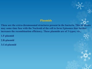
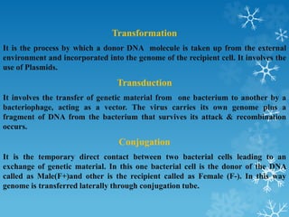
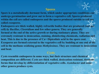
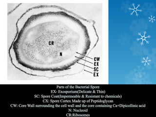
Bacteria have several external and internal structures. External structures include flagella, pili, and the cell wall. Flagella are hair-like projections that help bacteria move. Pili are involved in genetic transfer during bacterial mating. The cell wall maintains shape and protects the cell. Internally, bacteria have a cytoplasmic membrane, mesosomes in some gram-positive bacteria, ribosomes, plasmids, and a nucleoid containing genetic material. Bacteria can also take up genetic material through transformation, transduction, and conjugation. Some bacteria form spores or cysts as dormant, resistant structures.
This is a summary of a presentation our CEO has given at Northwestern University, Chicago - U.S.A. in 2003. Many aspects of protein targeting and membrane trafficking in eukaryotic cells are discussed.
Tags: Gokay-BIOTECH, K.Erden Gokay, Cell Biology, protein sorting, endotubin
Structure and function of plasma membrane 2ICHHA PURAK
The presentation consists of 72 slides,describes following heads
DEFINITION : STRUCTURE OF PLASMA MEMBRANE
COMPONENTS OF PLASMA MEMBRANE ( (BIOCHEMICAL PROPERTIES)
LIPID BILAYER
PROTEINS
CARBOHYDRATES
CHOLESTEROL
MODELS EXPLAINING STRUCTURE OF BIO MEMBRANE
FLUID MOSAIC MODEL
MOBILITY OF MEMBRANE
GLYCOCALYX : GLYCOPROTEINS AND GLYCOLIPIDS
TRANSPORT OF IONS AND MOLECULES ACROSS PLASMA MEMBRANE
FUNCTIONS OF PLASMA MEMBRANE
DIVERSITY OF CELL MEMBRANES
SITE OF ATPASE ION CARRIER CHANNELS AND PUMPS-RECEPTORS
contains detailed information about classification of life system
in particular three domains of classification sytem of living organism
into prokarya archea eukarya
Ultrastructure and characterstic features of bacteria.Archana Shaw
Ultrastructure and characterstic features of bacteria: BACTERIA AS A MODEL ORGANISM
THIS WAS MY PRESENTATION TOPIC IN CLASS. THOUGHT OF SHARING IT AND HOPE IT HELPS.
this is the overall information about the bacterial cell organization and ultra structure of bacteria and types of bacteria on the basis of cell wall. structure of bacterial plasmid and types of bacterial plasmid
Cell biology is the study of cell structure and function, and it revolves around the concept that the cell is the fundamental unit of life. Focusing on the cell permits a detailed understanding of the tissues and organisms that cells compose.
EubacteriaDefinitionBacteria are prokaryotic single-celled or BetseyCalderon89
Eubacteria
Definition
Bacteria are prokaryotic single-celled or colonial microorganisms
Characteristics of Bacteria
Lack Green Pigment Chlorophyll
Reproduce by Transverse Fission
Morphology
Bacteria display a wide diversity of shapes and sizes.
Size
0.5 µm diameter
Length 0.5 µm - 80 µm
Bacterial cells are about one-tenth the size of eukaryotic cells and are typically 0.5–5.0 micrometers in length. However, a few species are visible to the unaided eye—for example, Thiomargarita namibiensis is up to half a millimeter long] and Epulopiscium fishelsoni reaches 0.7 mm.] Among the smallest bacteria are members of the genus Mycoplasma, which measure only 0.3 micrometers, as small as the largest viruses.] Some bacteria may be even smaller, but these ultramicrobacteria are not well-studied.
Shape
Spherical – coccus
Rod-shaped - bacillus
Vibrio - Comma shaped
Spiral-shaped -spirillum
Spherical bacteria are known as cocci (singular coccus, Rod-shaped bacteria are called bacilli. Some bacteria, called vibrio, are shaped like slightly curved rods or comma-shaped; others can be spiral-shaped, called spirilla, or tightly coiled, called spirochaetes. A small number of other unusual shapes have been described, such as star-shaped bacteria.
Arrangements of Bacterial Cells
Bacteria are unicellular or colonial
Colonial – cells remain together after division
Colony type – depends on plane of cleavage and planes of successive cleavage.
Bacillus – can only divide in one plane, at right angles to the long axis of the cell.
Streptobacillus
Diplobacillus – remain in pairs following division. after 4 chain fragments
Spirillum- (spiral) divides in one plane
2 types:
Strepto spirillum
Diplo spirillum
Spherical (coccus) can initially divide in any plane. Great variation in colony types.
Streptococcus
Cells divide simultaneously
Diplococcus
If after 4 unit, chain fragments into chains of 2 organisms each – diplococcus
Tetrad Gaffkya
Cells divide at right angles to the preceding division
Sarcina – 3 planes of division. Successive planes are at right angles
Sarcina colonies are cuboidal. All dimensions are the same.
Staphylococcus – irregular cluster of spherical cells. Cells divide in any plane. No pattern
Coccus organism –the type of colony is a species characteristic. It can be used to identify an organism. The colony type is often indicated by the generic name. This is not true of bacillus or spirillum. The colony type can be varied by environment or temperature.
Many bacterial species exist simply as single cells; others associate in characteristic patterns: Neisseria forms diploids (pairs), streptococci form chains, and staphylococci group together in "bunch of grapes" clusters. Bacteria can also group to form larger multicellular structures, such as the elongated filaments of Actinobacteria species, the aggregates of Myxobacteria species, and the complex hyphae of Streptomyces species. These multicellular structures are often only seen in cert ...
What is bacteria?(Structures Present in Bacteria And their Functions | Prokar...sehriqayyum
Explains what bacteria is and where it exists.
A key feature of nearly all prokaryotic cells is the cell wall, which maintains cell shape, protects the cell, and prevents it from bursting in a hypotonic environment.
The cell walls of prokaryotes differ in structure from those of eukaryotes. In eukaryotes that have cell walls, such as plants and fungi, the walls are usually made of cellulose or chitin. In contrast, most bacterial cell walls contain peptidoglycan, a polymer composed of modified sugars cross-linked by short polypeptides.
Using a technique called the Gram stain, developed by the 19th-century Danish physician Hans Christian Gram, scientists can categorize many bacterial species according to differences in cell wall composition.
Gram-positive bacteria have simpler walls with a relatively large amount of peptidoglycan. Gram-negative bacteria have less peptidoglycan
and are structurally more complex, with an outer membrane
that contains lipopolysaccharides (carbohydrates bonded
to lipids).
LEARN ABOUT:
- Bacteria
- The number of viruses on earth is staggering
- Pathogenic yeasts
- Helminths
- Harnessing bacteria
- Microbes on the tree of life
- Living and working together
- Archaea
- Protozoa
LEARN ABOUT:
- Bacteria
- The number of viruses on earth is staggering
- Pathogenic yeasts
- Helminths
- Harnessing bacteria
- Microbes on the tree of life
- Living and working together
- Archaea
- Protozoa
The bacterial flagellum has three main parts (the motor, hook, and filament) that are themselves composed of 42 different kinds of proteins.The cells of prokaryotes are simpler than those of eukaryotes
in both their internal structure and the physical arrangement
of their DNA. The genome of a prokaryote is structurally different from
a eukaryotic genome and in most cases has considerably less DNA. Prokaryotes generally have circular chromosomes, whereas eukaryotes have linear chromosomes.
The Contingency plans cover contingency strategies to be taken up by farmers in response to major weather related aberrations such as delay in onset and breaks in monsoon causing early, mid and late season droughts, floods, unusual rains, extreme weather events such as heat wave, cold wave, frost, hailstorm and cyclone.
The contract farming system should be seen as a partnership between agribusiness and farmers’. To be successful it requires a long-term commitment from both parties.
This presentation covers the production technology of the arhar crop. It also includes the latest data with respect to the area and production in India and a little about its origin and botany of the plant.
This presentation is only with respect to the Parasitic Weed and their management tactics, falling under the category of Specificity while classifying weeds.
The increased availability of biomedical data, particularly in the public domain, offers the opportunity to better understand human health and to develop effective therapeutics for a wide range of unmet medical needs. However, data scientists remain stymied by the fact that data remain hard to find and to productively reuse because data and their metadata i) are wholly inaccessible, ii) are in non-standard or incompatible representations, iii) do not conform to community standards, and iv) have unclear or highly restricted terms and conditions that preclude legitimate reuse. These limitations require a rethink on data can be made machine and AI-ready - the key motivation behind the FAIR Guiding Principles. Concurrently, while recent efforts have explored the use of deep learning to fuse disparate data into predictive models for a wide range of biomedical applications, these models often fail even when the correct answer is already known, and fail to explain individual predictions in terms that data scientists can appreciate. These limitations suggest that new methods to produce practical artificial intelligence are still needed.
In this talk, I will discuss our work in (1) building an integrative knowledge infrastructure to prepare FAIR and "AI-ready" data and services along with (2) neurosymbolic AI methods to improve the quality of predictions and to generate plausible explanations. Attention is given to standards, platforms, and methods to wrangle knowledge into simple, but effective semantic and latent representations, and to make these available into standards-compliant and discoverable interfaces that can be used in model building, validation, and explanation. Our work, and those of others in the field, creates a baseline for building trustworthy and easy to deploy AI models in biomedicine.
Bio
Dr. Michel Dumontier is the Distinguished Professor of Data Science at Maastricht University, founder and executive director of the Institute of Data Science, and co-founder of the FAIR (Findable, Accessible, Interoperable and Reusable) data principles. His research explores socio-technological approaches for responsible discovery science, which includes collaborative multi-modal knowledge graphs, privacy-preserving distributed data mining, and AI methods for drug discovery and personalized medicine. His work is supported through the Dutch National Research Agenda, the Netherlands Organisation for Scientific Research, Horizon Europe, the European Open Science Cloud, the US National Institutes of Health, and a Marie-Curie Innovative Training Network. He is the editor-in-chief for the journal Data Science and is internationally recognized for his contributions in bioinformatics, biomedical informatics, and semantic technologies including ontologies and linked data.
Cancer cell metabolism: special Reference to Lactate PathwayAADYARAJPANDEY1
Normal Cell Metabolism:
Cellular respiration describes the series of steps that cells use to break down sugar and other chemicals to get the energy we need to function.
Energy is stored in the bonds of glucose and when glucose is broken down, much of that energy is released.
Cell utilize energy in the form of ATP.
The first step of respiration is called glycolysis. In a series of steps, glycolysis breaks glucose into two smaller molecules - a chemical called pyruvate. A small amount of ATP is formed during this process.
Most healthy cells continue the breakdown in a second process, called the Kreb's cycle. The Kreb's cycle allows cells to “burn” the pyruvates made in glycolysis to get more ATP.
The last step in the breakdown of glucose is called oxidative phosphorylation (Ox-Phos).
It takes place in specialized cell structures called mitochondria. This process produces a large amount of ATP. Importantly, cells need oxygen to complete oxidative phosphorylation.
If a cell completes only glycolysis, only 2 molecules of ATP are made per glucose. However, if the cell completes the entire respiration process (glycolysis - Kreb's - oxidative phosphorylation), about 36 molecules of ATP are created, giving it much more energy to use.
IN CANCER CELL:
Unlike healthy cells that "burn" the entire molecule of sugar to capture a large amount of energy as ATP, cancer cells are wasteful.
Cancer cells only partially break down sugar molecules. They overuse the first step of respiration, glycolysis. They frequently do not complete the second step, oxidative phosphorylation.
This results in only 2 molecules of ATP per each glucose molecule instead of the 36 or so ATPs healthy cells gain. As a result, cancer cells need to use a lot more sugar molecules to get enough energy to survive.
introduction to WARBERG PHENOMENA:
WARBURG EFFECT Usually, cancer cells are highly glycolytic (glucose addiction) and take up more glucose than do normal cells from outside.
Otto Heinrich Warburg (; 8 October 1883 – 1 August 1970) In 1931 was awarded the Nobel Prize in Physiology for his "discovery of the nature and mode of action of the respiratory enzyme.
WARNBURG EFFECT : cancer cells under aerobic (well-oxygenated) conditions to metabolize glucose to lactate (aerobic glycolysis) is known as the Warburg effect. Warburg made the observation that tumor slices consume glucose and secrete lactate at a higher rate than normal tissues.
Richard's entangled aventures in wonderlandRichard Gill
Since the loophole-free Bell experiments of 2020 and the Nobel prizes in physics of 2022, critics of Bell's work have retreated to the fortress of super-determinism. Now, super-determinism is a derogatory word - it just means "determinism". Palmer, Hance and Hossenfelder argue that quantum mechanics and determinism are not incompatible, using a sophisticated mathematical construction based on a subtle thinning of allowed states and measurements in quantum mechanics, such that what is left appears to make Bell's argument fail, without altering the empirical predictions of quantum mechanics. I think however that it is a smoke screen, and the slogan "lost in math" comes to my mind. I will discuss some other recent disproofs of Bell's theorem using the language of causality based on causal graphs. Causal thinking is also central to law and justice. I will mention surprising connections to my work on serial killer nurse cases, in particular the Dutch case of Lucia de Berk and the current UK case of Lucy Letby.
Professional air quality monitoring systems provide immediate, on-site data for analysis, compliance, and decision-making.
Monitor common gases, weather parameters, particulates.
This pdf is about the Schizophrenia.
For more details visit on YouTube; @SELF-EXPLANATORY;
https://www.youtube.com/channel/UCAiarMZDNhe1A3Rnpr_WkzA/videos
Thanks...!
6. Bacteria are typical members of the Kingdom Monera that
includes prokaryotic organisms. The structures present in
the bacteria may be internal or External. Firstly, the
external structures are:
Flagella
They are hair like projections coming out of the cell wall and help in the
movement of the bacteria. It has 3 parts; viz.,
1. Basal Body: Associated with plasma membrane and cell wall.
2. Hook: It connects the basal body to the filament.
3. Filament: It is long & slender. It is made up of protein called Flagellin.
These bacteria are divided on the basis of number of bacteria present.
1. Monotrichous
2. Amphitrichous
3. Cephalotrichous
4. Lophotrichous
5. Peritrichous
8. Pilli
Also called as fimbrae. They are small hollow tubular structure and are not
involved in movement. They are more numerous than Flagella. They are also made
up of protein called Pillin. They generally are involved in transfer of genetic
material during bacterial mating.
Cell Wall
It is a rigid structure that surrounds the bacterial membrane. It maintains the
shape of the cell and even protects the cell from lysis. The pathogenicity of the cell
depends on the presence or absence of capsule. The cell wall is made up of
peptidoglycan (N-Acetylglucosamine) or murein
(N-Acetylmuramic Acid). The proteins are of 4 types and are present besides
peptidoglycan or murein: L-alanine, D-alanine, D-glutamic acid and L-lysine or
DAP (Diaminopimellic Acid). The capsule protects the bacteria against desiccation
and attack of Bacteriophage by preventing their attachment.
Further the bacteria can be classified into two on the basis of Gram staining. They
are:
1.Gram Positive bacteria: eg. Bacillus, Staphylococcus
2.Gram Negative bacteria: eg. E.coli
9. Character Gram Positive
Bacteria
Gram Negative Bacteria
Thickness Thicker layer of
about 20-25 Nano-
meter
Thinner layer of 10-15 Nano-
meter
Layer Single layer made
of peptidoglycan
Double layer. Presence of an
outer-membrane
Peptidoglycan Accounts for 50%
of dry weight
Accounts for only 10% of the dry
weight. It has presence of
oligosaccharides & phospholipids
on outer-membrane
Techoic acid Present and act as
receptor
absent
Susceptibility More susceptible to
antibiotics and less
to mechanical
disintegration
Less susceptible to antibiotics but
more to mechanical disintegration.
12. The structures internally present in the cell are as
follows:
Cytoplasmic membrane
This is about 7.5 ηm thick and is immediately beneath the cell wall. This is
primarily composed of phospholipids (20-30%) and proteins (60-70%). On
the inner side of this layer are present respiratory enzymes that help in
ATP synthesis and also are present enzymes that help in formation of cell
wall component and capsule. Damage to this membrane may result in the
death of the cell due to unavailability of energy and hence, no movement.
Mesosomes
It is a structure that is commonly found in Gram positive bacteria, formed
by the in-folding of the plasma membrane and nearly touches the nucleoid.
This structure helps in division and DNA replication. Its absence in Gram
negative is mainly due to the presence of thick layer of outer-membrane.
13. Bacterial Chromosome
Bacteria lacks the presence of true nucleus. The bacterial nucleus, present, is not
enclosed in a well-defined membranous structure. The nuclear material is
generally confined to the centre of the cell. It consists of single circular double
stranded DNA molecule in which all the genes are linked. This nuclear material is
generally designated as nucleoid or chromatin body or nuclear equivalent.
Ribosomes
Ribosomes are 70 S type consisting of 50 S and 30 S sub-units. Some ribosomes are
free in the cytoplasm and some are attached to inner surface of the cytoplasmic
membrane. There are some ribosomes that attach themselves to the RNA strand
called as Polyribosomes.
Cytoplasm
It’s a dense semi-liquid substance contained inside the cell membrane. All the
organelles are present inside cytoplasmic fluid. It provides them with the food
present in the form of fat and Volutin granules (reserve source of phosphate),
poly-β-hydroxybutyrate (PHB) and glycogen (both serving as source of carbon and
energy) are some of the granules present in the cytoplasm of some bacteria. Gas
vesicles are present in bacteria that grow in aquatic habitat.
14. Plasmids
These are the extra-chromosomal structures present in the bacteria. This structure
may some time fuse with the Nucleoid of the cell to form Episomes that further
increases the recombination efficiency. These plasmids are of 3 types; viz.,
1.F-plasmid
2.R-plasmid
3.Col-plasmid
15. Transformation
It is the process by which a donor DNA molecule is taken up from the external
environment and incorporated into the genome of the recipient cell. It involves the
use of Plasmids.
Transduction
It involves the transfer of genetic material from one bacterium to another by a
bacteriophage, acting as a vector. The virus carries its own genome plus a
fragment of DNA from the bacterium that survives its attack & recombination
occurs.
Conjugation
It is the temporary direct contact between two bacterial cells leading to an
exchange of genetic material. In this one bacterial cell is the donor of the DNA
called as Male(F+)and other is the recipient called as Female (F-). In this way
genome is transferred laterally through conjugation tube.
16. Spores
Spore is a metabolically dormant form, which under appropriate conditions can
undergo germination and grow out to form a vegetative cell. Spores produced
within the cell are called endospores and the spores produced outside to cell are
called exospores.
Endospores are thick walled, highly refractile bodies that are produced (one per
cell) by Bacillus, Clostridium and few other genera. They are generally
formed at the end of the active growth or during stationary phase. They are
extremely resistant to desiccation, staining, disinfecting chemicals, radiation and
heat. This is due to the presence of Ca+ Dipicolinic acid in the spore coat.
Exospores are formed external to the vegetative cell by budding at one end of the
cell in the methane oxidizing genus Methylosinus. They are resistant to desiccation
and heat.
Cysts
Cysts resemble endospores in some ways, but their structure and chemical
composition are different. Cysts are thick walled, desiccation resistant, dormant
forms that develop by differentiation of vegetative cells. Azotobacter and some
other genera produce cysts.