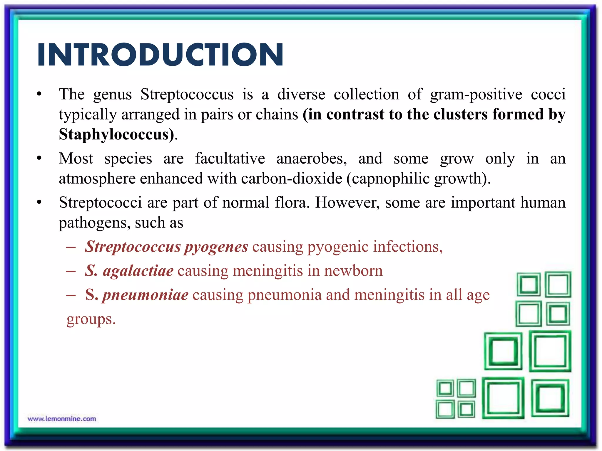
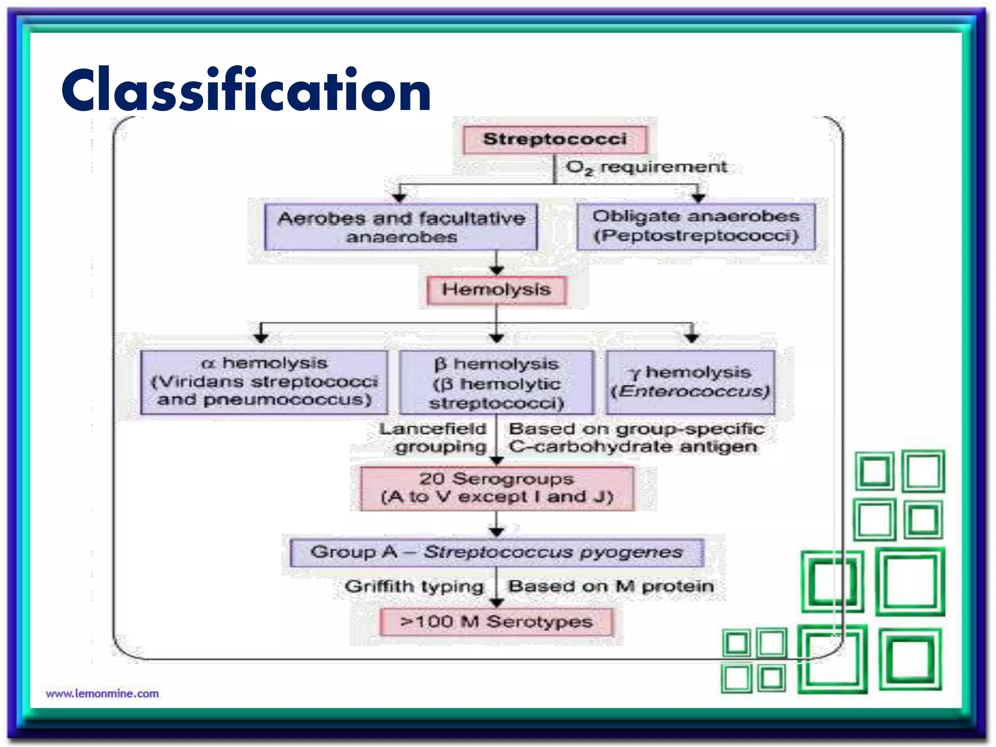
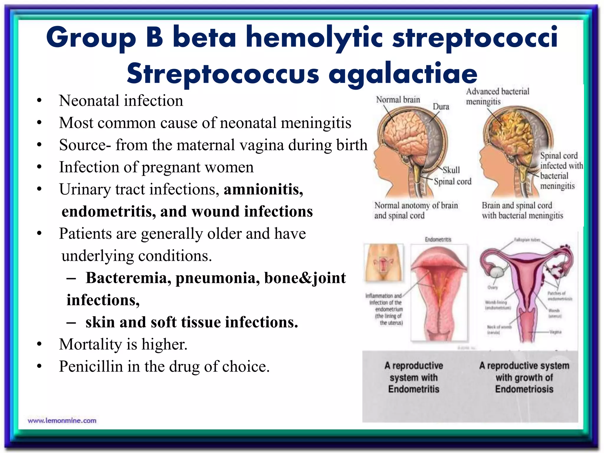
STREPTOCOCCUS
By
THENARUVI S
INTRODUCTION
• The genus Streptococcus is a diverse collection of gram-positive cocci
typically arranged in pairs or chains (in contrast to the clusters formed by
Staphylococcus).
• Most species are facultative anaerobes, and some grow only in an
atmosphere enhanced with carbon-dioxide (capnophilic growth).
• Streptococci are part of normal flora. However, some are important human
pathogens, such as
– Streptococcus pyogenes causing pyogenic infections,
– S. agalactiae causing meningitis in newborn
– S. pneumoniae causing pneumonia and meningitis in all age
groups.
Classification
Streptococcus pyogenes(group A)
• Rapidly growing gram-positive cocci arranged in chains, and Catalase-
negative.
• S. pyogenes is the only species under Lancefield' s group A Streptococcus
(GAS).
• Grow better in media enriched with blood or tissue fluid. Most are
facultative anaerobic and some are capnophilic.
• For most species growth and hemolysis are aided by incubation in 10%
CO2.
• After 24 hours of incubation at 37˚C, 1- to 2-mm white colonies with large
zones of β-hemolysis are observed
• It is associated with a variety of suppurative infections and can also trigger
post infectious nonsuppurative complications such as acute rheumatic
fever and acute glomerulonephritis.
Antigenic structure:
• Virulence factors of S. pyogenes can be categorized into
• Cell wall antigens,
• Toxins
• Enzymes
Cell Wall Antigens:
• Cell wall of S. pyogenes is composed of :
– Inner thick peptidoglycan layer- it confers cell wall rigidity, induces
inflammatory response and has thrombolytic activity.
– Middle layer of group specific C-carbohydrate antigen.
– Outer layer of protein and lipoteichoic acid
(helps in adhesion).
Toxins:
• Hemolysins:
β hemolytic streptococci produces two hemolysins-
streptolysin-O and streptolysin- S.
They cause RBC membrane lysis that leads to complete
β hemolysis surrounding the colonies.
• Streptococcal pyogenic exotoxin(SPE):
Induces fever (pyrogenic). It is responsible for the
pathogenesis of certain streptococcal infections such as scarlet fever,
necrotizing fasciitis and toxic shock syndrome.
Enzymes:
• Streptokinase(Fibrinolysin):
– It activates plasminogen to plasmin, thus breaks down the fibrin barrier
around the infected site, there by facilitating the spread of infection.
• Streptodornase(DNase):
– It breaks down the DNA, thus helps in liquefying the thick pus
(containing large amount of DNA derived from nuclei of necrotic cells)
and may be responsible for the serous nature of streptococcal exudates
• Other enzymes:
– Hyaluronidase(spreading factor).
– NADase
– Serum opacity factor
– SpyCEP
– C5a pepdidase
Epidemiology
• S. pyogenes can transiently colonize the oropharynx and skin.
• Diseases are caused by recently acquired strains that can establish an
infection of the pharynx or skin.
• S. pyogenes causes pharyngitis mainly in children of 5 to 15 years old.
• The pathogen is spread mainly by respiratory droplets.
• Crowding increases the opportunity for the pathogen to spread, particularly
during the winter months.
• Soft tissue infections are preceded by skin colonization and the organisms
are introduced into the superficial or deep tissue through a break in the
skin.
Pathogenesis:
• Pathogenesis (via invasiveness and
production of toxins)
– Adherence to the epithelial cells;
• >10 adhesion molecules
– invasion into the epithelial cells;
• mediated by M protein and
protein F important for
persistent infections and
invasion into deep tissues
– avoiding opsonization and
phagocytosis;
• M protein, M-like proteins, and
C5a peptidase
– producing enzymes and toxins
Clinical manifestation
SUPPURATIVE:
 Respiratory infections
• Pharyngitis/sore throat
• Pneumonia
• Empyema
 Scarlet fever
 Skin and soft tissue infection
• Impetigo (pyoderma)
• Cellulitis and erysipelas
 Deep soft tissue infections
• Necrotizing fascitis
• Streptococcal myositis
• Toxic shock syndrome
 Bacteremia leading to toxic shock
syndrome, osteomyelitls,
meningitis, etc.
 Complications: Puerperal sepsis,
otitis media, quinsy, Ludwig's
angina, pneumonia (post viral), etc.
NON- SUPPURATIVE:
 Acute rheumatic fever
 Acute glomerulonephritis
 Guttate psoriasis
 Reactive arthritis
 PANDAS-(PediatricAutoimmune Neuropsychiatric Disorders Associated
with Streptococcal infections)
c
Streptococcal skin infections.
A. Impetigo;
B. Erysipelas on face (peau d'orange skin)
C. Necrotizing fasciitis of hand
Lab diagnosis
 Specimen collection and transport:
 Depends on the site of the infection
 Transport medium:
 Pike's medium
 Direct smear microscopy:
 Pus cells with gram-positive cocci in short chains
 Culture:
 Blood agar: Pinpoint colony with a wide zone of beta hemolysis
 Selective media: Crystal violet blood agar and PNF media
 Liquid media: Granular turbidity with powdery deposit
 Culture smear microscopy:
 Gram-positive cocci in short chains
• Biochemical identification:
– Catalase negative
– Bacitracin sensitive
– PYR test positive
• Typing:
– Lancefield grouping: Shows group A Streptococcus
– Typing of group A Streptococcus: Griffith typing and emm typing
• Serology:
– ASO antibodies
– Anti-DNase B antibodies
• Antimcrobial susceptibility testing
Treatment , prevention and
control
• Penicillin V or amoxicillin used to treat pharyngitis; oral cephalosporin
or macrolide for penicillin-allergic patients; intravenous penicillin plus
clindamycin used for systemic infections
• Oropharyngeal carriage occurring after treatment can be re-treated;
treatment is not indicated for prolonged asymptomatic carriage because
antibiotics disrupt normal protective flora
• Starting antibiotic therapy within 10 days in patients with pharyngitis
prevents rheumatic fever
• For glomerulonephritis, no specific antibiotic treatment or prophylaxis is
indicated
Group B beta hemolytic streptococci
Streptococcus agalactiae
• Neonatal infection
• Most common cause of neonatal meningitis
• Source- from the maternal vagina during birth
• Infection of pregnant women
• Urinary tract infections, amnionitis,
endometritis, and wound infections
• Patients are generally older and have
underlying conditions.
– Bacteremia, pneumonia, bone&joint
infections,
– skin and soft tissue infections.
• Mortality is higher.
• Penicillin in the drug of choice.
Diagnostic markers- hippurate hydrolysis,
CAMP test
CAMP TEST
• When S.agalactiae is inoculated perpendicular to a streak of S.aureus
grown on blood agar.
• An accentuated zone of hemolysis occurs
Viridans group streptococci
• Streptococci normally resident in the mouth and upper respiratory tract
• Shows alpha hemolysis on blood agar
• Cannot be categorized under lancefield antigenic group
• Types:
– S. mitis
– S. mutans
– S. salivarius
– S. sanguis
• Diseases:
– Subacute endocarditis (group: Mitis)
– Intra-abdominal infections (group: Anginosus)
– Dental caries (group: Mutans)
– Cariogenicity of S. mutans is related to its ability to synthesize
glucan from fermentable carbohydrates (e.g. sucrose) as well
as to modify glucan in promoting increased adhesiveness.
• Diagnosis:
– On Gram stain, they appear as long chains of gram positive cocci
– They produce minute alpha or green hemolytic (rarely non hemolytic)
colonies on blood agar ("viridis' means green).
– They can be differentiated from S. pneumoniae( which is also alpha
hemolytic) by a number of tests
• Gram-positive cocci in long chains, Round/oval noncapsulated,
Convex shaped colony on blood agar.
• Granular turbidity in liquid broth, Insoluble in bile, Insulin Non
fermenter
• Resistant to optochin
• Treatment:
– They are usually sensitive to penicillin
except in neutropenic patients with bacteremia,
where vancomycin is given.
Streptococcol pneumoniae
(pneumococcus)
(α-hemolytic)
• Streptococcus pneumoniae ( commonly referred to as pneumococcus) is the
leading cause of lobar pneumonia, olitis media in children and meningitis
in all ages.
• They are α-hemolytic and may present as commensals in human upper
respiratory tract. They differ from α -hemolytic viridans streptococci in
many ways such as
– their shape (lanceolate shaped diplococci),
– bile solubility, optochin sensitivity and
– presence of a polysaccharide capsule
Virulence factors:
– Capsular polysaccharide
– C Carbohydrate antigen
– Pneumolysins
– Autolysins
• Other virulence factors
– Pneumococcol surface protein A(PspA), IgA protease ,Pnuemococcol
protein C(PspC), Adhesins
Pathogenesis:
Pneumococci colonize the human nasopharynx at an early age.
From the nasopharynx, the bacteria spread either via
the bloodstream to distant sites (e.g. brain, Joint, bones
and peritoneal cavity) or spread locally to cause olitis
media or pneumonia
Clinical manifestation:
– Pneumonia
– Sinusitis and otitis media
– Meningitis
– Bacterimia
Lab diagnosis:
• Specimen collection: Sputum. CSF, pleural fluid
• Direct smear microscopy: Reveals pus cells and lanceolate shaped gram
positive cocci in pairs, surrounded by a clear halo (due to capsule)
• Capsular antigen detection in CSF: By latex agglutination
• Culture
– Blood agar: It shows draughtsman or carrom coin appearance to colonies
– Chocolate agar: It produces greenish discoloration (bleaching e ffect)
– In liquid media: It shows uniform turbidity
• Culture smear: Reveals lanceolate shaped gram—positive cocci in pairs.
• Biochemical identification
– Bile soluble
– Optochin sensitive
– Inulin fermented
• Serotyping: By Quellung reaction or latex agglutination test
• Mouse pathogenicity
• Molecular methods: Such as multiplex PCR
• Antimicrobial susceptibility testing
A. a hemolytic
draughtsman shaped colonies on blood agar;
B. Bleaching effect
on chocolate agar
A. Bile solubility test (left-Viridans
streptococci, not soluble in bile; right-
pneumococcus, soluble in bile);
B. Optochin sensitivity;
C. Quellung reaction
Treatment, Prevention, and Control:
– Penicillin is the drug of choice for susceptible strains, although
resistance is increasingly common
– Vancomycin combined with ceftriaxone is used for empiric therapy;
monotherapy with a cephalosporin, fluoroquinolone, or vancomycin
can be used in patients with susceptible isolates
– Immunization with 13-valent conjugated vaccine is recommended for
all children younger than 2 years of age; a 23-valent polysaccharide
vaccine is recommended for adults at risk for disease
Enterococcus
(γ hemolysis )
E. faecalis and E. faecium
• Physiological properties are similar to the streptococci.
• Form large colonies on blood plate; most are non hemolytic.
• Microscopic morphology is similar to S. pneumoniae.
• Resistant to 6.5% NaCl, 0.1% methyl blue and grow in bile-esculin agar.
• More resistant to antibiotics than the streptococci.
• Colonize the large intestine of humans and animals.
Laboratory Diagnosis:
• Enterococci can be differentiated by simple biochemical tests (e.g.,
resistant to optochin and bile, hydrolyze PYR, etc.)
Clinlcal Manifestation:
• Enterococci produce various infections such as:
– Urinary tract infections (cystitis, urethritis, pyelonephritis and
prostatitis)
– Bacteremia and mitral valve endocarditis (in intravenous drug abusers)
– lntra-abdominal, pelvic and soft tissue infections
– Late-onset neonatal sepsis and meningitis
– Infection on burn surface
Treatment:
– Penicillin + Aminoglycoside
– Vancomycin
Streptococcus

Streptococcus

  • 1.
  • 2.
    INTRODUCTION • The genusStreptococcus is a diverse collection of gram-positive cocci typically arranged in pairs or chains (in contrast to the clusters formed by Staphylococcus). • Most species are facultative anaerobes, and some grow only in an atmosphere enhanced with carbon-dioxide (capnophilic growth). • Streptococci are part of normal flora. However, some are important human pathogens, such as – Streptococcus pyogenes causing pyogenic infections, – S. agalactiae causing meningitis in newborn – S. pneumoniae causing pneumonia and meningitis in all age groups.
  • 3.
  • 4.
    Streptococcus pyogenes(group A) •Rapidly growing gram-positive cocci arranged in chains, and Catalase- negative. • S. pyogenes is the only species under Lancefield' s group A Streptococcus (GAS). • Grow better in media enriched with blood or tissue fluid. Most are facultative anaerobic and some are capnophilic. • For most species growth and hemolysis are aided by incubation in 10% CO2. • After 24 hours of incubation at 37˚C, 1- to 2-mm white colonies with large zones of β-hemolysis are observed • It is associated with a variety of suppurative infections and can also trigger post infectious nonsuppurative complications such as acute rheumatic fever and acute glomerulonephritis.
  • 5.
    Antigenic structure: • Virulencefactors of S. pyogenes can be categorized into • Cell wall antigens, • Toxins • Enzymes Cell Wall Antigens: • Cell wall of S. pyogenes is composed of : – Inner thick peptidoglycan layer- it confers cell wall rigidity, induces inflammatory response and has thrombolytic activity. – Middle layer of group specific C-carbohydrate antigen. – Outer layer of protein and lipoteichoic acid (helps in adhesion).
  • 6.
    Toxins: • Hemolysins: β hemolyticstreptococci produces two hemolysins- streptolysin-O and streptolysin- S. They cause RBC membrane lysis that leads to complete β hemolysis surrounding the colonies. • Streptococcal pyogenic exotoxin(SPE): Induces fever (pyrogenic). It is responsible for the pathogenesis of certain streptococcal infections such as scarlet fever, necrotizing fasciitis and toxic shock syndrome.
  • 7.
    Enzymes: • Streptokinase(Fibrinolysin): – Itactivates plasminogen to plasmin, thus breaks down the fibrin barrier around the infected site, there by facilitating the spread of infection. • Streptodornase(DNase): – It breaks down the DNA, thus helps in liquefying the thick pus (containing large amount of DNA derived from nuclei of necrotic cells) and may be responsible for the serous nature of streptococcal exudates • Other enzymes: – Hyaluronidase(spreading factor). – NADase – Serum opacity factor – SpyCEP – C5a pepdidase
  • 8.
    Epidemiology • S. pyogenescan transiently colonize the oropharynx and skin. • Diseases are caused by recently acquired strains that can establish an infection of the pharynx or skin. • S. pyogenes causes pharyngitis mainly in children of 5 to 15 years old. • The pathogen is spread mainly by respiratory droplets. • Crowding increases the opportunity for the pathogen to spread, particularly during the winter months. • Soft tissue infections are preceded by skin colonization and the organisms are introduced into the superficial or deep tissue through a break in the skin.
  • 9.
    Pathogenesis: • Pathogenesis (viainvasiveness and production of toxins) – Adherence to the epithelial cells; • >10 adhesion molecules – invasion into the epithelial cells; • mediated by M protein and protein F important for persistent infections and invasion into deep tissues – avoiding opsonization and phagocytosis; • M protein, M-like proteins, and C5a peptidase – producing enzymes and toxins
  • 10.
    Clinical manifestation SUPPURATIVE:  Respiratoryinfections • Pharyngitis/sore throat • Pneumonia • Empyema  Scarlet fever  Skin and soft tissue infection • Impetigo (pyoderma) • Cellulitis and erysipelas  Deep soft tissue infections • Necrotizing fascitis • Streptococcal myositis • Toxic shock syndrome  Bacteremia leading to toxic shock syndrome, osteomyelitls, meningitis, etc.  Complications: Puerperal sepsis, otitis media, quinsy, Ludwig's angina, pneumonia (post viral), etc.
  • 11.
    NON- SUPPURATIVE:  Acuterheumatic fever  Acute glomerulonephritis  Guttate psoriasis  Reactive arthritis  PANDAS-(PediatricAutoimmune Neuropsychiatric Disorders Associated with Streptococcal infections) c Streptococcal skin infections. A. Impetigo; B. Erysipelas on face (peau d'orange skin) C. Necrotizing fasciitis of hand
  • 12.
    Lab diagnosis  Specimencollection and transport:  Depends on the site of the infection  Transport medium:  Pike's medium  Direct smear microscopy:  Pus cells with gram-positive cocci in short chains  Culture:  Blood agar: Pinpoint colony with a wide zone of beta hemolysis  Selective media: Crystal violet blood agar and PNF media  Liquid media: Granular turbidity with powdery deposit  Culture smear microscopy:  Gram-positive cocci in short chains
  • 13.
    • Biochemical identification: –Catalase negative – Bacitracin sensitive – PYR test positive • Typing: – Lancefield grouping: Shows group A Streptococcus – Typing of group A Streptococcus: Griffith typing and emm typing • Serology: – ASO antibodies – Anti-DNase B antibodies • Antimcrobial susceptibility testing
  • 14.
    Treatment , preventionand control • Penicillin V or amoxicillin used to treat pharyngitis; oral cephalosporin or macrolide for penicillin-allergic patients; intravenous penicillin plus clindamycin used for systemic infections • Oropharyngeal carriage occurring after treatment can be re-treated; treatment is not indicated for prolonged asymptomatic carriage because antibiotics disrupt normal protective flora • Starting antibiotic therapy within 10 days in patients with pharyngitis prevents rheumatic fever • For glomerulonephritis, no specific antibiotic treatment or prophylaxis is indicated
  • 15.
    Group B betahemolytic streptococci Streptococcus agalactiae • Neonatal infection • Most common cause of neonatal meningitis • Source- from the maternal vagina during birth • Infection of pregnant women • Urinary tract infections, amnionitis, endometritis, and wound infections • Patients are generally older and have underlying conditions. – Bacteremia, pneumonia, bone&joint infections, – skin and soft tissue infections. • Mortality is higher. • Penicillin in the drug of choice.
  • 16.
    Diagnostic markers- hippuratehydrolysis, CAMP test CAMP TEST • When S.agalactiae is inoculated perpendicular to a streak of S.aureus grown on blood agar. • An accentuated zone of hemolysis occurs
  • 17.
    Viridans group streptococci •Streptococci normally resident in the mouth and upper respiratory tract • Shows alpha hemolysis on blood agar • Cannot be categorized under lancefield antigenic group • Types: – S. mitis – S. mutans – S. salivarius – S. sanguis • Diseases: – Subacute endocarditis (group: Mitis) – Intra-abdominal infections (group: Anginosus) – Dental caries (group: Mutans) – Cariogenicity of S. mutans is related to its ability to synthesize glucan from fermentable carbohydrates (e.g. sucrose) as well as to modify glucan in promoting increased adhesiveness.
  • 18.
    • Diagnosis: – OnGram stain, they appear as long chains of gram positive cocci – They produce minute alpha or green hemolytic (rarely non hemolytic) colonies on blood agar ("viridis' means green). – They can be differentiated from S. pneumoniae( which is also alpha hemolytic) by a number of tests • Gram-positive cocci in long chains, Round/oval noncapsulated, Convex shaped colony on blood agar. • Granular turbidity in liquid broth, Insoluble in bile, Insulin Non fermenter • Resistant to optochin • Treatment: – They are usually sensitive to penicillin except in neutropenic patients with bacteremia, where vancomycin is given.
  • 19.
    Streptococcol pneumoniae (pneumococcus) (α-hemolytic) • Streptococcuspneumoniae ( commonly referred to as pneumococcus) is the leading cause of lobar pneumonia, olitis media in children and meningitis in all ages. • They are α-hemolytic and may present as commensals in human upper respiratory tract. They differ from α -hemolytic viridans streptococci in many ways such as – their shape (lanceolate shaped diplococci), – bile solubility, optochin sensitivity and – presence of a polysaccharide capsule
  • 20.
    Virulence factors: – Capsularpolysaccharide – C Carbohydrate antigen – Pneumolysins – Autolysins • Other virulence factors – Pneumococcol surface protein A(PspA), IgA protease ,Pnuemococcol protein C(PspC), Adhesins Pathogenesis: Pneumococci colonize the human nasopharynx at an early age. From the nasopharynx, the bacteria spread either via the bloodstream to distant sites (e.g. brain, Joint, bones and peritoneal cavity) or spread locally to cause olitis media or pneumonia
  • 21.
    Clinical manifestation: – Pneumonia –Sinusitis and otitis media – Meningitis – Bacterimia Lab diagnosis: • Specimen collection: Sputum. CSF, pleural fluid • Direct smear microscopy: Reveals pus cells and lanceolate shaped gram positive cocci in pairs, surrounded by a clear halo (due to capsule)
  • 22.
    • Capsular antigendetection in CSF: By latex agglutination • Culture – Blood agar: It shows draughtsman or carrom coin appearance to colonies – Chocolate agar: It produces greenish discoloration (bleaching e ffect) – In liquid media: It shows uniform turbidity • Culture smear: Reveals lanceolate shaped gram—positive cocci in pairs. • Biochemical identification – Bile soluble – Optochin sensitive – Inulin fermented • Serotyping: By Quellung reaction or latex agglutination test • Mouse pathogenicity • Molecular methods: Such as multiplex PCR • Antimicrobial susceptibility testing
  • 23.
    A. a hemolytic draughtsmanshaped colonies on blood agar; B. Bleaching effect on chocolate agar A. Bile solubility test (left-Viridans streptococci, not soluble in bile; right- pneumococcus, soluble in bile); B. Optochin sensitivity; C. Quellung reaction
  • 24.
    Treatment, Prevention, andControl: – Penicillin is the drug of choice for susceptible strains, although resistance is increasingly common – Vancomycin combined with ceftriaxone is used for empiric therapy; monotherapy with a cephalosporin, fluoroquinolone, or vancomycin can be used in patients with susceptible isolates – Immunization with 13-valent conjugated vaccine is recommended for all children younger than 2 years of age; a 23-valent polysaccharide vaccine is recommended for adults at risk for disease
  • 25.
    Enterococcus (γ hemolysis ) E.faecalis and E. faecium • Physiological properties are similar to the streptococci. • Form large colonies on blood plate; most are non hemolytic. • Microscopic morphology is similar to S. pneumoniae. • Resistant to 6.5% NaCl, 0.1% methyl blue and grow in bile-esculin agar. • More resistant to antibiotics than the streptococci. • Colonize the large intestine of humans and animals.
  • 26.
    Laboratory Diagnosis: • Enterococcican be differentiated by simple biochemical tests (e.g., resistant to optochin and bile, hydrolyze PYR, etc.) Clinlcal Manifestation: • Enterococci produce various infections such as: – Urinary tract infections (cystitis, urethritis, pyelonephritis and prostatitis) – Bacteremia and mitral valve endocarditis (in intravenous drug abusers) – lntra-abdominal, pelvic and soft tissue infections – Late-onset neonatal sepsis and meningitis – Infection on burn surface Treatment: – Penicillin + Aminoglycoside – Vancomycin