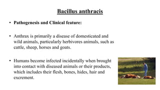
GRAM POSITIVE SPORE FORMING
RODS
SAMIRA FATTAH HAMID
Ph.D. Medical Bacteriology
College of Health Sciences
Hawler Medical University
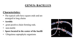
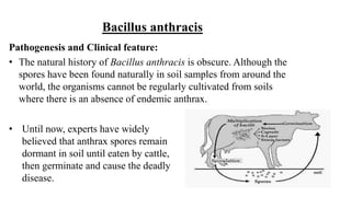
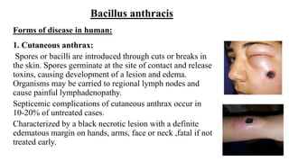
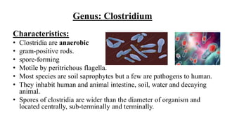
Scientists divide Gram-positive bacilli (rod-shaped cells) into
spore-forming and non-spore-forming genera. The spore-forming
genera are:
1. Genus Bacillus
2. Genus Clostridium
GENUS: BACILLUS
Characteristics:
• The typical cells have square ends and are
arranged in long chains
• Aerobic
• gram-positive chain forming rods.
• non-motile
• Spore located in the center of the bacilli
• Ubiquitous saprophytic organisms
GENUS: BACILLUS
• Many species of Bacillus do not cause disease, few species
cause important diseases in human:
1. B. anthracis
2. B. cereus
Bacillus anthracis
• large rod-shaped
• facultatively anaerobic
• normally dwells in soil.
• The tough external coat and the internal
chemicals of endospores make theme
resistant to harsh environmental conditions,
enabling Bacillus to survive in the
environment for centuries or perhaps even
longer.
Antigenic structure:
• Capsule:
Polypeptide of D-glutamic acid
All virulent strains of B. anthracis form this
capsule.
The poly-D-glutamyl capsule is itself
nontoxic, but functions to protect the
organism against complement and the
bactericidal components of serum and
phagocytes.
Bacillus anthracis
Bacillus anthracis
Antigenic structure:
• Anthrax toxin :
The toxin consists of three distinct antigenic:
1. Factor I: is the edema factor (EF) composed of adenylate cyclase which is
necessary for the edema producing activity of the toxin.
2. Factor II: is the protective antigen (PA): is the binding domain of the anthrax
toxin. Is a protein that binds to specific cell receptors, and after proteolytic
activation, it forms a membrane channel that mediates entry of EF and LF into the
cell.
3. Factor III is known as the lethal factor (LF) because it is essential for the lethal
effects of the anthrax toxin. is a major virulence factor and cause of death in
infected animals and humans.
Bacillus anthracis
• Pathogenesis and Clinical feature:
• Anthrax is primarily a disease of domesticated and
wild animals, particularly herbivores animals, such as
cattle, sheep, horses and goats.
• Humans become infected incidentally when brought
into contact with diseased animals or their products,
which includes their flesh, bones, hides, hair and
excrement.
Pathogenesis and Clinical feature:
• The natural history of Bacillus anthracis is obscure. Although the
spores have been found naturally in soil samples from around the
world, the organisms cannot be regularly cultivated from soils
where there is an absence of endemic anthrax.
• Until now, experts have widely
believed that anthrax spores remain
dormant in soil until eaten by cattle,
then germinate and cause the deadly
disease.
Bacillus anthracis
Forms of disease in human:
1. Cutaneous anthrax:
Spores or bacilli are introduced through cuts or breaks in
the skin. Spores germinate at the site of contact and release
toxins, causing development of a lesion and edema.
Organisms may be carried to regional lymph nodes and
cause painful lymphadenopathy.
Septicemic complications of cutaneous anthrax occur in
10-20% of untreated cases.
Characterized by a black necrotic lesion with a definite
edematous margin on hands, arms, face or neck ,fatal if not
treated early.
Bacillus anthracis
Forms of disease in human:
2. Pulmonary anthrax (Wool sorter’s disease):
Respiratory infection in humans is relatively rare and presents as two stages.
1. It infects the lymph nodes of mediastinal in the chest first, rather than
the lungs themselves, spores start to germinates and produce toxin
causing hemorrhagic mediastinitis, bloody fluid accumulate in the
chest cavity, causing shortness of breath.
The first stage causes cold and flu-like symptoms. Symptoms include fever,
shortness of breath, cough, fatigue, and chills. Often, many fatalities from
inhalational anthrax are when the first stage is mistaken for the cold or flu
and the victim does not seek treatment until the second stage, which is 90%
fatal.
Bacillus anthracis
Bacillus anthracis
Forms of disease in human:
2. Pulmonary anthrax (Wool sorter’s disease):
2. The second (pneumonia) stage occurs when
infection spreads from the lymph nodes to the
lungs. Symptoms of the second stage develop
suddenly after hours or days of the first stage.
Symptoms include high fever, extreme shortness
of breath, shock, and rapid death within 48
hours in fatal cases.
Bacillus anthracis
Forms of disease in human:
3. Intestinal anthrax:
Gastrointestinal anthrax is rare, the organisms probably invade
the mucosa through a preexisting lesion. The bacteria spread
from the mucosal lesion to the lymphatic system.
It results from the ingestion of poorly cooked meat from
infected animals. The incubation period for GI anthrax is 1-7
days, symptoms are fever and abdominal pain, and has an
extremely high mortality rate.
Bacillus anthracis and Bioterrorism
• Major agent of bioterrorism and biological warfare
• Anthrax is considered a potential threat as a biological
weapon because B. anthracis forms spores easily and can be
produced in a dry form that can be spread through the air or
as a powder.
• Notable bioterrorism uses include the 2001 anthrax attacks -
through sending letters containing anthrax spores by mail -
resulted in 22 cases of anthrax—11 inhalation and 11
cutaneous. Five of the patients with inhalation anthrax died.
Bacillus anthracis
Treatment
• Ciprofloxacin is recommended for treatment.
• penicillin G, doxycycline, erythromycin, and vancomycin are also
active .
• In the setting of potential exposure to B. anthracis as an agent of
biologic warfare, prophylaxis with ciprofloxacin or doxycycline should
be given for 60 days.
Bacillus cereus
General characteristics:
• beta-hemolytic
• commonly found in soil and food.
Bacillus cereus
It is the cause of "fried rice syndrome".
B. cereus is a soil organism that
commonly contaminates rice. When large
amounts of rice are cooked and allowed to
cool slowly, the B. cereus spores germinate,
and the vegetative cells produce the toxin
Bacillus cereus
Clinical features
B. cereus causes two types of foodborne illness :
1. Emetic type
2. Diarrheal type
• The emetic syndrome is an intoxication that is caused by ingestion of a toxin
called cereulide that is pre-formed in the food during growth by B. cereus.
This syndrome has a short incubation period and recovery time. The symptoms
of nausea, vomiting and abdominal cramping occur within 1–5 hours of
ingestion, with self-limiting recovery usually within 6–24 hours
Bacillus cereus
• Clinical features
• The diarrhoeal syndrome is caused by enterotoxins produced by B. cereus
inside the host which induce fluid accumulation in the small intestine. The
incubation period before onset of disease is 1–24 hours and the illness
usually lasts for 12–14 hours, although it can continue for several days.
Symptoms are watery diarrhea ,abdominal pain and cramp.
Bacillus cereus
B. cereus has also been associated with localized infections,
such as wound infections, and with systemic infections including:
• Endocarditis
• catheter-associated bacteremia
• Central nervous system infections
• Osteomyelitis
• pneumonia;
the presence of a medical device or intravenous drug use predisposes
to these infections.
Bacillus cereus
Treatment
• Serious nonfoodborne infections should be treated with vancomycin or
clindamycin with or without an aminoglycoside.
• Ciprofloxacin has been useful for the treatment of wound infections.
Genus: Clostridium
Characteristics:
• Clostridia are anaerobic
• gram-positive rods.
• spore-forming
• Motile by peritrichous flagella.
• Most species are soil saprophytes but a few are pathogens to human.
• They inhabit human and animal intestine, soil, water and decaying
animal.
• Spores of clostridia are wider than the diameter of organism and
located centrally, sub-terminally and terminally.
Genus: Clostridium
•Species of medical importance:
1. C. perfringens
2. C. tetani
3. C. botulinum
Clostridium perfringens
• formerly known as C. welchii, or Bacillus welchii.
• form central or sub-terminal spore.
• present in nature and can be found as a normal component of
decaying vegetation, marine sediment, the intestinal tract of
humans and soil.
• can produce invasive infection.
Clostridium perfringens
Antigenic Structure:
1. Enzymes:
• Collagenase
• Proteinase
• Hyaluronidase
• DNase
Clostridium perfringens
Antigenic Structure:
2. Toxins
A. Alpha toxin
It has lethal, necrotizing and hemolytic effect on tissue. It causes cell lysis due to
lecithinase action on the lecithin which is found in mammalian cell membrane.
Alpha toxin also aggregates platelets, thereby leading to formation of thrombi in
small blood vessels.
B. Theta toxin
It has hemolytic and necrotic effect on tissue but not lecithinase.
C. Enterotoxin
induces intense diarrhea but it is self-limiting.
Clostridium perfringens
Pathogenesis and clinical manifestation:
1. Clostridial myonecrosis:
(Gas gangrene)
IP=1-3 days
Colonization of wound by C. perfringens spores, and
organism germination and release of toxins.
Presentation: Muscle and subcutaneous tissue necrosis
and crepitation, foul smelling wound discharge, fever,
hemolysis, toxemia, shock and death.
Clostridium perfringens
Pathogenesis and clinical manifestation:
2. Clostridial food poisoning
Follows the ingestion of large numbers of clostridia that have grown in
warmed meat dishes. It causes diarrhea due to release of enterotoxin from
germinated spores in the intestine, Self-limiting diarrhea similar to that
produced by B. cereus.
Clostridium perfringens
Treatment
• Extensive surgical debridement of the involved area.
• Penicillin
• Hyperbaric oxygen
Clostridium tetani
General characteristics:
• World wide in distribution in the soil and in animal
feces
• Longer and thinner gram-positive rods with round
terminal spores giving characteristic “drum-stick”
appearance.
Clostridium tetani
Antigenic structure:
There are ten antigenic types of C. tetani but all produce the same
neurotoxin.
• The toxin has two components:
1. Tetanospasmin: Neurotoxic property
2. Tetanolysin: Hemolytic property
Pathogenesis and Clinical manifestation:
• C. tetani is not an invasive organism.
• The infection remains strictly localized in the area of
devitalized tissue (wound, burn, injury, umbilical stump,
surgical suture) into which the spores have been
introduced.
• The volume of infected tissue is small, and the disease is
almost entirely a toxemia.
• tetanospasmin is one of the most potent toxins known.
The estimated minimum human lethal dose is 2.5
nanograms per kilogram of body weight, or 175
nanograms in a 70 kg human.
Clostridium tetani
Clostridium tetani
Pathogenesis and Clinical manifestation:
• The tetanus toxin enters the nerve terminals through the
lower motor neurons, which are usually responsible for
activating voluntary muscular movements. It is then
transported via the axons to the spinal cord and brainstem.
• The toxin then moves throughout the nervous system trans-
synaptically into the nerve terminals responsible for the
release of inhibitory neurotransmitters. If this vesicular
process becomes blocked, the ability to inhibit motor
neurons is disrupted, which can result in muscle rigidity and
spasm.
Pathogenesis and Clinical manifestation:
IP= 4-5 days to several weeks
Tetanus classical presentation:
• Spasms and stiffness in jaw muscles (trismus). so that the mouth cannot
be opened.
• Stiffness of neck muscles.
• Difficulty swallowing
• Stiffness of abdominal muscles
• Painful body spasms lasting for several minutes.
• Death usually results from interference with the mechanics of respiration
• Mortality rate is very high
Clostridium tetani
Clostridium tetani
Treatment
The results of treatment of tetanus are not satisfactory. Therefore,
prevention is all important.
Prevention of tetanus depends on :
(1) active immunization with toxoids
(2) Surgical debridement is vitally important because it removes the
necrotic tissue that is essential for proliferation of the organisms.
(3) prophylactic use of antitoxin
(4) administration of penicillin to stops further growth and toxin
production.
Clostridium botulinum
General characteristics:
• produces oval, sub-terminal endospores
• Spores are highly resistant to heat, withstanding 100°C for several hours
• widely distributed in soil, they often contaminate vegetables, fruits and
other materials.
Toxins
• Types of C. botulinum are distinguished by the
antigenic type of toxin.
• There are eight serotypes(A-G) of which A,B,E and F
are the principal causes of human illness.
Clostridium botulinum
Pathogenesis and Clinical manifestation
1. Food botulism
• Route of entry is under cooked consumption of C. botulinum toxin
contaminated spiced, smoked, vaccum-packed or canned food.
The toxin is absorbed from the gut, enters the blood
circulation, and binds to receptors of presynaptic membranes
of motor neurons of the peripheral nervous system and acts
by blocking the release of acetylcholine at synapses and
neuromuscular junction and resulting in lack of muscle
contraction and paralysis. Death is secondary to respiratory
failure or cardiac arrest.
The lethal dose for a human is probably about 1–2 µg/kg. The toxins are destroyed by
heating for 20 minutes at 100°C.
Clostridium botulinum
Pathogenesis and Clinical manifestation
2. Infantile botulism
• Infant botulism is a rare condition, honey is the most frequent vehicle of
infection.
• infant ingests spores , and the spores germinate within the intestinal
tract. The vegetative cells produce toxin as they multiply;The enteric
toxin causes intestinal immobility and progressive descending paralysis
due to the effect on acetylcholine release at the neuromuscular junction
and other cholinergic nerve terminals, particularly in the gut.
Clostridium botulinum
Signs and symptoms include:
Constipation, which is often the first sign
Floppy movements due to muscle weakness and trouble controlling the
head
Weak cry
Drooping eyelids
Tiredness
Difficulty sucking or feeding
Paralysis
floppy baby
Clostridium botulinum
Pathogenesis and Clinical manifestation
3. Wound botulism
Wound botulism is the result of tissue contamination with spores. signs and
symptoms include:
Difficulty swallowing or speaking
Facial weakness on both sides of the face
Blurred or double vision
Drooping eyelids
Trouble breathing
Paralysis
Clostridium botulinum
Botulinum neurotoxin poses a major bioweapon threat because of:
• its extreme potency and lethality
• its ease of production, transport, and misuse
• the need for prolonged intensive care among affected persons.
• A single gram of crystalline toxin, evenly dispersed and inhaled, can
kill more than one million people.
Clostridium botulinum
Treatment
• Antitoxin administration intravenously.
• Antitoxin does not reverse the paralysis, but if administered early, it can
prevent its advancement.
Use of botulinum toxin for cosmetic purpose
It can be used to smooth out facial lines and
wrinkles, such as those that form between the
eyebrows, on the forehead, and around the eyes . It
gives skin a smoother appearance by relaxing the
muscles in the area where it was injected.
Botulinum Toxin Use In Pain Management
• Chronic back pain
• Headaches/Migraines
• Arthralgia
• Chronic pelvic pain
• Neuropathic pain
An example of how it assists with pain, is in those with
chronic back pain. One study utilized 40-50 units injected in
muscles next to the spine. That study showed that patients
had relief for 3-4 months.

Gram Positive Spore Forming Rods

  • 1.
    GRAM POSITIVE SPOREFORMING RODS SAMIRA FATTAH HAMID Ph.D. Medical Bacteriology College of Health Sciences Hawler Medical University
  • 2.
    Scientists divide Gram-positivebacilli (rod-shaped cells) into spore-forming and non-spore-forming genera. The spore-forming genera are: 1. Genus Bacillus 2. Genus Clostridium
  • 3.
    GENUS: BACILLUS Characteristics: • Thetypical cells have square ends and are arranged in long chains • Aerobic • gram-positive chain forming rods. • non-motile • Spore located in the center of the bacilli • Ubiquitous saprophytic organisms
  • 4.
    GENUS: BACILLUS • Manyspecies of Bacillus do not cause disease, few species cause important diseases in human: 1. B. anthracis 2. B. cereus
  • 5.
    Bacillus anthracis • largerod-shaped • facultatively anaerobic • normally dwells in soil. • The tough external coat and the internal chemicals of endospores make theme resistant to harsh environmental conditions, enabling Bacillus to survive in the environment for centuries or perhaps even longer.
  • 6.
    Antigenic structure: • Capsule: Polypeptideof D-glutamic acid All virulent strains of B. anthracis form this capsule. The poly-D-glutamyl capsule is itself nontoxic, but functions to protect the organism against complement and the bactericidal components of serum and phagocytes. Bacillus anthracis
  • 7.
    Bacillus anthracis Antigenic structure: •Anthrax toxin : The toxin consists of three distinct antigenic: 1. Factor I: is the edema factor (EF) composed of adenylate cyclase which is necessary for the edema producing activity of the toxin. 2. Factor II: is the protective antigen (PA): is the binding domain of the anthrax toxin. Is a protein that binds to specific cell receptors, and after proteolytic activation, it forms a membrane channel that mediates entry of EF and LF into the cell. 3. Factor III is known as the lethal factor (LF) because it is essential for the lethal effects of the anthrax toxin. is a major virulence factor and cause of death in infected animals and humans.
  • 8.
    Bacillus anthracis • Pathogenesisand Clinical feature: • Anthrax is primarily a disease of domesticated and wild animals, particularly herbivores animals, such as cattle, sheep, horses and goats. • Humans become infected incidentally when brought into contact with diseased animals or their products, which includes their flesh, bones, hides, hair and excrement.
  • 9.
    Pathogenesis and Clinicalfeature: • The natural history of Bacillus anthracis is obscure. Although the spores have been found naturally in soil samples from around the world, the organisms cannot be regularly cultivated from soils where there is an absence of endemic anthrax. • Until now, experts have widely believed that anthrax spores remain dormant in soil until eaten by cattle, then germinate and cause the deadly disease. Bacillus anthracis
  • 10.
    Forms of diseasein human: 1. Cutaneous anthrax: Spores or bacilli are introduced through cuts or breaks in the skin. Spores germinate at the site of contact and release toxins, causing development of a lesion and edema. Organisms may be carried to regional lymph nodes and cause painful lymphadenopathy. Septicemic complications of cutaneous anthrax occur in 10-20% of untreated cases. Characterized by a black necrotic lesion with a definite edematous margin on hands, arms, face or neck ,fatal if not treated early. Bacillus anthracis
  • 11.
    Forms of diseasein human: 2. Pulmonary anthrax (Wool sorter’s disease): Respiratory infection in humans is relatively rare and presents as two stages. 1. It infects the lymph nodes of mediastinal in the chest first, rather than the lungs themselves, spores start to germinates and produce toxin causing hemorrhagic mediastinitis, bloody fluid accumulate in the chest cavity, causing shortness of breath. The first stage causes cold and flu-like symptoms. Symptoms include fever, shortness of breath, cough, fatigue, and chills. Often, many fatalities from inhalational anthrax are when the first stage is mistaken for the cold or flu and the victim does not seek treatment until the second stage, which is 90% fatal. Bacillus anthracis
  • 12.
    Bacillus anthracis Forms ofdisease in human: 2. Pulmonary anthrax (Wool sorter’s disease): 2. The second (pneumonia) stage occurs when infection spreads from the lymph nodes to the lungs. Symptoms of the second stage develop suddenly after hours or days of the first stage. Symptoms include high fever, extreme shortness of breath, shock, and rapid death within 48 hours in fatal cases.
  • 13.
    Bacillus anthracis Forms ofdisease in human: 3. Intestinal anthrax: Gastrointestinal anthrax is rare, the organisms probably invade the mucosa through a preexisting lesion. The bacteria spread from the mucosal lesion to the lymphatic system. It results from the ingestion of poorly cooked meat from infected animals. The incubation period for GI anthrax is 1-7 days, symptoms are fever and abdominal pain, and has an extremely high mortality rate.
  • 14.
    Bacillus anthracis andBioterrorism • Major agent of bioterrorism and biological warfare • Anthrax is considered a potential threat as a biological weapon because B. anthracis forms spores easily and can be produced in a dry form that can be spread through the air or as a powder. • Notable bioterrorism uses include the 2001 anthrax attacks - through sending letters containing anthrax spores by mail - resulted in 22 cases of anthrax—11 inhalation and 11 cutaneous. Five of the patients with inhalation anthrax died.
  • 15.
    Bacillus anthracis Treatment • Ciprofloxacinis recommended for treatment. • penicillin G, doxycycline, erythromycin, and vancomycin are also active . • In the setting of potential exposure to B. anthracis as an agent of biologic warfare, prophylaxis with ciprofloxacin or doxycycline should be given for 60 days.
  • 16.
    Bacillus cereus General characteristics: •beta-hemolytic • commonly found in soil and food.
  • 17.
    Bacillus cereus It isthe cause of "fried rice syndrome". B. cereus is a soil organism that commonly contaminates rice. When large amounts of rice are cooked and allowed to cool slowly, the B. cereus spores germinate, and the vegetative cells produce the toxin
  • 18.
    Bacillus cereus Clinical features B.cereus causes two types of foodborne illness : 1. Emetic type 2. Diarrheal type • The emetic syndrome is an intoxication that is caused by ingestion of a toxin called cereulide that is pre-formed in the food during growth by B. cereus. This syndrome has a short incubation period and recovery time. The symptoms of nausea, vomiting and abdominal cramping occur within 1–5 hours of ingestion, with self-limiting recovery usually within 6–24 hours
  • 19.
    Bacillus cereus • Clinicalfeatures • The diarrhoeal syndrome is caused by enterotoxins produced by B. cereus inside the host which induce fluid accumulation in the small intestine. The incubation period before onset of disease is 1–24 hours and the illness usually lasts for 12–14 hours, although it can continue for several days. Symptoms are watery diarrhea ,abdominal pain and cramp.
  • 20.
    Bacillus cereus B. cereushas also been associated with localized infections, such as wound infections, and with systemic infections including: • Endocarditis • catheter-associated bacteremia • Central nervous system infections • Osteomyelitis • pneumonia; the presence of a medical device or intravenous drug use predisposes to these infections.
  • 21.
    Bacillus cereus Treatment • Seriousnonfoodborne infections should be treated with vancomycin or clindamycin with or without an aminoglycoside. • Ciprofloxacin has been useful for the treatment of wound infections.
  • 22.
    Genus: Clostridium Characteristics: • Clostridiaare anaerobic • gram-positive rods. • spore-forming • Motile by peritrichous flagella. • Most species are soil saprophytes but a few are pathogens to human. • They inhabit human and animal intestine, soil, water and decaying animal. • Spores of clostridia are wider than the diameter of organism and located centrally, sub-terminally and terminally.
  • 23.
    Genus: Clostridium •Species ofmedical importance: 1. C. perfringens 2. C. tetani 3. C. botulinum
  • 24.
    Clostridium perfringens • formerlyknown as C. welchii, or Bacillus welchii. • form central or sub-terminal spore. • present in nature and can be found as a normal component of decaying vegetation, marine sediment, the intestinal tract of humans and soil. • can produce invasive infection.
  • 25.
    Clostridium perfringens Antigenic Structure: 1.Enzymes: • Collagenase • Proteinase • Hyaluronidase • DNase
  • 26.
    Clostridium perfringens Antigenic Structure: 2.Toxins A. Alpha toxin It has lethal, necrotizing and hemolytic effect on tissue. It causes cell lysis due to lecithinase action on the lecithin which is found in mammalian cell membrane. Alpha toxin also aggregates platelets, thereby leading to formation of thrombi in small blood vessels. B. Theta toxin It has hemolytic and necrotic effect on tissue but not lecithinase. C. Enterotoxin induces intense diarrhea but it is self-limiting.
  • 27.
    Clostridium perfringens Pathogenesis andclinical manifestation: 1. Clostridial myonecrosis: (Gas gangrene) IP=1-3 days Colonization of wound by C. perfringens spores, and organism germination and release of toxins. Presentation: Muscle and subcutaneous tissue necrosis and crepitation, foul smelling wound discharge, fever, hemolysis, toxemia, shock and death.
  • 28.
    Clostridium perfringens Pathogenesis andclinical manifestation: 2. Clostridial food poisoning Follows the ingestion of large numbers of clostridia that have grown in warmed meat dishes. It causes diarrhea due to release of enterotoxin from germinated spores in the intestine, Self-limiting diarrhea similar to that produced by B. cereus.
  • 29.
    Clostridium perfringens Treatment • Extensivesurgical debridement of the involved area. • Penicillin • Hyperbaric oxygen
  • 30.
    Clostridium tetani General characteristics: •World wide in distribution in the soil and in animal feces • Longer and thinner gram-positive rods with round terminal spores giving characteristic “drum-stick” appearance.
  • 31.
    Clostridium tetani Antigenic structure: Thereare ten antigenic types of C. tetani but all produce the same neurotoxin. • The toxin has two components: 1. Tetanospasmin: Neurotoxic property 2. Tetanolysin: Hemolytic property
  • 32.
    Pathogenesis and Clinicalmanifestation: • C. tetani is not an invasive organism. • The infection remains strictly localized in the area of devitalized tissue (wound, burn, injury, umbilical stump, surgical suture) into which the spores have been introduced. • The volume of infected tissue is small, and the disease is almost entirely a toxemia. • tetanospasmin is one of the most potent toxins known. The estimated minimum human lethal dose is 2.5 nanograms per kilogram of body weight, or 175 nanograms in a 70 kg human. Clostridium tetani
  • 33.
    Clostridium tetani Pathogenesis andClinical manifestation: • The tetanus toxin enters the nerve terminals through the lower motor neurons, which are usually responsible for activating voluntary muscular movements. It is then transported via the axons to the spinal cord and brainstem. • The toxin then moves throughout the nervous system trans- synaptically into the nerve terminals responsible for the release of inhibitory neurotransmitters. If this vesicular process becomes blocked, the ability to inhibit motor neurons is disrupted, which can result in muscle rigidity and spasm.
  • 35.
    Pathogenesis and Clinicalmanifestation: IP= 4-5 days to several weeks Tetanus classical presentation: • Spasms and stiffness in jaw muscles (trismus). so that the mouth cannot be opened. • Stiffness of neck muscles. • Difficulty swallowing • Stiffness of abdominal muscles • Painful body spasms lasting for several minutes. • Death usually results from interference with the mechanics of respiration • Mortality rate is very high Clostridium tetani
  • 36.
    Clostridium tetani Treatment The resultsof treatment of tetanus are not satisfactory. Therefore, prevention is all important. Prevention of tetanus depends on : (1) active immunization with toxoids (2) Surgical debridement is vitally important because it removes the necrotic tissue that is essential for proliferation of the organisms. (3) prophylactic use of antitoxin (4) administration of penicillin to stops further growth and toxin production.
  • 37.
    Clostridium botulinum General characteristics: •produces oval, sub-terminal endospores • Spores are highly resistant to heat, withstanding 100°C for several hours • widely distributed in soil, they often contaminate vegetables, fruits and other materials. Toxins • Types of C. botulinum are distinguished by the antigenic type of toxin. • There are eight serotypes(A-G) of which A,B,E and F are the principal causes of human illness.
  • 38.
    Clostridium botulinum Pathogenesis andClinical manifestation 1. Food botulism • Route of entry is under cooked consumption of C. botulinum toxin contaminated spiced, smoked, vaccum-packed or canned food. The toxin is absorbed from the gut, enters the blood circulation, and binds to receptors of presynaptic membranes of motor neurons of the peripheral nervous system and acts by blocking the release of acetylcholine at synapses and neuromuscular junction and resulting in lack of muscle contraction and paralysis. Death is secondary to respiratory failure or cardiac arrest.
  • 39.
    The lethal dosefor a human is probably about 1–2 µg/kg. The toxins are destroyed by heating for 20 minutes at 100°C.
  • 40.
    Clostridium botulinum Pathogenesis andClinical manifestation 2. Infantile botulism • Infant botulism is a rare condition, honey is the most frequent vehicle of infection. • infant ingests spores , and the spores germinate within the intestinal tract. The vegetative cells produce toxin as they multiply;The enteric toxin causes intestinal immobility and progressive descending paralysis due to the effect on acetylcholine release at the neuromuscular junction and other cholinergic nerve terminals, particularly in the gut.
  • 41.
    Clostridium botulinum Signs andsymptoms include: Constipation, which is often the first sign Floppy movements due to muscle weakness and trouble controlling the head Weak cry Drooping eyelids Tiredness Difficulty sucking or feeding Paralysis floppy baby
  • 42.
    Clostridium botulinum Pathogenesis andClinical manifestation 3. Wound botulism Wound botulism is the result of tissue contamination with spores. signs and symptoms include: Difficulty swallowing or speaking Facial weakness on both sides of the face Blurred or double vision Drooping eyelids Trouble breathing Paralysis
  • 43.
    Clostridium botulinum Botulinum neurotoxinposes a major bioweapon threat because of: • its extreme potency and lethality • its ease of production, transport, and misuse • the need for prolonged intensive care among affected persons. • A single gram of crystalline toxin, evenly dispersed and inhaled, can kill more than one million people.
  • 44.
    Clostridium botulinum Treatment • Antitoxinadministration intravenously. • Antitoxin does not reverse the paralysis, but if administered early, it can prevent its advancement.
  • 45.
    Use of botulinumtoxin for cosmetic purpose It can be used to smooth out facial lines and wrinkles, such as those that form between the eyebrows, on the forehead, and around the eyes . It gives skin a smoother appearance by relaxing the muscles in the area where it was injected.
  • 46.
    Botulinum Toxin UseIn Pain Management • Chronic back pain • Headaches/Migraines • Arthralgia • Chronic pelvic pain • Neuropathic pain An example of how it assists with pain, is in those with chronic back pain. One study utilized 40-50 units injected in muscles next to the spine. That study showed that patients had relief for 3-4 months.