A dyeis a colured substance which has an
affinity to the substrate to which it is being
applied
May be natural or synthetic (majority)
Hematoxylin – natural – heartwood of tree
Hematoxylon campechianum
Eosin – synthetic – xanthene
DYES
4.
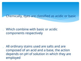
Chemically, dyesare classified as acidic or basic
Which combine with basic or acidic
components respectively
All ordinary stains used are salts and are
composed of an acid and a base, the action
depends on pH of solution in which they are
employed
5.
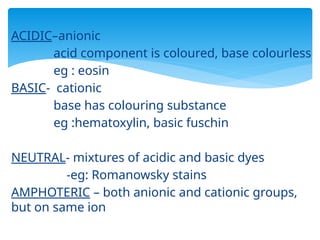
ACIDIC–anionic
acid component iscoloured, base colourless
eg : eosin
BASIC- cationic
base has colouring substance
eg :hematoxylin, basic fuschin
NEUTRAL- mixtures of acidic and basic dyes
-eg: Romanowsky stains
AMPHOTERIC – both anionic and cationic groups,
but on same ion
6.
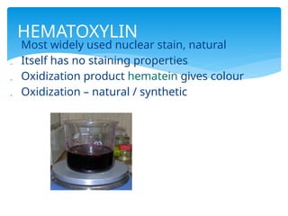
Most widelyused nuclear stain, natural
Itself has no staining properties
Oxidization product hematein gives colour
Oxidization – natural / synthetic
HEMATOXYLIN
7.
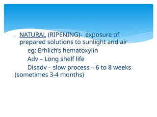
NATURAL (RIPENING)-exposure of
prepared solutions to sunlight and air
eg: Erhlich’s hematoxylin
Adv – Long shelf life
Disadv – slow process – 6 to 8 weeks
(sometimes 3-4 months)
8.
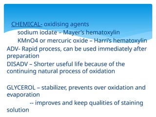
CHEMICAL- oxidisingagents
sodium iodate – Mayer’s hematoxylin
KMnO4 or mercuric oxide – Harri’s hematoxylin
ADV- Rapid process, can be used immediately after
preparation
DISADV – Shorter useful life because of the
continuing natural process of oxidation
GLYCEROL – stabilizer, prevents over oxidation and
evaporation
-- improves and keep qualities of staining
solution
9.
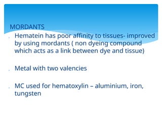
MORDANTS
Hematein haspoor affinity to tissues- improved
by using mordants ( non dyeing compound
which acts as a link between dye and tissue)
Metal with two valencies
MC used for hematoxylin – aluminium, iron,
tungsten
10.
CLASSIFICATION BASED ONMORDANT
(1) Alum H
(2) Iron H
(3) Tungsten H
(4) Molybdenum H
(5) Lead H
(6) H without mordant
Most mordants are incorporated into the H
But in few, they are used before staining
eg- Heidenhain’s iron H
11.
Used commonlyin routine H and E stain
Alums are double sulphates with active trivalent
metal ions such as Fe, Al, Cr together with K or
ammonium as second cation
NATURAL
Erhlich’s ,Delafield’s
CHEMICAL
Mayer’s, Harri’s, Cole’s,
Gill’s,Delafeld’s,Carazzi’s
ALUM HEMATOXYLINS
12.
Mordant used– aluminium as alum. Pot.
Sulphate or alum. Ammon. Sulphate
Produces good nuclear staining
It stains nucleus red – converted to blue black
when washed in a weak alkali solution –blueing
Regressive or progressive stain
Time required depends type and age of alum H
and type of tissue
13.
1.ERHLICH’S HEMATOXYLIN
Erhlichin 1886
Naturally ripened, 2months(emergency – add
sodium iodate), last for years
ADV AND USES
Stains nuclei intensely and crisply
Staining bone and cartilage
For tissues exposed to acid (fixed long in
formalin)
Stains mucin in salivary gland & goblet cells
Fades much more slowly
Used for regressive staining
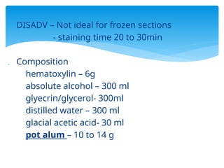
14.
DISADV –Not ideal for frozen sections
- staining time 20 to 30min
Composition
hematoxylin – 6g
absolute alcohol – 300 ml
glyecrin/glycerol- 300ml
distilled water – 300 ml
glacial acetic acid- 30 ml
pot alum – 10 to 14 g
15.
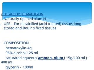
2.DELAFIELD’S HEMATOXYLIN
Naturallyripened alum H
USE – For decalcified (acid treated) tissue, long
stored and Bouin’s fixed tissues
COMPOSITION
hematoxylin-4g
95% alcohol-125 ml
saturated aqueous ammon. Alum ( 15g/100 ml ) –
400 ml
glycerin - 100ml
16.
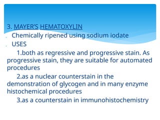
3. MAYER’S HEMATOXYLIN
Chemically ripened using sodium iodate
USES
1.both as regressive and progressive stain. As
progressive stain, they are suitable for automated
procedures
2.as a nuclear counterstain in the
demonstration of glycogen and in many enzyme
histochemical procedures
3.as a counterstain in immunohistochemistry
17.
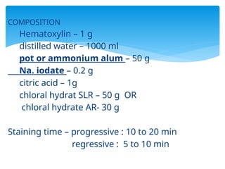
COMPOSITION
Hematoxylin – 1g
distilled water – 1000 ml
pot or ammonium alum – 50 g
Na. iodate – 0.2 g
citric acid – 1g
chloral hydrat SLR – 50 g OR
chloral hydrate AR- 30 g
Staining time – progressive : 10 to 20 min
regressive : 5 to 10 min
18.
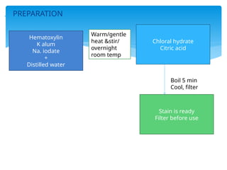
PREPARATION
Hematoxylin
K alum
Na.iodate
+
Distilled water
Chloral hydrate
Citric acid
Stain is ready
Filter before use
Warm/gentle
heat &stir/
overnight
room temp
Boil 5 min
Cool, filter
19.
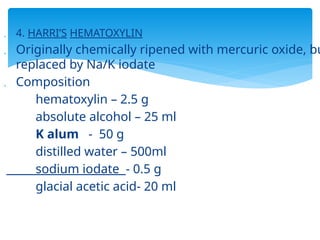
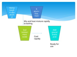
4. HARRI’SHEMATOXYLIN
Originally chemically ripened with mercuric oxide, bu
replaced by Na/K iodate
Composition
hematoxylin – 2.5 g
absolute alcohol – 25 ml
K alum - 50 g
distilled water – 500ml
sodium iodate - 0.5 g
glacial acetic acid- 20 ml
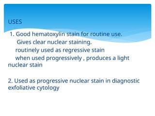
USES
1. Good hematoxylinstain for routine use.
Gives clear nuclear staining.
routinely used as regressive stain
when used progressively , produces a light
nuclear stain
2. Used as progressive nuclear stain in diagnostic
exfoliative cytology
22.
Staining time- 10 min
Disadvantage
quality of nuclear stain and speed
of staining deteriorates after few months –
formation of precipitate in stored solution
should be filtered and also increase
the staining time
hence, preparation of a fresh
batch of stain every month is advised
23.
5.COLE’S HEMATOXYLIN
alum H , artificially ripened with alcoholic
iodine solution
Staining time – 20-45 min
Composition
hematoxylin – 1.5 g
aquous K/Ammon alum- 700 ml
1% iodine in 95% alcohol- 50 ml
distilled water – 250 ml
24.
6 CARAZZI’S HEMATOXYLIN
Chemicallyripened by pot iodate
Staining time 1-2min
USES :
progressive nuclear counterstain- pale and
precise nuclear staining and doesn’t stain any
cytoplasmic components
COMPOSITION
hematoxylin – 5g k. alum – 25g
glycerol- 100 ml distilled H2O- 400 ml
potassium iodate – 0.1 g
25.
7. GILL’SHEMATOXYLIN
Chemically ripened by sodium iodate
ADV :-
1.Fast in action
2.stable for 12 months , more stable than
Harri’s
3.little or no surface precipitation
Staining time regressive – 5 to 15 min
26.
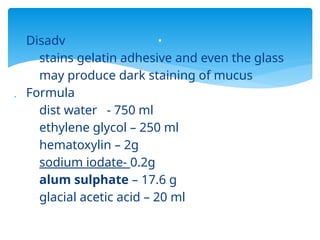
Disadv
stains gelatinadhesive and even the glass
may produce dark staining of mucus
Formula
dist water - 750 ml
ethylene glycol – 250 ml
hematoxylin – 2g
sodium iodate- 0.2g
alum sulphate – 17.6 g
glacial acetic acid – 20 ml
.
27.
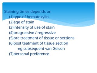
Staining times dependson
(1)type of hematoxylin
(2)age of stain
(3)intensity of use of stain
(4)progressive / regressive
(5)pre treatment of tissue or sections
(6)post teatment of tissue section
eg subsequent van Geison
(7)personal preference
28.
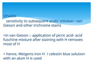
DISADV
sensitivity to subsequentacidic solution : van
Gieson and other trichrome stains
>in van Gieson :- application of picric acid- acid
fuschine mixture after staining with H removes
most of H
> hence, Weigerts iron H / celestin blue solution
with an alum H is used
29.
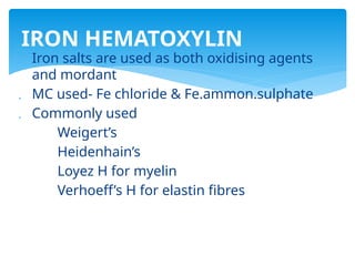
Iron saltsare used as both oxidising agents
and mordant
MC used- Fe chloride & Fe.ammon.sulphate
Commonly used
Weigert’s
Heidenhain’s
Loyez H for myelin
Verhoeff’s H for elastin fibres
IRON HEMATOXYLIN
30.
PRECAUTION :
Over oxidationof H – hence H and Fe
solutions are prepared separately and
either mix immediately before use/ use
consecutively
31.
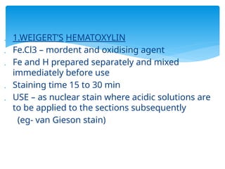
1.WEIGERT’S HEMATOXYLIN
Fe.Cl3 – mordent and oxidising agent
Fe and H prepared separately and mixed
immediately before use
Staining time 15 to 30 min
USE – as nuclear stain where acidic solutions are
to be applied to the sections subsequently
(eg- van Gieson stain)
32.
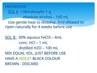
PREPARATION
SOL A :Hematoxylin 1 g
absolute alcohol – 100 mL
Use gentle heat to dissolve. And allowed to
ripen naturally for 4 weeks before use
SOL B : 30% aquous FeCl3 – 4mL
conc. HCl – 1 mL
distilled H2O – 100 mL
MIX EQUAL VOL. JUST BEFORE USE
HAVE A VIOLET BLACK COLOUR
BROWN - DISCARD
33.
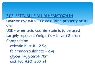
2.CELESTIN BLUEALUM HEMATOXYLIN
Oxazine dye with little colouring property on its
own
USE – when acid counterstain is to be used
Largely replaced Weigert’s H in van Gieson
Composition
celestin blue B – 2.5g
fe.ammon.sulphate – 25g
glycerin/glycerol- 70ml
distilled H2O- 500 ml
34.
3.HEIDENHAIN’S IRONHEMATOXYLIN
FE. AMMON. SULPHATE mordant and oxidising agent
Used as regressive stain and same solution used as
differentiating fluid
Good cellular details and good photomicrography
SOL A – hematoxylin – 0.5 g
abs alcohol – 10 ml
distilled water- 90 ml
SOL B – fe. ammon.sulphate- 5 g
dist water- 100ml
HERE, IRON ALUM APPLIED FIRST, F/B HEMATOXYLIN
35.
mitochondria, chromosomes,muscle striation,
myelin, nuclear chromatin – GREY BLACK
Time reqd in mordant – varies acc to fixative
formalin, formalin sublimtes, Bouin’, carnoy-
1hr
Helly’s or Zenker’s- 3 hrs
osmium tetroxide, flemings fluid – upto 24hrs
36.
Differentiation –difficult to judge. So, dip
slide in and out of mordant until slide is
clear & check under microscope
Cytoplasmic counterstains usually not
needed
Fading – resistant to fading only if washed
well after differentiation
Staining time :- reduced at 60 deg C
37.
4.LOYEZ HEMATOXYLIN
Fe.ammon.SO4
Use – to demonstrate myelin , and may be
applied to paraffin, frozen or nitrocellulose
section
5. VERHOEFF’S HEMATOXYLIN
for demonstarting elastin fibres
Stains coarse elastic fibres black
FeCl3 – differentiator
Provides excellent contrast
But requires careful differentiation
38.
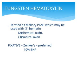
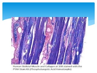
Termed asMallory PTAH which may be
used with (1) hematin
(2)chemical oxdn,
(3)Natural oxdn
FIXATIVE – Zenker’s – preferred
10% BNF
TUNGSTEN HEMATOXYLIN
39.
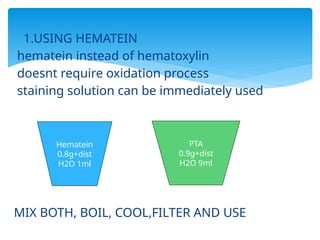
1.USING HEMATEIN
hemateininstead of hematoxylin
doesnt require oxidation process
staining solution can be immediately used
MIX BOTH, BOIL, COOL,FILTER AND USE
Hematein
0.8g+dist
H2O 1ml
PTA
0.9g+dist
H2O 9ml
40.
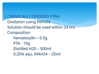
2.CHEMICALLY OXIDISED PTAH
Oxidation using KMnO4
Solution should be used within 24 hrs
Composition
hematoxylin – 0.5g
PTA- 10g
Distilled H20 – 500ml
0.25% aqu. KMnO4 – 25ml
41.
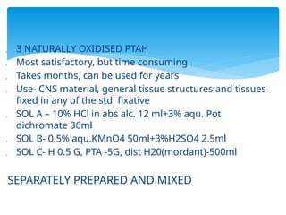
3 NATURALLYOXIDISED PTAH
Most satisfactory, but time consuming
Takes months, can be used for years
Use- CNS material, general tissue structures and tissues
fixed in any of the std. fixative
SOL A – 10% HCl in abs alc. 12 ml+3% aqu. Pot
dichromate 36ml
SOL B- 0.5% aqu.KMnO4 50ml+3%H2SO4 2.5ml
SOL C- H 0.5 G, PTA -5G, dist H20(mordant)-500ml
SEPARATELY PREPARED AND MIXED
42.
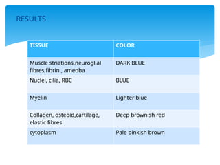
RESULTS
TISSUE COLOR
Musclestriations,neuroglial
fibres,fibrin , ameoba
DARK BLUE
Nuclei, cilia, RBC BLUE
Myelin Lighter blue
Collagen, osteoid,cartilage,
elastic fibres
Deep brownish red
cytoplasm Pale pinkish brown
44.
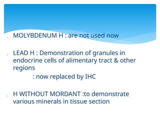
MOLYBDENUM H: are not used now
LEAD H : Demonstration of granules in
endocrine cells of alimentary tract & other
regions
: now replaced by IHC
H WITHOUT MORDANT :to demonstrate
various minerals in tissue section
45.
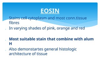
Stains cellcytoplasm and most conn.tissue
fibres
In varying shades of pink, orange and red
Most suitable stain that combine with alum
H
Also demonstartes general histologic
architecture of tissue
EOSIN
46.
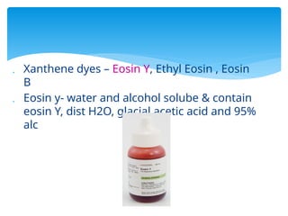
Xanthene dyes– Eosin Y, Ethyl Eosin , Eosin
B
Eosin y- water and alcohol solube & contain
eosin Y, dist H2O, glacial acetic acid and 95%
alc
47.
USES
Counterstainin routine H & E – pink – orange
colour to cytoplasm
RBC – intensley red
To distinguish b/w cytoplasm of diff types of
cells and conn tissue
PHLOXINE & BIEBRICH SCARLET – SUBSTITUTES
FOR EOSIN
48.
Glasswares shouldbe clean &dry
Weigh accurately & use correct solvent
Keep silver & osmic acid stains in dark bottles
Dilute ammonia should be freshly prepared
Alcoholic solutions to be kept in glass stoppered
bottle-to prevent evaporation of alcohol
PREPARATION OF STAINS
Should allow easyflow of work from one
procedure to another without any crossover
Staining bench should face window &well
illuminated
Sinks –atleast 2 in number with atleast 2 cold
water taps -1 for routine and 1 for special stain
Staining rack
Adequate supply of distilled water
Microscope
51.
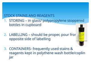
STOCK STAINS ANDREAGENTS
1. STORING – in glass/ polypropylene stoppered
bottles in cupboard
2. LABELLING – should be proper, pour from
opposite side of labelling
3. CONTAINERS- frequently used stains &
reagents kept in polythene wash bottle/coplin
jar
52.
MANUAL/ AUTOMATIC
MANUAL
Basic staining equipments :
*glass troughs and racks of 12,25 or 50
slides
*xylene-at start and end of staining
STAINING PROCEDURE
54.
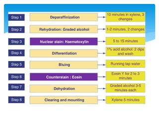
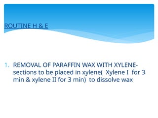
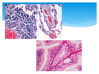
ROUTINE H &E
1. REMOVAL OF PARAFFIN WAX WITH XYLENE-
sections to be placed in xylene( Xylene I for 3
min & xylene II for 3 min) to dissolve wax
55.
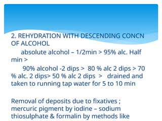
2. REHYDRATION WITHDESCENDING CONCN
OF ALCOHOL
absolute alcohol – 1/2min > 95% alc. Half
min >
90% alcohol -2 dips > 80 % alc 2 dips > 70
% alc. 2 dips> 50 % alc 2 dips > drained and
taken to running tap water for 5 to 10 min
Removal of deposits due to fixatives ;
mercuric pigment by iodine – sodium
thiosulphate & formalin by methods like
56.
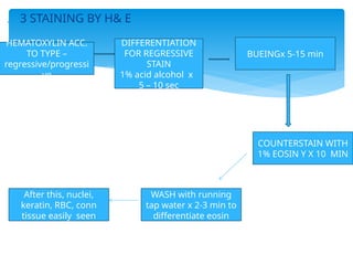
3 STAININGBY H& E
DIFFERENTIATION
FOR REGRESSIVE
STAIN
1% acid alcohol x
5 – 10 sec
HEMATOXYLIN ACC.
TO TYPE –
regressive/progressi
ve
BUEINGx 5-15 min
COUNTERSTAIN WITH
1% EOSIN Y X 10 MIN
WASH with running
tap water x 2-3 min to
differentiate eosin
After this, nuclei,
keratin, RBC, conn
tissue easily seen
57.
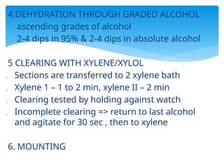
4.DEHYDRATION THROUGH GRADEDALCOHOL
ascending grades of alcohol
2-4 dips in 95% & 2-4 dips in absolute alcohol
5 CLEARING WITH XYLENE/XYLOL
Sections are transferred to 2 xylene bath
Xylene 1 – 1 to 2 min, xylene II – 2 min
Clearing tested by holding against watch
Incomplete clearing => return to last alcohol
and agitate for 30 sec , then to xylene
6. MOUNTING
58.
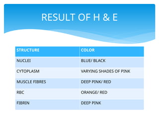
STRUCTURE COLOR
NUCLEI BLUE/BLACK
CYTOPLASM VARYING SHADES OF PINK
MUSCLE FIBRES DEEP PINK/ RED
RBC ORANGE/ RED
FIBRIN DEEP PINK
RESULT OF H & E
60.
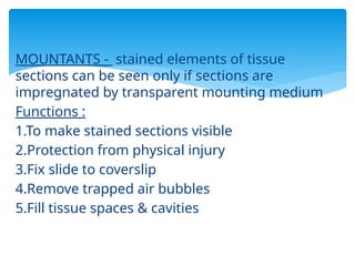
MOUNTANTS - stainedelements of tissue
sections can be seen only if sections are
impregnated by transparent mounting medium
Functions :
1.To make stained sections visible
2.Protection from physical injury
3.Fix slide to coverslip
4.Remove trapped air bubbles
5.Fill tissue spaces & cavities
61.
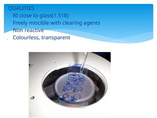
QUALITIES
RI closeto glass(1.518)
Freely miscible with clearing agents
Non reactive
Colourless, transparent
62.
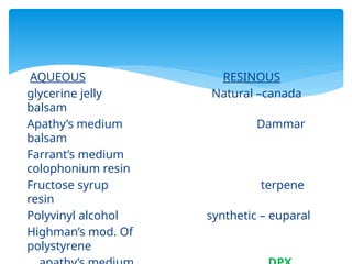
AQUEOUS RESINOUS
glycerine jellyNatural –canada
balsam
Apathy’s medium Dammar
balsam
Farrant’s medium
colophonium resin
Fructose syrup terpene
resin
Polyvinyl alcohol synthetic – euparal
Highman’s mod. Of
polystyrene
63.
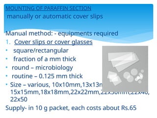
MOUNTING OF PARAFFINSECTION
manually or automatic cover slips
Manual method: - equipments required
1. Cover slips or cover glasses
• square/rectangular
• fraction of a mm thick
• round – microbiology
• routine – 0.125 mm thick
• Size – various, 10x10mm,13x13mm,
15x15mm,18x18mm,22x22mm,22x30mm,22x40,
22x50
Supply- in 10 g packet, each costs about Rs.65
64.
2.DISSECTING NEEDLES
to adjustcoverslip during & after mounting
3.MOUNTANTS
depends on the type of section
MOUNTING PROCEDURE
1. clean the coverslips
2.lay coverslips in a row
3.bring sections to xylene
4.removal of excess xylene
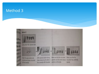
FOR URGENTFROZEN SECTIONS – RAPID H & E
Procedure
1.Freeze the required tissue block in freezing
microtome/ cryostat.
cut sections 3-6 micometre
2.Fix sections in AAF x 30 -60 sec
alternative – 10% BNF x 20 sec
3.Rinse rapidly in water
4.Hematoxylin – harri’s x 2min/ carazzi’s x 1min
69.
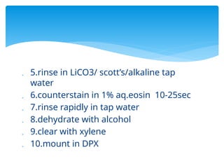
5.rinse inLiCO3/ scott’s/alkaline tap
water
6.counterstain in 1% aq.eosin 10-25sec
7.rinse rapidly in tap water
8.dehydrate with alcohol
9.clear with xylene
10.mount in DPX
70.
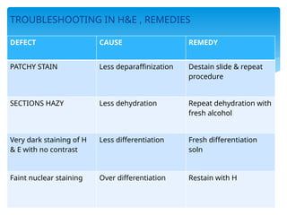
TROUBLESHOOTING IN H&E, REMEDIES
DEFECT CAUSE REMEDY
PATCHY STAIN Less deparaffinization Destain slide & repeat
procedure
SECTIONS HAZY Less dehydration Repeat dehydration with
fresh alcohol
Very dark staining of H
& E with no contrast
Less differentiation Fresh differentiation
soln
Faint nuclear staining Over differentiation Restain with H
71.
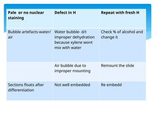
Pale or nonuclear
staining
Defect in H Repeat with fresh H
Bubble artefacts-water/
air
Water bubble- d/t
improper dehydration
because xylene wont
mix with water
Check % of alcohol and
change it
Air bubble due to
improper mounting
Remount the slide
Sections floats after
differentiation
Not well embedded Re embedd
72.
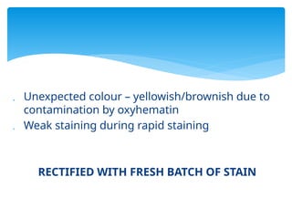
Unexpected colour– yellowish/brownish due to
contamination by oxyhematin
Weak staining during rapid staining
RECTIFIED WITH FRESH BATCH OF STAIN
73.
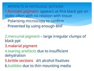
ARTEFACTS INHISTOLOGIC SECTIONS
1.formalin pigment- appears as fine black ppt on
slides often with no relation with tissue
Polarising microscopy to confirm
Prevented by using enough BNF
2.mercurial pigment – large irregular clumps of
black ppt
3.malarial pigment
4.tearing artefacts due to insufficient
dehydration
5.brittle sections d/t alcohol fixatives
6.bubbles due to thin mounting media