Downloaded 344 times
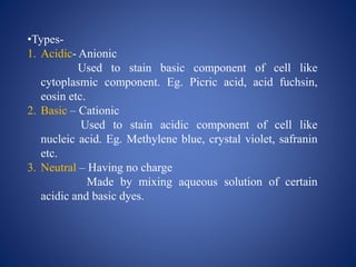


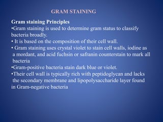


1. Staining techniques are used to increase visibility and provide extra information about cells and tissues in pathogen diagnosis. Stains are chemical substances that bind to specific components of cells. 2. There are different types of stains including acidic, basic, neutral, simple, differential and special stains. Simple stains use one dye while differential stains use multiple dyes to categorize cells. Special stains highlight specific structures. 3. Common staining techniques discussed are Gram staining, acid-fast staining, capsule staining, endospore staining and lactophenol cotton blue staining for fungi diagnosis and analysis. Staining aids in visualizing various cell components and classifying pathogens.


