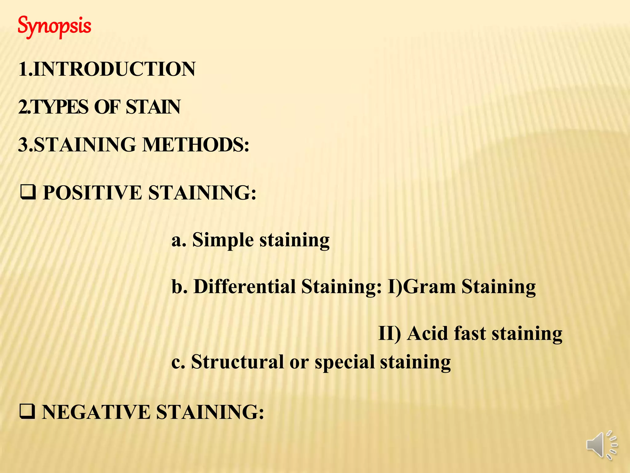
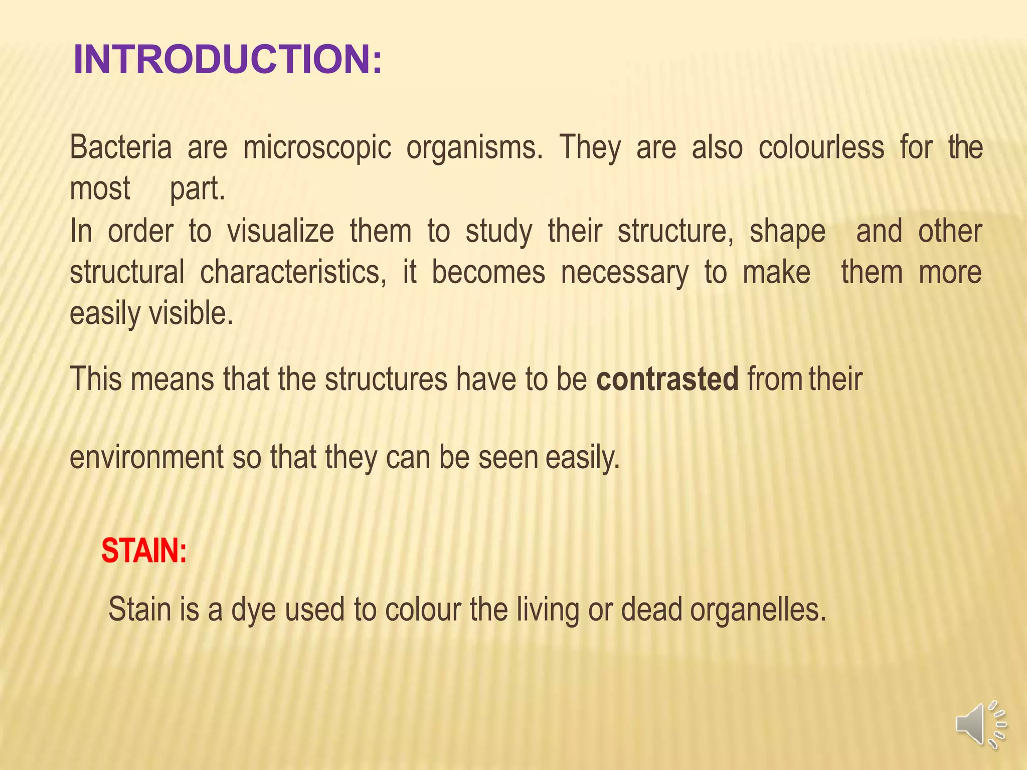
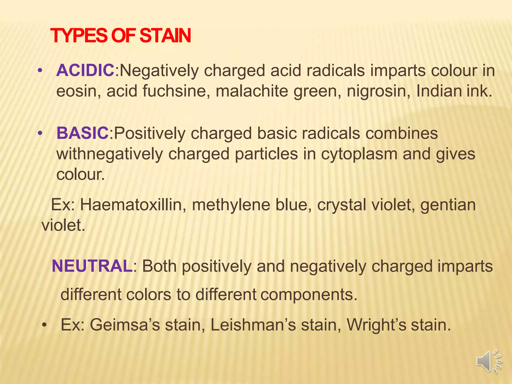
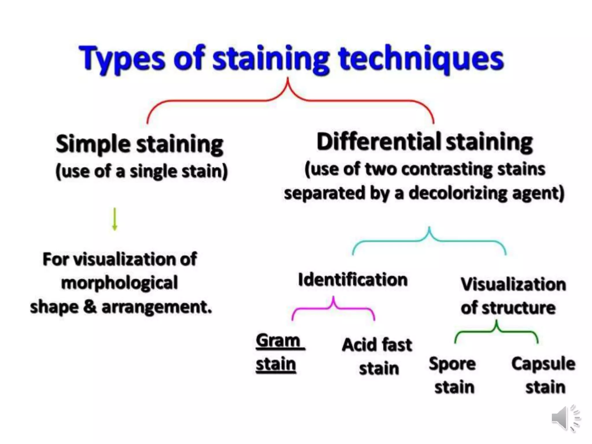
STAINING TECHNIQUES
Dr. Rachana Choudhary
Shri Shankaracharya Mahavidyalaya Junwani Bhilai
Synopsis
1.INTRODUCTION
2.TYPES OF STAIN
3.STAINING METHODS:
 POSITIVE STAINING:
a. Simple staining
b. Differential Staining: I)Gram Staining
II) Acid fast staining
c. Structural or special staining
 NEGATIVE STAINING:
INTRODUCTION:
Bacteria are microscopic organisms. They are also colourless for the
most part.
In order to visualize them to study their structure, shape and other
structural characteristics, it becomes necessary to make them more
easily visible.
This means that the structures have to be contrasted fromtheir
environment so that they can be seen easily.
STAIN:
Stain is a dye used to colour the living or dead organelles.
TYPESOFSTAIN
• ACIDIC:Negatively charged acid radicals imparts colour in
eosin, acid fuchsine, malachite green, nigrosin, Indian ink.
• BASIC:Positively charged basic radicals combines
withnegatively charged particles in cytoplasm and gives
colour.
Ex: Haematoxillin, methylene blue, crystal violet, gentian
violet.
NEUTRAL: Both positively and negatively charged imparts
different colors to different components.
• Ex: Geimsa’s stain, Leishman’s stain, Wright’s stain.
STAINING METHODS:
A.POSITIVE STAINING: - where the actual cells are
themselves coloured and appear in a clear background.
1. Simple staining
2. Differential Staining:
3. Structural or special staining
B.NEGATIVE STAINING:
where the cells remain clear (uncoloured) and the
background is coloured to create a contrast to aid in the
better visualization of the image.
(a) Indian ink
(b) Nigrosine .
1.Simple Staining
•Simple staining is one step method using only one dye.
•Basic dyes are used in direct stain and acidic dye is used in
negative stain.
•Used to study the morphology better, to show the nature of
the cellular contents of the exudates and also to study the
intracellular location of the bacteria
Commonly used simple stains are
Methylene blue ( Loeffler’s methylene blue )
Dilute Carbol fuchsin
Polychrome methylene blue
SIMPLE STAINING: Loffler's Methylene Blue
2.Differential staining
Differential stains use two or more stains and allow the
cells to be categorized into various groups or types. It
usually provides more information about the
characteristics of the cell wall (thickness).
Two step method.
•Gram staining
•Acid fast staining
GRAM STAINING
The Gram stain was devised by
the Danish physician, Hans
Christian Joachim Gram, while
working in Berlin in 1883. He
later published this procedure
in 1884. At the time, Dr. Gram
was studying lung tissue sections
from patients who had died of
pneumonia.
16
Gram staining Principles
•Gram staining is used to determine gram status to classify
bacteria broadly.
• It is based on the composition of their cell wall.
•Gram staining uses crystal violet to stain cell walls, iodine as a
mordant, and acid fuchsin or safranin counterstain to mark all
bacteria
•Gram-positive bacteria stain dark blue or violet.
•Their cell wall is typically rich with peptidoglycan and lacks
the secondary membrane and lipopolysaccharide layer found
in Gram-negative bacteria
METHOD CONSISTS OFFOURCOMPONENTS:
• Primary stain—Crystal violet, Methyl violet &
Gentian violet.
• Mordant—Gram Iodine, Rarely Lugol’s Iodine.
• Decolourizer—Alcohol, Acetone, Alcohol: Acetone
(1:1) mixture.
• Counter stain—Dilute Carbol fuchsin, Safranin,
Neutral red, Sandi ford stain for Gonococci.
Procedure
13
Observation
14
Gram negative bacilli
Gram Positive Cocci in Chain
ACID – FAST STAINING OR ZIEHL-NEELSENSTAINING
• Thismethod is amodification of Ehrlich’s (1882)method.
• The Ziehl-Neelsen acid fast staining method has proved to
be most useful for staining acid fast bacilli belonging to
the genus Mycobacterium especially Mycobacterium
tuberculosis and Mycobacterium leprae, and also for
Nocardia.
PRINCIPLE OF ZIEHL–NEELSEN STAIN
• Acid fastness of acid-fast bacilli is attributed to the presence of large
quantities of unsaponifiable wax fraction called mycolic acid in their
cell wall and also the intactness of the cell wall . The degree of acid
fastness varies in different bacteria.
• In this staining method, application of heat helps the dye to penetrate
the tubercle bacillus.
• Once stained, the stain cannot be easily removed. The tubercle bacilli
resist the decolorizing action of acid-alcohol which confers acid
fastness to the bacteria.
• The other microorganisms, which are easily decolorized by acid-
alcohol, are considered non-acid fast . The non-acid fast bacilli readily
absorb the colour of the counter stain appearing blue, while the acid
fast cells retain the red colour of primary stain.
ACID - FAST STAIN BASIC REQUIREMENTS
1. Primary And Mordant Staining with Strong Carbol fuchsin
2.Decolourization with Acid Alcohol : The acid alcohol contains
3% HCl and 95% ethanol or 20% H2 SO4.
3. Counterstain with Methylene Blue.
Acid - Fast Cells Red
NonAcid – Fast Blue
MICROSCOPIC OBSERVATION:
• The stained smear are contains
pink coloured slender rod shaped
structures are seen with curved
ends acid fast bacilli seen among
the blue coloured multilobed pus
cells.
• The smear is positive for acid fast
bacilli.
DIFFERENTMODIFICATION OF ACID FASTSTAIN
1) 5% Sulphuric acid is used as a decolourizing agent for staining
Mycobacterium leprae.
2) 1% Sulphuric acid is used as a decolourizing agent for staining
Nocardia species, Cryptosporidium and Isospora oocysts (Kinyoun’s
modification of acid fast stain).
3) 0.25% Sulphuric acid is used as a decolourizing agent for staining
spores.
3.Structural or special staining
• Stain for endospores
• Stain for capsules
• Stain for flagella
•Stain for bacterial nucleus
STAINING FOR ENDOSPORES
Spores are highly resistant and metabolically inactive forms .
 The morphology of bacterial endospores is best observed in
unstained wet films under the phase contrast microscope.
Different staining techniques are available for staining of
spores.
 A modified Ziehl-Neelsen stain in which weak, 0.25%
sulphuric acid is used as decolourizer, yields red spores in
blue-stained bacteria. Lipid granules also stain red,
appearing like small spherical spores.
MALACHITE GREEN STAIN FOR SPORES (METHOD OF
SCHAEFFER AND FULTON, MODIFIED BY ASHBY, 1938).
• When, within several seconds, large droplets have condensed on the
underside of the slide, flood it with 5% aqueous solution of
malachite green and leave to act for 1 min while the water continues
to boil.
• Wash in cold water.
• Treat with 0.5% safranine or 0.05% basic fuchsin for 30 seconds.
• Wash and dry.
• This method colours the spores green and the vegetative bacilli red.
Lipid granules are unstained.
Procedure
MALACHITE GREEN STAIN FOR SPORES
The Microscopy picture shows presence of green endospores stained
with malachite green and vegetative cells stained with safranin red in
color.
CAPSULE STAINING
 The capsules serves as protective material by slowing down or preventing
penetration of chemicals and body juices.
PRINCIPLE:
• Chemically, the capsular material is a polysaccharide, a glycoprotein or a
polypeptide.
• Capsule staining is more difficult than other types of differential staining
procedures because the capsular materials are water soluble and may be
dislodged and removed with vigorous washing.
• Bacterial smears should not be heated, because the resultant cell shrinkage
may create a clear zone around the organism, an artifact that can be mistake for
capsule.
•
The capsule is non-ionic, so that the dyes commonly used will not bind to it.
Two dyes, one acidic and one basic, are used to stain the background and the
cell wall, respectively.
METHODS:
1) Negative staining. India ink
2) Positive staining. crystal violet 20% copper sulphate ,
3) Mc Fdyean reaction : which uses the Loefflers polychrome
methylene blue to demonstrate the capsule of the Bacillus
anthracis.
CAPSULESTAINING:
Cryptococcus neoformans india
Capsulated Bacteria In India ink.
FLAGELLASTAINING
• Flagella is the organ of locomotion, which is one or
more unbrached, long filament.
• The presence or absence of flagella and their number and
arrangement are characteristic of different genera of
bacteria.
• The flagella can be visualized by the special staining
techniques in which their thickness is increased by
mordanting, or by electronic microscopy.
• Here staining is preceded by using
of some precipitating agent like tannic acid or iron
chloride
• Liefson’s stain, Carbol fuchsin or Fontana’s solution is used to
demonstrate the flagella
WET- MOUNT FLAGELLASTAIN
• The wet mount flagellar stain has been described by
Heimbrook et al in 1989.
• It is easier, cleaner and more reliable method.
Procedure:
• Grow bacteria for 16-24 h on a non-inhibitory medium,
e.g. tryptic soy agar or blood agar. Touch a loopful of
water onto the edge of a colony and let motile bacteria
swim into it.
FLAGELLASTAIN BYRYU STAIN: MICROSCOPY
Salmonella typhimurium stained with a modified Ryu stain to show
their flagella that are peritrichously arranged around the cell.
Nucleus staining
Bacteria
•Here nuclear material is present in a region called
nucleoid, devoid of nuclear membrane.
•Since cytoplasm has a strong affinity for most stains and it
may interfere with observation of nuclear material.
•It should be hydrolysed first with HCl.
•Later stained with Giemsa stain.
•Nuclear bodies will appear purple coloured.
Fungi Lactophenol Cotton Blue(LPCB)
Nuclear staining in fungi
• Fungi are eukaryotic
•Got organized nuclei bounded by nuclear membrane
with characteristics pores, a nucleolus and chromatin
strands.
•Fungal nuclei are oftenly stained with Hematoxylin,
Giemsa, Feulgen, or Acetocarmine
Scotch TapePreparation:
LACTOPHENOL COTTON BLUEMICROSCOPY
Thank you

Staining techniques

  • 1.
    STAINING TECHNIQUES Dr. RachanaChoudhary Shri Shankaracharya Mahavidyalaya Junwani Bhilai
  • 2.
    Synopsis 1.INTRODUCTION 2.TYPES OF STAIN 3.STAININGMETHODS:  POSITIVE STAINING: a. Simple staining b. Differential Staining: I)Gram Staining II) Acid fast staining c. Structural or special staining  NEGATIVE STAINING:
  • 3.
    INTRODUCTION: Bacteria are microscopicorganisms. They are also colourless for the most part. In order to visualize them to study their structure, shape and other structural characteristics, it becomes necessary to make them more easily visible. This means that the structures have to be contrasted fromtheir environment so that they can be seen easily. STAIN: Stain is a dye used to colour the living or dead organelles.
  • 4.
    TYPESOFSTAIN • ACIDIC:Negatively chargedacid radicals imparts colour in eosin, acid fuchsine, malachite green, nigrosin, Indian ink. • BASIC:Positively charged basic radicals combines withnegatively charged particles in cytoplasm and gives colour. Ex: Haematoxillin, methylene blue, crystal violet, gentian violet. NEUTRAL: Both positively and negatively charged imparts different colors to different components. • Ex: Geimsa’s stain, Leishman’s stain, Wright’s stain.
  • 5.
    STAINING METHODS: A.POSITIVE STAINING:- where the actual cells are themselves coloured and appear in a clear background. 1. Simple staining 2. Differential Staining: 3. Structural or special staining B.NEGATIVE STAINING: where the cells remain clear (uncoloured) and the background is coloured to create a contrast to aid in the better visualization of the image. (a) Indian ink (b) Nigrosine .
  • 7.
    1.Simple Staining •Simple stainingis one step method using only one dye. •Basic dyes are used in direct stain and acidic dye is used in negative stain. •Used to study the morphology better, to show the nature of the cellular contents of the exudates and also to study the intracellular location of the bacteria Commonly used simple stains are Methylene blue ( Loeffler’s methylene blue ) Dilute Carbol fuchsin Polychrome methylene blue
  • 8.
  • 9.
    2.Differential staining Differential stainsuse two or more stains and allow the cells to be categorized into various groups or types. It usually provides more information about the characteristics of the cell wall (thickness). Two step method. •Gram staining •Acid fast staining
  • 10.
    GRAM STAINING The Gramstain was devised by the Danish physician, Hans Christian Joachim Gram, while working in Berlin in 1883. He later published this procedure in 1884. At the time, Dr. Gram was studying lung tissue sections from patients who had died of pneumonia. 16
  • 11.
    Gram staining Principles •Gramstaining is used to determine gram status to classify bacteria broadly. • It is based on the composition of their cell wall. •Gram staining uses crystal violet to stain cell walls, iodine as a mordant, and acid fuchsin or safranin counterstain to mark all bacteria •Gram-positive bacteria stain dark blue or violet. •Their cell wall is typically rich with peptidoglycan and lacks the secondary membrane and lipopolysaccharide layer found in Gram-negative bacteria
  • 12.
    METHOD CONSISTS OFFOURCOMPONENTS: •Primary stain—Crystal violet, Methyl violet & Gentian violet. • Mordant—Gram Iodine, Rarely Lugol’s Iodine. • Decolourizer—Alcohol, Acetone, Alcohol: Acetone (1:1) mixture. • Counter stain—Dilute Carbol fuchsin, Safranin, Neutral red, Sandi ford stain for Gonococci.
  • 13.
  • 14.
  • 15.
    ACID – FASTSTAINING OR ZIEHL-NEELSENSTAINING • Thismethod is amodification of Ehrlich’s (1882)method. • The Ziehl-Neelsen acid fast staining method has proved to be most useful for staining acid fast bacilli belonging to the genus Mycobacterium especially Mycobacterium tuberculosis and Mycobacterium leprae, and also for Nocardia.
  • 16.
    PRINCIPLE OF ZIEHL–NEELSENSTAIN • Acid fastness of acid-fast bacilli is attributed to the presence of large quantities of unsaponifiable wax fraction called mycolic acid in their cell wall and also the intactness of the cell wall . The degree of acid fastness varies in different bacteria. • In this staining method, application of heat helps the dye to penetrate the tubercle bacillus. • Once stained, the stain cannot be easily removed. The tubercle bacilli resist the decolorizing action of acid-alcohol which confers acid fastness to the bacteria. • The other microorganisms, which are easily decolorized by acid- alcohol, are considered non-acid fast . The non-acid fast bacilli readily absorb the colour of the counter stain appearing blue, while the acid fast cells retain the red colour of primary stain.
  • 17.
    ACID - FASTSTAIN BASIC REQUIREMENTS 1. Primary And Mordant Staining with Strong Carbol fuchsin 2.Decolourization with Acid Alcohol : The acid alcohol contains 3% HCl and 95% ethanol or 20% H2 SO4. 3. Counterstain with Methylene Blue. Acid - Fast Cells Red NonAcid – Fast Blue
  • 19.
    MICROSCOPIC OBSERVATION: • Thestained smear are contains pink coloured slender rod shaped structures are seen with curved ends acid fast bacilli seen among the blue coloured multilobed pus cells. • The smear is positive for acid fast bacilli.
  • 20.
    DIFFERENTMODIFICATION OF ACIDFASTSTAIN 1) 5% Sulphuric acid is used as a decolourizing agent for staining Mycobacterium leprae. 2) 1% Sulphuric acid is used as a decolourizing agent for staining Nocardia species, Cryptosporidium and Isospora oocysts (Kinyoun’s modification of acid fast stain). 3) 0.25% Sulphuric acid is used as a decolourizing agent for staining spores.
  • 21.
    3.Structural or specialstaining • Stain for endospores • Stain for capsules • Stain for flagella •Stain for bacterial nucleus
  • 22.
    STAINING FOR ENDOSPORES Sporesare highly resistant and metabolically inactive forms .  The morphology of bacterial endospores is best observed in unstained wet films under the phase contrast microscope. Different staining techniques are available for staining of spores.  A modified Ziehl-Neelsen stain in which weak, 0.25% sulphuric acid is used as decolourizer, yields red spores in blue-stained bacteria. Lipid granules also stain red, appearing like small spherical spores.
  • 23.
    MALACHITE GREEN STAINFOR SPORES (METHOD OF SCHAEFFER AND FULTON, MODIFIED BY ASHBY, 1938). • When, within several seconds, large droplets have condensed on the underside of the slide, flood it with 5% aqueous solution of malachite green and leave to act for 1 min while the water continues to boil. • Wash in cold water. • Treat with 0.5% safranine or 0.05% basic fuchsin for 30 seconds. • Wash and dry. • This method colours the spores green and the vegetative bacilli red. Lipid granules are unstained.
  • 24.
  • 25.
    MALACHITE GREEN STAINFOR SPORES The Microscopy picture shows presence of green endospores stained with malachite green and vegetative cells stained with safranin red in color.
  • 26.
    CAPSULE STAINING  Thecapsules serves as protective material by slowing down or preventing penetration of chemicals and body juices. PRINCIPLE: • Chemically, the capsular material is a polysaccharide, a glycoprotein or a polypeptide. • Capsule staining is more difficult than other types of differential staining procedures because the capsular materials are water soluble and may be dislodged and removed with vigorous washing. • Bacterial smears should not be heated, because the resultant cell shrinkage may create a clear zone around the organism, an artifact that can be mistake for capsule. • The capsule is non-ionic, so that the dyes commonly used will not bind to it. Two dyes, one acidic and one basic, are used to stain the background and the cell wall, respectively.
  • 27.
    METHODS: 1) Negative staining.India ink 2) Positive staining. crystal violet 20% copper sulphate , 3) Mc Fdyean reaction : which uses the Loefflers polychrome methylene blue to demonstrate the capsule of the Bacillus anthracis.
  • 28.
  • 29.
    FLAGELLASTAINING • Flagella isthe organ of locomotion, which is one or more unbrached, long filament. • The presence or absence of flagella and their number and arrangement are characteristic of different genera of bacteria. • The flagella can be visualized by the special staining techniques in which their thickness is increased by mordanting, or by electronic microscopy. • Here staining is preceded by using of some precipitating agent like tannic acid or iron chloride • Liefson’s stain, Carbol fuchsin or Fontana’s solution is used to demonstrate the flagella
  • 30.
    WET- MOUNT FLAGELLASTAIN •The wet mount flagellar stain has been described by Heimbrook et al in 1989. • It is easier, cleaner and more reliable method. Procedure: • Grow bacteria for 16-24 h on a non-inhibitory medium, e.g. tryptic soy agar or blood agar. Touch a loopful of water onto the edge of a colony and let motile bacteria swim into it.
  • 31.
    FLAGELLASTAIN BYRYU STAIN:MICROSCOPY Salmonella typhimurium stained with a modified Ryu stain to show their flagella that are peritrichously arranged around the cell.
  • 32.
    Nucleus staining Bacteria •Here nuclearmaterial is present in a region called nucleoid, devoid of nuclear membrane. •Since cytoplasm has a strong affinity for most stains and it may interfere with observation of nuclear material. •It should be hydrolysed first with HCl. •Later stained with Giemsa stain. •Nuclear bodies will appear purple coloured.
  • 33.
    Fungi Lactophenol CottonBlue(LPCB) Nuclear staining in fungi • Fungi are eukaryotic •Got organized nuclei bounded by nuclear membrane with characteristics pores, a nucleolus and chromatin strands. •Fungal nuclei are oftenly stained with Hematoxylin, Giemsa, Feulgen, or Acetocarmine Scotch TapePreparation:
  • 34.
  • 35.