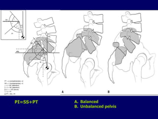
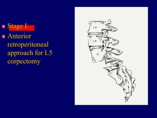
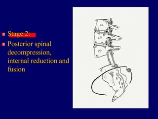
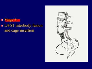
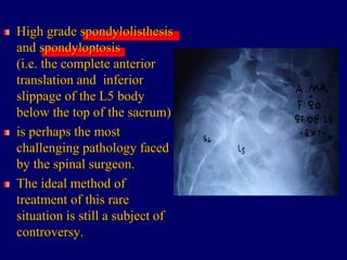
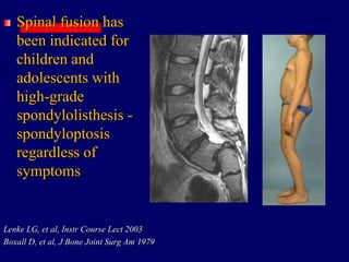
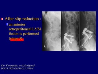
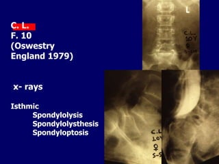
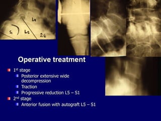
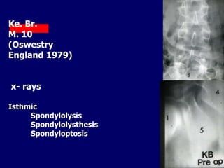
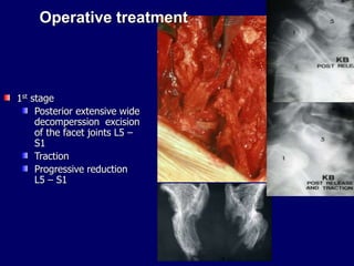
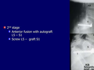
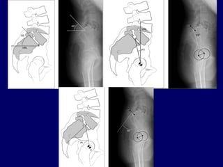
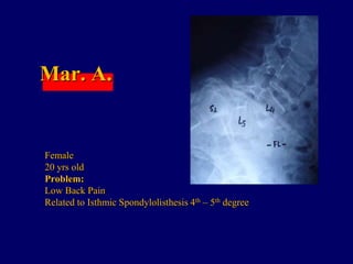
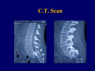
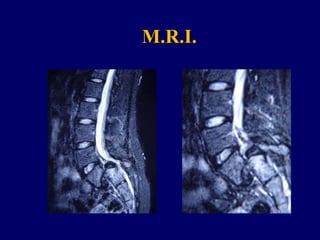
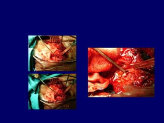
Spondyloptosis
GEORGE SAPKAS
PROFESSOR OF ORTHOPAEDICS
Metropolitan Hospital Athens
Spinal Disorders and
Musculo-sceletal Unit
High grade spondylolisthesis
and spondyloptosis
(i.e. the complete anterior
translation and inferior
slippage of the L5 body
below the top of the sacrum)
is perhaps the most
challenging pathology faced
by the spinal surgeon.
The ideal method of
treatment of this rare
situation is still a subject of
controversy.
Non surgical
management of the
symptomatic patient
with high grade
spondylolisthesis is
generally less
successful than with
low grade
spondylolisthesis.
Frennered AK, et al, Spine1991
Spinal fusion has
been indicated for
children and
adolescents with
high-grade
spondylolisthesis -
spondyloptosis
regardless of
symptoms
Lenke LG, et al, Instr Course Lect 2003
Boxall D, et al, J Bone Joint Surg Am 1979
The goals of surgery
include :
neural decompression,
partial reduction and
correction of the
lumbo-sacral slip
angle and
kyphosis when
present. Dysplastic type of
spondyloptosis
Many surgical
techniques have been
proposed in the
literature including:
complete or partial
reduction and
instrumented fusion,
in situ fusion,
the Gaines procedure
and
posterior osteotomies.
Stoker et al are in favor of
a single all-posterior
operation consisting of :
wide decompression,
discectomy with or without
sacral dome osteotomy,
postural reduction, and
posterior fusion with
pedicle screw
instrumentation.
G.E. Stoker, et al, ArgoSpine NEWS&JOURNAL-
quarterly march , 2011
Cont.
Both
in situ fusion and
complete anatomic
reduction and fusion
have lower fusion rates
and
higher associated
neurologic complication
rates comparing
with partial reduction and
fusion.
G.E. Stoker, et al, ArgoSpine NEWS&JOURNAL-quarterly
march , 2011
An. La.
Problem:
Low back pain – non neurologic
deficit
x- rays
Isthmic Spondylolysthesis
Extensive posterior
decompresion
(Isola system)
Allografts – Autografts
Postero-laterly
Lumbar - femoral Brace for 6 months
Many different
methods of reduction
of spondylolisthesis –
spondyloptosis
and fusion have been
described:
A.
Pre-operative
halo-femoral traction
with pelvic
suspension,
anterior posterior
fusion and
placement of a
pantaloon spica cast
in hyperextension
Dubousset J, Cl in Or thop Relat, 1997
Cont.
B.
Gradual
intra-operative closed
reduction
with instrumentation
and posterior fusion
Matthiass HH , et al, Clin Orthop Relat Res1986
Cont.
C.
Anterior release
with partial reduction
and anterior interbody
fusion
Muschik M, et al. Spine 1997
Cont.
D.
Posterior
decompression
with postero-lateral
fusion,
followed by halo-
skeletal traction
and then by a
second-stage anterior
interbody fusion
Bradford DS, et al, J BoneJoint Surg Am 1990
Karampalis et al,
proposed a new
method of :
staged reduction and
fusion of high-grade
spondylolisthesis
using Magerl’s
external fixator.
Chr. Karampalis, et al, EurSpineJ
DOI10.1007/s00586-012-2190-6
Cont.
The procedure is
carried out in three
stages:
Cont.
In stage 1
a wide L5/S1 posterior
decompression is first
carried out (Gill’s
procedure)
followed by discectomy.
Schanz pins are inserted
in the pedicles of L4
and iliac crests through
stab incisions
and Magerl’s external
fixator is assembled on
them.
Chr. Karampalis, et al, EurSpineJ
DOI10.1007/s00586-012-2190-6
Cont.
The second stage is:
the gradual and
progressive reduction of
the slip.
The amount of distraction
is monitored by daily
standing radiographs
and the whole process is
guided by patient’s
comfort and neurological
status.
Chr. Karampalis, et al, EurSpineJ
DOI10.1007/s00586-012-2190-6
Cont.
After slip reduction :
an anterior
retroperitoneal L5/S1
fusion is performed
(stage 3)
Chr. Karampalis, et al, EurSpineJ
DOI10.1007/s00586-012-2190-6
It is suggested that this
technique:
restores sagittal balance
and improves a severe
cosmetic deformity.
Chr. Karampalis, et al, EurSpineJ
DOI10.1007/s00586-012-2190-6
The main advantage of
this technique is
that gradual reduction
under subsequent
assessment of patients’
neurological status
allows the surgeon to
detect early any
neurological deterioration,
reverse reduction
procedure
and decrease the overall
incidence of dysfunction of
neural elements.
Chr. Karampalis, et al, EurSpineJ
DOI10.1007/s00586-012-2190-6
C. L.
F. 10
(Oswestry
England 1979)
x- rays
Isthmic
Spondylolysis
Spondylolysthesis
Spondyloptosis
Operative treatment
1st stage
Posterior extensive wide
decompression
Traction
Progressive reduction L5 – S1
2nd stage
Anterior fusion with autograft L5 – S1
Ke. Br.
M. 10
(Oswestry
England 1979)
x- rays
Isthmic
Spondylolysis
Spondylolysthesis
Spondyloptosis
Operative treatment
1st stage
Posterior extensive wide
decomperssion excision
of the facet joints L5 –
S1
Traction
Progressive reduction
L5 – S1
2nd stage
Anterior fusion with autograft
L5 – S1
Screw L5 – graft S1
Ruf et al suggest that
complete reduction
should be the goal of
any surgical
procedure for the
treatment of
spondylolisthesis
Michael Ruf MD, et al, Spine Vol 31
Cont.
Complete correction of
the local deformity
ideally
corrects the overall
sagittal spinal profile,
reduces the loads at the
lumbosacral junction
and normalizes important
parameters such as
the gravity line,
the sacral inclination,
thoracic kyphosis
and lumbar lordosis.
Michael Ruf MD, et al, Spine Vol 31
Cont.
Lumbosacral angle (LSA)
For that reason they
propose
a method of
reduction of L5/S1
with temporary
instrumentation of
L4
and mono-segmental
fusion of L5/S1.
Michael Ruf MD, et al, Spine Vol 31
The complications associated
with reduction should be the
primary reason to question
its necessity.
Reduction and instrumented
fusion methods are
more technically demanding,
require longer surgical time
and result in higher amounts of
blood loss than in situ fusion.
Poussa M, et al, Spine1993
The most serious
complication of
spondylolisthesis
reduction is
iatrogenic neurologic
injury
and deficit
and it has been shown
that it correlates with the
degree of reduction
achieved
Matthiass HH, et al, Clin Orthop Relat Res1986
Dick WT, et al, Clin Orthop Relat Res 1988
Molinari RW, et al, Spine 1999
Petraco et al,
demonstrated the
extent of L5 nerve
stretch in
spondylolisthesis
reduction.
Petraco DM, et al, Spine 1996
Cont.
Stretch injury of the
L5 nerve with the
reduction maneuver
for high-grade
spondylolisthesis –
spondyloptosis
is not linear;
75% of the total nerve
strain occurs
during the second half
of reduction.
Petraco DM, et al, Spine 1996
Cont.
Given the severity and
high rate of
complications of
spondylolisthesis
reduction,
a number of safer
alternatives to reduction
are available.
The main principal
among them is
improvement of
slip angle alone.
Sacral slope
PI=SS+PT A. Balanced
B. Unbalanced pelvis
I.
In situ fusion is performed
with excision of the facets
and decortication of the pars,
transverse processes
and alae.
Then cortico-cancellous and
cancellous autogenous iliac
crest grafts are placed
at the level of or slightly
anterior
to the transverse processes
extending
to the sacral alae.
Cont.
This graft placement
creates
a large posterolateral
fusion mass
that is able to
counteract the
significant shear
stresses
applied at the lumbo-
sacral junction.
Pizzutillo PD, et al, J Pediatr Orthop 1986
II.
Alternatively the
fibular dowel technique may
be used.
This technique involves
three-column fusion via the
insertion of a fibular dowel
graft
and/or elongated pedicle
screws through S1 and into L5.
Given the tendency for
progression of an unreduced
slip, the graft is beneficially
subjected to compression.
Bohlman HH, et al, J Bone Joint Surg, 1990
Mar. A.
Female
20 yrs old
Problem:
Low Back Pain
Related to Isthmic Spondylolisthesis 4th – 5th degree
C.T. Scan
M.R.I.
Post – Op X – rays
Brace
Long-term follow-up of
patients with high-grade
spondylolisthesis
spondyloptosis treated
with
in situ fusion
demonstrates that
symptom relief persists
and that development of
accelerated degenerative
spinal arthrosis
does not occur at the
cephalad mobile segments
Johnson JR , et al, J BoneJoint Surg Br 1983
Grzegorzewski A, et al, J Pediatr Orthop 2000
Direct comparison of
in situ fusion
and
fusion with reduction
suggests that
outcomes are
similarly satisfactory,
with potentially lower
risk for the patient
without reduction
Poussa M, et al, Spine 1993
Gaines and Nicols have
described:
The treatment of grade V
spondylolisthesis
spondyloptosis,
the technique of L5
vertebrectomy
with subsequent reduction
and fusion of L4 on to the
sacrum.
Gaines RW, et al, Spine 1985
The first stage of this
technique involves
anterior corpectomy
and adjacent two-level
discectomy.
With corpectomy
complete,
the patient is turned
prone for
removal of the
corresponding posterior
elements.
Gaines RW, et al, Spine 1985
Cont.
Prolonged duration and
increased blood loss during
the anterior procedure may
mandate a delay of several
days to weeks before the
posterior stage.
Gaines RW, et al, Spine 1985
Cont.
Second stage
The posterior operation
closely resembles those
indicated for lower-grade
deformities;
L4 is reduced onto the sacrum
and fused circumferentially.
Gaines RW, et al, Spine 1985
Cont.
According to a review
of Gaines at 2005
mild to moderate
clinical deficit in the
L5 nerve root
occurs to many of the
patients.
Gaines RW, et al, Spine 2005
Cont.
The problems from
L5 root dissection
generally recover,
however,
and only very rarely
leave the patient with
permanent need for
bracing.
Gaines RW, et al, Spine 2005
Cont.
Relief of
back pain,
leg pain,
and functional
rehabilitation
and for several, marked
and gratifying cosmetic
improvement
uniformly occurs and
has been permanent
over the follow-up.
Gaines RW, et al, Spine 2005
Cont.
Iatrogenic
bowel,
bladder,
or sexual dysfunction
does not occur with this
procedure, since it does
not lengthen the spine.
Gaines RW, et al, Spine 2005
A Modified Gaines Approach for
Lumbosacral Traumatic
Spondyloptosis:
A Historical Review and Case
Illustration
Eisha Christian, et al, J. Spine 2014
Unlike dysplastic
spondyloptosis,
traumatic
spondyloptosis
can be associated with
variable neurologic
deficits and is often
times
complicated by
polytrauma due to the
high impact force of
the injury.
Even if there is no
neurological deficit
secondary to
spondyloptosis,
patients eventually have
difficulty maintaining their
sagittal balance, and
further verticalization of
the sacrum leads to
difficulty with gait and
maintaining posture
(hyperlordosis).
The natural history of
traumatic spondyloptosis
is unknown given its rare
clinical manifestation.
In addition, the clinical
picture is not uniform
where some patients are
almost asymptomatic
whereas others have
severe functional
disability
from posture
and gait imbalances
and others are complete
ASIA A spinal cord
injuries.
Given such a variable clinical
presentation, there is
considerable controversy about
surgical management of
spondyloptosis.
In general, the goals of
management are
to treat symptoms, preserve and
improve neurologic status,
restore and maintain sagittal
balance,
and obtain a solid arthrodesis while
fusing as few segments as possible.
Stage I:
Anterior
retroperitoneal
approach for L5
corpectomy
Stage 2:
Posterior spinal
decompression,
internal reduction and
fusion
Stage 3:
L4-S1 interbody fusion
and cage insertion
Conclusions
Treatment of high grade
Spondylolisthesis –
Spondyloptosis
is one of the most
controversial in all
orthopaedics, with the
amount,
timing
and technique of reduction
producing the controversy.
Satisfactory clinical
outcomes may be
achieved by
many surgical methods,
including
in situ fusion with or
without postural reduction,
instrumented reduction and
fusion,
and combined anterior
posterior fusion techniques.
Objectives that
reduction of the slip
angle
and L5 incidence
correlate with better
clinical outcomes
suggest that this is
becoming the
treatment of choice.
Current thinking
appears
to favor
partial reduction
maintained
by internal fixation as
the appropriate choice
to achieve neurologic
safety,
sagittal realignment
and a high fusion rate.
The best decision for an
individual patient is
based on
careful analysis of the
presenting symptoms,
clinical deformity,
neurologic function,
and spinal imaging,
together
with the operating
surgeon’s preference and
experience.
Spondyloptosis

Spondyloptosis

  • 1.
    Spondyloptosis GEORGE SAPKAS PROFESSOR OFORTHOPAEDICS Metropolitan Hospital Athens Spinal Disorders and Musculo-sceletal Unit
  • 2.
    High grade spondylolisthesis andspondyloptosis (i.e. the complete anterior translation and inferior slippage of the L5 body below the top of the sacrum) is perhaps the most challenging pathology faced by the spinal surgeon. The ideal method of treatment of this rare situation is still a subject of controversy.
  • 3.
    Non surgical management ofthe symptomatic patient with high grade spondylolisthesis is generally less successful than with low grade spondylolisthesis. Frennered AK, et al, Spine1991
  • 4.
    Spinal fusion has beenindicated for children and adolescents with high-grade spondylolisthesis - spondyloptosis regardless of symptoms Lenke LG, et al, Instr Course Lect 2003 Boxall D, et al, J Bone Joint Surg Am 1979
  • 5.
    The goals ofsurgery include : neural decompression, partial reduction and correction of the lumbo-sacral slip angle and kyphosis when present. Dysplastic type of spondyloptosis
  • 6.
    Many surgical techniques havebeen proposed in the literature including: complete or partial reduction and instrumented fusion, in situ fusion, the Gaines procedure and posterior osteotomies.
  • 7.
    Stoker et alare in favor of a single all-posterior operation consisting of : wide decompression, discectomy with or without sacral dome osteotomy, postural reduction, and posterior fusion with pedicle screw instrumentation. G.E. Stoker, et al, ArgoSpine NEWS&JOURNAL- quarterly march , 2011 Cont.
  • 8.
    Both in situ fusionand complete anatomic reduction and fusion have lower fusion rates and higher associated neurologic complication rates comparing with partial reduction and fusion. G.E. Stoker, et al, ArgoSpine NEWS&JOURNAL-quarterly march , 2011
  • 9.
    An. La. Problem: Low backpain – non neurologic deficit x- rays Isthmic Spondylolysthesis
  • 10.
  • 11.
    Lumbar - femoralBrace for 6 months
  • 12.
    Many different methods ofreduction of spondylolisthesis – spondyloptosis and fusion have been described:
  • 13.
    A. Pre-operative halo-femoral traction with pelvic suspension, anteriorposterior fusion and placement of a pantaloon spica cast in hyperextension Dubousset J, Cl in Or thop Relat, 1997 Cont.
  • 14.
    B. Gradual intra-operative closed reduction with instrumentation andposterior fusion Matthiass HH , et al, Clin Orthop Relat Res1986 Cont.
  • 15.
    C. Anterior release with partialreduction and anterior interbody fusion Muschik M, et al. Spine 1997 Cont.
  • 16.
    D. Posterior decompression with postero-lateral fusion, followed byhalo- skeletal traction and then by a second-stage anterior interbody fusion Bradford DS, et al, J BoneJoint Surg Am 1990
  • 17.
    Karampalis et al, proposeda new method of : staged reduction and fusion of high-grade spondylolisthesis using Magerl’s external fixator. Chr. Karampalis, et al, EurSpineJ DOI10.1007/s00586-012-2190-6 Cont.
  • 18.
    The procedure is carriedout in three stages: Cont.
  • 19.
    In stage 1 awide L5/S1 posterior decompression is first carried out (Gill’s procedure) followed by discectomy. Schanz pins are inserted in the pedicles of L4 and iliac crests through stab incisions and Magerl’s external fixator is assembled on them. Chr. Karampalis, et al, EurSpineJ DOI10.1007/s00586-012-2190-6 Cont.
  • 20.
    The second stageis: the gradual and progressive reduction of the slip. The amount of distraction is monitored by daily standing radiographs and the whole process is guided by patient’s comfort and neurological status. Chr. Karampalis, et al, EurSpineJ DOI10.1007/s00586-012-2190-6 Cont.
  • 21.
    After slip reduction: an anterior retroperitoneal L5/S1 fusion is performed (stage 3) Chr. Karampalis, et al, EurSpineJ DOI10.1007/s00586-012-2190-6
  • 22.
    It is suggestedthat this technique: restores sagittal balance and improves a severe cosmetic deformity. Chr. Karampalis, et al, EurSpineJ DOI10.1007/s00586-012-2190-6
  • 23.
    The main advantageof this technique is that gradual reduction under subsequent assessment of patients’ neurological status allows the surgeon to detect early any neurological deterioration, reverse reduction procedure and decrease the overall incidence of dysfunction of neural elements. Chr. Karampalis, et al, EurSpineJ DOI10.1007/s00586-012-2190-6
  • 24.
    C. L. F. 10 (Oswestry England1979) x- rays Isthmic Spondylolysis Spondylolysthesis Spondyloptosis
  • 25.
    Operative treatment 1st stage Posteriorextensive wide decompression Traction Progressive reduction L5 – S1 2nd stage Anterior fusion with autograft L5 – S1
  • 26.
    Ke. Br. M. 10 (Oswestry England1979) x- rays Isthmic Spondylolysis Spondylolysthesis Spondyloptosis
  • 27.
    Operative treatment 1st stage Posteriorextensive wide decomperssion excision of the facet joints L5 – S1 Traction Progressive reduction L5 – S1
  • 28.
    2nd stage Anterior fusionwith autograft L5 – S1 Screw L5 – graft S1
  • 29.
    Ruf et alsuggest that complete reduction should be the goal of any surgical procedure for the treatment of spondylolisthesis Michael Ruf MD, et al, Spine Vol 31 Cont.
  • 30.
    Complete correction of thelocal deformity ideally corrects the overall sagittal spinal profile, reduces the loads at the lumbosacral junction and normalizes important parameters such as the gravity line, the sacral inclination, thoracic kyphosis and lumbar lordosis. Michael Ruf MD, et al, Spine Vol 31 Cont. Lumbosacral angle (LSA)
  • 31.
    For that reasonthey propose a method of reduction of L5/S1 with temporary instrumentation of L4 and mono-segmental fusion of L5/S1. Michael Ruf MD, et al, Spine Vol 31
  • 32.
    The complications associated withreduction should be the primary reason to question its necessity. Reduction and instrumented fusion methods are more technically demanding, require longer surgical time and result in higher amounts of blood loss than in situ fusion. Poussa M, et al, Spine1993
  • 33.
    The most serious complicationof spondylolisthesis reduction is iatrogenic neurologic injury and deficit and it has been shown that it correlates with the degree of reduction achieved Matthiass HH, et al, Clin Orthop Relat Res1986 Dick WT, et al, Clin Orthop Relat Res 1988 Molinari RW, et al, Spine 1999
  • 34.
    Petraco et al, demonstratedthe extent of L5 nerve stretch in spondylolisthesis reduction. Petraco DM, et al, Spine 1996 Cont.
  • 35.
    Stretch injury ofthe L5 nerve with the reduction maneuver for high-grade spondylolisthesis – spondyloptosis is not linear; 75% of the total nerve strain occurs during the second half of reduction. Petraco DM, et al, Spine 1996 Cont.
  • 36.
    Given the severityand high rate of complications of spondylolisthesis reduction, a number of safer alternatives to reduction are available. The main principal among them is improvement of slip angle alone.
  • 37.
  • 38.
    PI=SS+PT A. Balanced B.Unbalanced pelvis
  • 39.
    I. In situ fusionis performed with excision of the facets and decortication of the pars, transverse processes and alae. Then cortico-cancellous and cancellous autogenous iliac crest grafts are placed at the level of or slightly anterior to the transverse processes extending to the sacral alae. Cont.
  • 40.
    This graft placement creates alarge posterolateral fusion mass that is able to counteract the significant shear stresses applied at the lumbo- sacral junction. Pizzutillo PD, et al, J Pediatr Orthop 1986
  • 41.
    II. Alternatively the fibular doweltechnique may be used. This technique involves three-column fusion via the insertion of a fibular dowel graft and/or elongated pedicle screws through S1 and into L5. Given the tendency for progression of an unreduced slip, the graft is beneficially subjected to compression. Bohlman HH, et al, J Bone Joint Surg, 1990
  • 42.
    Mar. A. Female 20 yrsold Problem: Low Back Pain Related to Isthmic Spondylolisthesis 4th – 5th degree
  • 43.
  • 44.
  • 46.
    Post – OpX – rays
  • 47.
  • 48.
    Long-term follow-up of patientswith high-grade spondylolisthesis spondyloptosis treated with in situ fusion demonstrates that symptom relief persists and that development of accelerated degenerative spinal arthrosis does not occur at the cephalad mobile segments Johnson JR , et al, J BoneJoint Surg Br 1983 Grzegorzewski A, et al, J Pediatr Orthop 2000
  • 49.
    Direct comparison of insitu fusion and fusion with reduction suggests that outcomes are similarly satisfactory, with potentially lower risk for the patient without reduction Poussa M, et al, Spine 1993
  • 50.
    Gaines and Nicolshave described: The treatment of grade V spondylolisthesis spondyloptosis, the technique of L5 vertebrectomy with subsequent reduction and fusion of L4 on to the sacrum. Gaines RW, et al, Spine 1985
  • 51.
    The first stageof this technique involves anterior corpectomy and adjacent two-level discectomy. With corpectomy complete, the patient is turned prone for removal of the corresponding posterior elements. Gaines RW, et al, Spine 1985 Cont.
  • 52.
    Prolonged duration and increasedblood loss during the anterior procedure may mandate a delay of several days to weeks before the posterior stage. Gaines RW, et al, Spine 1985 Cont.
  • 53.
    Second stage The posterioroperation closely resembles those indicated for lower-grade deformities; L4 is reduced onto the sacrum and fused circumferentially. Gaines RW, et al, Spine 1985 Cont.
  • 54.
    According to areview of Gaines at 2005 mild to moderate clinical deficit in the L5 nerve root occurs to many of the patients. Gaines RW, et al, Spine 2005 Cont.
  • 55.
    The problems from L5root dissection generally recover, however, and only very rarely leave the patient with permanent need for bracing. Gaines RW, et al, Spine 2005 Cont.
  • 56.
    Relief of back pain, legpain, and functional rehabilitation and for several, marked and gratifying cosmetic improvement uniformly occurs and has been permanent over the follow-up. Gaines RW, et al, Spine 2005 Cont.
  • 57.
    Iatrogenic bowel, bladder, or sexual dysfunction doesnot occur with this procedure, since it does not lengthen the spine. Gaines RW, et al, Spine 2005
  • 58.
    A Modified GainesApproach for Lumbosacral Traumatic Spondyloptosis: A Historical Review and Case Illustration Eisha Christian, et al, J. Spine 2014
  • 59.
    Unlike dysplastic spondyloptosis, traumatic spondyloptosis can beassociated with variable neurologic deficits and is often times complicated by polytrauma due to the high impact force of the injury.
  • 60.
    Even if thereis no neurological deficit secondary to spondyloptosis, patients eventually have difficulty maintaining their sagittal balance, and further verticalization of the sacrum leads to difficulty with gait and maintaining posture (hyperlordosis).
  • 61.
    The natural historyof traumatic spondyloptosis is unknown given its rare clinical manifestation.
  • 62.
    In addition, theclinical picture is not uniform where some patients are almost asymptomatic whereas others have severe functional disability from posture and gait imbalances and others are complete ASIA A spinal cord injuries.
  • 63.
    Given such avariable clinical presentation, there is considerable controversy about surgical management of spondyloptosis. In general, the goals of management are to treat symptoms, preserve and improve neurologic status, restore and maintain sagittal balance, and obtain a solid arthrodesis while fusing as few segments as possible.
  • 64.
  • 65.
  • 66.
    Stage 3: L4-S1 interbodyfusion and cage insertion
  • 67.
  • 68.
    Treatment of highgrade Spondylolisthesis – Spondyloptosis is one of the most controversial in all orthopaedics, with the amount, timing and technique of reduction producing the controversy.
  • 69.
    Satisfactory clinical outcomes maybe achieved by many surgical methods, including in situ fusion with or without postural reduction, instrumented reduction and fusion, and combined anterior posterior fusion techniques.
  • 70.
    Objectives that reduction ofthe slip angle and L5 incidence correlate with better clinical outcomes suggest that this is becoming the treatment of choice.
  • 71.
    Current thinking appears to favor partialreduction maintained by internal fixation as the appropriate choice to achieve neurologic safety, sagittal realignment and a high fusion rate.
  • 72.
    The best decisionfor an individual patient is based on careful analysis of the presenting symptoms, clinical deformity, neurologic function, and spinal imaging, together with the operating surgeon’s preference and experience.