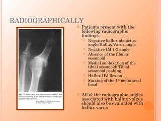
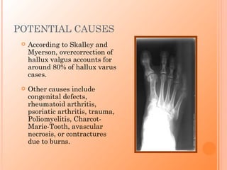
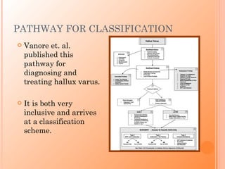
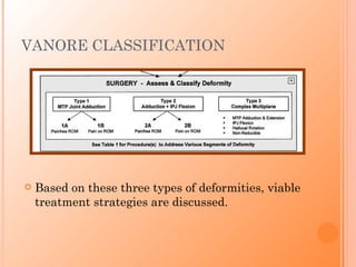
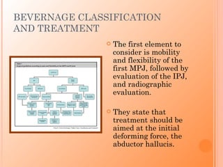
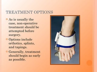
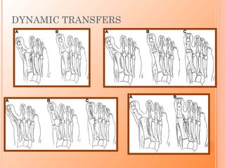
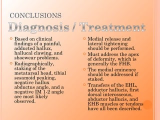
Hallux varus is a deformity of the great toe characterized by three key radiographic findings: a negative hallux abductus angle, negative IM 1-2 angle, and tibial sesamoid peaking. Patients typically present with pain, difficulty with shoes, and decreased push off ability. Treatment begins with orthotics and splinting but often requires surgery involving medial soft tissue release and lateral tightening, with tendon transfers used to correct deformity and maintain correction. Classification systems aim to guide surgical decision making by considering joint flexibility and deforming forces at work.
Deformity: It’s the position of a limb/Joint, from which it cannot be brought back to its normal anatomical position.
Described as abnormalities of :
Length
Angulation
Rotation
Translation
Combination
Deformity: It’s the position of a limb/Joint, from which it cannot be brought back to its normal anatomical position.
Described as abnormalities of :
Length
Angulation
Rotation
Translation
Combination
Hallux rigidus:
A condition characterized by loss of motion of first MTP joint in adults due to degenerative arthritis
second most common condition affecting the big toe after hallux valgus
most common arthritic condition in the foot.
Hoffa's Fracture: Diagnosis, management & New Classification System by BAGARI...Vaibhav Bagaria
Hoffa's Fracture - coronal split fracture of distal femur, its diagnosis, management strategy, a new classification and tips and tricks of management. First described Hoffa, a new classification system by Bagaria et al helps plan the surgery for these tricky fracture. The most crucial step is not to miss these fractures in ER.
Disorders of the Great toe (hallux) are very important as they are very painful, causes many clinical symptoms,and very difficult to treat.The presentation compiled from various important orthopedic textbooks and international journals.
Hallux valgus contiene; definición, etiología, anatomía, características, diagnóstico, tratamento, cirugía, anatomía patológica y sus características. De igual forma, incluye los factores desencadenantes, clasificación roger mann y sus complicaciones.
Bursitis and tendinitis are both common conditions that involve inflammation of the soft tissue around muscles and bones, most often in the shoulder, elbow, wrist, hip, knee, or ankle.
A bursa is a small, fluid-filled sac that acts as a cushion between a bone and other moving parts: muscles, tendons, or skin. Bursae are found throughout the body. Bursitis occurs when a bursa becomes inflamed (redness and increased fluid in the bursa).
A tendon is a flexible band of fibrous tissue that connects muscles to bones. Tendinitis is inflammation of a tendon. Tendons transmit the pull of the muscle to the bone to cause movement. They are found throughout the body, including the hands, wrists, elbows, shoulders, hips, knees, ankles, and feet. Tendons can be small, like those found in the hand, or large, like the Achilles tendon in the heel.
NIAMS
i present this lovely topic at Notional Guard Hospital in Al-Ahsa in the Orthopedic department.
hope you enjoy
Fahad Al Hulaibi
Orthopedic Resident
NGH-A
MANAGEMENT OF ATRIOVENTRICULAR CONDUCTION BLOCK.pdfJim Jacob Roy
Cardiac conduction defects can occur due to various causes.
Atrioventricular conduction blocks ( AV blocks ) are classified into 3 types.
This document describes the acute management of AV block.
Ozempic: Preoperative Management of Patients on GLP-1 Receptor Agonists Saeid Safari
Preoperative Management of Patients on GLP-1 Receptor Agonists like Ozempic and Semiglutide
ASA GUIDELINE
NYSORA Guideline
2 Case Reports of Gastric Ultrasound
- Video recording of this lecture in English language: https://youtu.be/lK81BzxMqdo
- Video recording of this lecture in Arabic language: https://youtu.be/Ve4P0COk9OI
- Link to download the book free: https://nephrotube.blogspot.com/p/nephrotube-nephrology-books.html
- Link to NephroTube website: www.NephroTube.com
- Link to NephroTube social media accounts: https://nephrotube.blogspot.com/p/join-nephrotube-on-social-media.html
New Drug Discovery and Development .....NEHA GUPTA
The "New Drug Discovery and Development" process involves the identification, design, testing, and manufacturing of novel pharmaceutical compounds with the aim of introducing new and improved treatments for various medical conditions. This comprehensive endeavor encompasses various stages, including target identification, preclinical studies, clinical trials, regulatory approval, and post-market surveillance. It involves multidisciplinary collaboration among scientists, researchers, clinicians, regulatory experts, and pharmaceutical companies to bring innovative therapies to market and address unmet medical needs.
New Directions in Targeted Therapeutic Approaches for Older Adults With Mantl...i3 Health
i3 Health is pleased to make the speaker slides from this activity available for use as a non-accredited self-study or teaching resource.
This slide deck presented by Dr. Kami Maddocks, Professor-Clinical in the Division of Hematology and
Associate Division Director for Ambulatory Operations
The Ohio State University Comprehensive Cancer Center, will provide insight into new directions in targeted therapeutic approaches for older adults with mantle cell lymphoma.
STATEMENT OF NEED
Mantle cell lymphoma (MCL) is a rare, aggressive B-cell non-Hodgkin lymphoma (NHL) accounting for 5% to 7% of all lymphomas. Its prognosis ranges from indolent disease that does not require treatment for years to very aggressive disease, which is associated with poor survival (Silkenstedt et al, 2021). Typically, MCL is diagnosed at advanced stage and in older patients who cannot tolerate intensive therapy (NCCN, 2022). Although recent advances have slightly increased remission rates, recurrence and relapse remain very common, leading to a median overall survival between 3 and 6 years (LLS, 2021). Though there are several effective options, progress is still needed towards establishing an accepted frontline approach for MCL (Castellino et al, 2022). Treatment selection and management of MCL are complicated by the heterogeneity of prognosis, advanced age and comorbidities of patients, and lack of an established standard approach for treatment, making it vital that clinicians be familiar with the latest research and advances in this area. In this activity chaired by Michael Wang, MD, Professor in the Department of Lymphoma & Myeloma at MD Anderson Cancer Center, expert faculty will discuss prognostic factors informing treatment, the promising results of recent trials in new therapeutic approaches, and the implications of treatment resistance in therapeutic selection for MCL.
Target Audience
Hematology/oncology fellows, attending faculty, and other health care professionals involved in the treatment of patients with mantle cell lymphoma (MCL).
Learning Objectives
1.) Identify clinical and biological prognostic factors that can guide treatment decision making for older adults with MCL
2.) Evaluate emerging data on targeted therapeutic approaches for treatment-naive and relapsed/refractory MCL and their applicability to older adults
3.) Assess mechanisms of resistance to targeted therapies for MCL and their implications for treatment selection
Knee anatomy and clinical tests 2024.pdfvimalpl1234
This includes all relevant anatomy and clinical tests compiled from standard textbooks, Campbell,netter etc..It is comprehensive and best suited for orthopaedicians and orthopaedic residents.
NVBDCP.pptx Nation vector borne disease control programSapna Thakur
NVBDCP was launched in 2003-2004 . Vector-Borne Disease: Disease that results from an infection transmitted to humans and other animals by blood-feeding arthropods, such as mosquitoes, ticks, and fleas. Examples of vector-borne diseases include Dengue fever, West Nile Virus, Lyme disease, and malaria.
TEST BANK for Operations Management, 14th Edition by William J. Stevenson, Ve...kevinkariuki227
TEST BANK for Operations Management, 14th Edition by William J. Stevenson, Verified Chapters 1 - 19, Complete Newest Version.pdf
3. HISTORY OF HALLUX VARUS
In 1900, Clarke described hallux varus simply,
as being the opposite of hallux valgus.
Hawkins, in 1971, offered the most in depth
paper on the cause, prevention, and correction
of the deformity.
Hawkins’ home: Toledo, OH
4. DIAGNOSIS OF HALLUX VARUS
Diagnosis is based
on both clinical
presentation and
radiographic
evaluation.
Cleveland Clinic Arnold and Sydell Miller Family Pavilion
As described by Boike, the deformity is triplanar, with
supination and hyperextension of the first MPJ and
hyperflexion of the hallucal IPJ.
5. PRESENTATION
Patients present with
hallucal pain, shoewear
difficulty, weakness with
pushoff, metatarso- From: The Institute for Foot and Ankle Reconstruction at Mercy
phalangeal joint instability,
or possiblymetatarsalgia.
Belczyk describes the
presentation as including
deformity, pain, decreased
range of motion, first MPJ
arthrosis, hallucal clawing,
and shoewear problems.
From: John Schuberth, DPM
6. RADIOGRAPHICALLY
Patients present with the
following radiographic
findings:
Negative hallux abductus
angle/Hallux Varus angle
Negative IM 1-2 angle
Absence of the fibular
sesamoid
Medial subluxation of the
tibial sesamoid/ Tibial
sesamoid peaking
Hallux IPJ flexion
Staking of the 1st metatarsal
head
All of the radiographic angles
associated with hallux valgus
should also be evaluated with
hallux varus
7. POTENTIAL CAUSES
According to Skalley and
Myerson, overcorrection of
hallux valgus accounts for
around 80% of hallux varus
cases.
Other causes include
congenital defects,
rheumatoid arthritis,
psoriatic arthritis, trauma,
Poliomyelitis, Charcot-
Marie-Tooth, avascular
necrosis, or contractures
due to burns.
8. PATHOANATOMY, ADAPTED FROM
DONLEY
Cleveland Clinic Main Campus
A brief review of the anatomy shows that four intrinsic
and two extrinsic muscles cross the first MPJ.
The crista, along with the medial and lateral sesamoid
ligaments help provide balance.
9. VARUS AND VALGUS FORCES
The adductor hallucis
exerts a valgus force on
the joint, while the
abductor exerts a varus
force.
Once the toe is rotated
out of a neutral position,
those varus and valgus
forces become deforming
forces.
10. PATHWAY FOR CLASSIFICATION
Vanore et. al.
published this
pathway for
diagnosing and
treating hallux varus.
It is both very
inclusive and arrives
at a classification
scheme.
11. VANORE CLASSIFICATION
Based on these three types of deformities, viable
treatment strategies are discussed.
12. BEVERNAGE CLASSIFICATION
AND TREATMENT
The first element to
consider is mobility
and flexibility of the
first MPJ, followed by
evaluation of the IPJ,
and radiographic
evaluation.
They state that
treatment should be
aimed at the initial
deforming force, the
abductor hallucis.
13. TREATMENT OPTIONS
As is usually the
case, non-operative
treatment should be
attempted before
surgery.
Options include
orthotics, splints,
and tapings.
Generally, treatment
should begin as early
as possible.
14. SURGICAL OPTIONS
The mainstay of
treatment is going to be
surgery, most of the time.
There are numerous
surgical options available
but the principles are all
the same.
Medial soft tissue release
Lateral soft tissue
tightening
Tendon transfer to correct
deformity/maintain
correction
Osseous repair
17. OSSEOUS BUTTRESSING
If the metatarsal head
was too aggressively
resected, a bone graft
should be used to
stabilize the MTP
joint.
Bevernage states that
this will help restore
the intrinsic/extrinsic Fig. 11. Hallux Varus: Classification and Treatment from
muscle balance to
Bernhard Devos Bevernage, MD, Thibaut Leemrijse, MD
(A) AP standing radiograph showing an excessive medial eminence resection,
neutral.
associated with an aggressive lateral release distal to the sesamoids, after failed
hallux valgus surgery. The first MTP joint is mobile, reducible, and painless.
(B) Intraoperative view with nice correction into a physiologic valgus position,
illustrating the combination of an osseous allograft buttress and a static reversed
abductor hallucis tendon transfer.
(C) Postoperative AP standing radiograph at 2-year follow-up.
(D) CT scan illustrating the persistence of the osseous trajectory of the tendinous
transplant as well as the osteo-integration of the allograft buttress with the
metatarsal head.
18. VANORE TREATMENT ALGORITHM
Vanore’s proposed treatment algorithm,
which is fairly similar to Bevernage’s
19. CONCLUSIONS
Based on clinical Medial release and
findings of a painful, lateral tightening
adducted hallux, should be performed.
hallucal clawing, and Must address the apex
shoewear problems. of deformity, which is
Radiographically, generally the FHB.
staking of the The medial eminence
metatarsal head, tibial should be addressed if
sesamoid peaking, staked.
negative hallux
abductus angle, and a
Transfers of the EHL,
negative IM 1-2 angle adductor hallucis, first
are most likely dorsal interosseous,
observed. abductor hallucis, and
EHB muscles or tendons
have all been described.
20. FINALLY,
Cleveland Clinic Las Vegas
A roundtable discussion between experienced
surgeons from the Cleveland Clinic, UPMC,
University of Connecticut School of Medicine, and
Belfast, Ireland revealed startling discrepancies.
I took this picture at the Denver Museum of Nature and Science on my first full day in town. I had been working on a presentation dealing with how evolution has shaped the foot, so I thought this would fit well in it. When Dr. Farrett gave me hallux varus, I knew it would make an appearance. In our ancestors and primate relatives, hallux varus is the norm. An adducted hallux, in relation to the midline of the body, allows for climbing trees. The more abducted our hallux, the better propulsion humans were able to obtain.
Clarke wrote in the Lancet, stating that hallux varus was largely a congenital problem. He had never seen it acquired, though he heard of it occurring with severe cases of genu valgum and thought it could occur with trauma or paralysis. It wasn’t until 1928 that McBride first described his “conservative operation for bunions,” which called for a transfer of the adductor hallucis to the distal metatarsal head, a fibular sesamoidectomy, and a medial eminence resection. This procedure has been described as having hallux varus complications up to 13% of the time. Hawkins wrote for Clinical Orthopedics and Related Research and astutely stated that “Once a post-operative hallux varus deformity occurs, the disability becomes greater than that of the original problem. He even described an abductor hallucis tendon transfer for treatment. He would offer the first comprehensive look at causes and correction of hallux varus.
Allan Boike is a former chair of the Cleveland Clinic Foundation residency and still a big deal around the area. He wrote a chapter on hallux varus for a forefoot surgery book by Dr. Hetherington, our dean of academics. Perhaps more notably, he recently discussed Steven Tyler’s toes for an MSNBC article.
Schuberth states that hallux varus requires two things: an incongruous first metatarsophalangeal joint and a negative hallux abductus angle. Other deformities are possible, but those two need to be present for it to be considered hallux varus. What patients will complain of, however, is hallucal pain, joint deformity, shoe gear problems, weak pushoff, MPJ instability, or possibly metatarsalgia.
I grabbed this radiograph from George Wallace’s chapter in Dr. Hetherington’s book. It shows several of the findings that you may expect in a patient with hallux varus. They can include a negative Hallux abductus angle, also described as the hallux varus angle, negative IM 1-2 angle, fibular sesamoid absence, tibial sesamoid peaking, flexion of the hallux IPJ, and staking or spiking of the first metatarsal head. The radiographic findings of a bunion that should be evaluated include the hallux abductus angle, hallux interphalangeus angle, tibial sesamoid position, metatarsal parabola angle, metatarsal elevation/Seiberg’s index, intermetatarsal angle 1-2, first metatarsal protrusion, proximal articular set angle, distal articular set angle,metatarsus adductus angle, and joint condition.
Hallux varus is a detrimental condition caused, usually, by surgical correction of hallux valgus or a bunion deformity. This accounts for roughly 80% of all Hallux Varus cases. Post operative complications of hallux valgus surgery that most often lead to hallux varus include fibular sesamoid removal, excessive lateral soft tissue release, excessive tightening of the medial capsule, excessive medial eminence removal, overcorrection of the intermetatarsal 1-2 angle, or excessive bandaging.
Donley, in an article for foot and ankle international, gives a current topic review of hallux varus. He reminds us that the first metatarsophalangeal joint is crossed by four intrinsic muscles, the extensor hallucis brevis, flexor hallucis brevis, abductor hallucis, and adductor hallucis, and two extrinsic muscles, the flexor hallucis longus and extensor hallucis longus. The crista, along with the medial and lateral sesamoid ligaments help provide balance between the pull of the two heads of the flexor hallucis brevis.
In the case of hallux varus, the abductor hallucis has the advantage. You can see that once the axis of motion is internally rotated, both the flexor hallucis brevis and abductor hallucis muscles gain more leverage. Usually, this is due to an overaggressive lateral release. Even Hawkins knew in the 1970s that release of both the adductor hallucis and the lateral head of the flexor hallucis brevis would lead to a varus deformity, though either could be sacrificed individually.
This flow chart came from a panel put together to develop treatment guidelines for hallux varus. Other big names on the panel include Christensen, Schuberth, Weil, and Mendocino. They take into account the most common histories, findings, onset times, and etiologies in arriving at a classification.
This is the last section of the previous flow chart. Basically, Vanore establishes three categories of hallux varus. Type one is the easiest to treat, with just metatarsophalangeal joint adduction. Type two has the addition of hallux malleus and type three is more complex, with rotation of the hallux and is non-reducible. Based on these three classifications, a separate chart would lead you to treatment options. I’ll discuss those findings in a few minutes.
This flowchart may be too small to read but it basically describes a treatment algorithm. Bevernage and Leemrijse first take into consideration the flexibility or mobility of the MPJ, followed by IPJ motion. From there, it goes through things like arthrosis of the joints involved, osseous positions, and ligament integrity. These authors state that the abductor hallucis muscle is the initial deforming force. Many other sources state that the flexor hallucis brevis muscle is the apex of the deformity. I think the take home message is that both of these muscles have an adductory effect when unchallenged by lateral soft tissues.
Bevernage and Leemrijse state that conservative treatment must begin within a few weeks of hallux valgus surgery. Aggressive taping in a valgus position, if a varus problem is immediately recognized, can be corrective for the problem. The taping will need to be maintained for three months, but this can correct the varus non-surgically. This dynasplint was used in a study by John, which stated that low-torque stretching for prolonged durations using the splint was effective in reducing both hallux varus and valgus without requiring surgery. There were no long term results, however. The longest patient follow up visit was around 6 months.
One or more of these techniques needs to be used to correct the problem. Bevernage and Leemrijse offer multiple treatment options in their 2009 article from the Foot and Ankle Clinics. They describe several options, which fall into dynamic or static treatments, all of which are performed with a medial soft tissue release.
So, this slide gets a little busy. Dynamic procedures seek to restore muscle balance by redirecting tendons and their direction of pull. These include adductor tendon transfer to the lateral sesamoid (Top left, B), abductor tendon transfer through the lateral aspect of the proximal phalanx (top right, C), EHL transfer to the lateral base of the proximal phalanx (bottom left, C), and transfer of the first dorsal interosseous muscle to the base of the proximal phalanx (bottom right, B).
Static procedures use muscle and tendon to simply hold a position. These include the reversed abductor hallucis tendon transfer (top left, C), which releases the tendon from the muscle and routes it through the first metatarsal and proximal phalanx, the split EHL tendon transfer (middle right, B), which allows motion to be conserved at the IPJ while routing the tendon, split distally, to be rerouted through the first metatarsal, and an EHB tenodesis (bottom left, C), which uses the tendon in a similar fashion as the abductor transfer. Those with Ehlers-Danlos or other ligamentous laxity conditions can require ligamentoplasties in addition to the other described treatments, which could be performed with autologous fascia lata or Ligapro suture.
Osseous buttressing is the treatment of choice if there is obvious overresection of the first metatarsal head. Again, staking of the head and tibial sesamoid peaking will be noted on the radiograph. This buttress will reapproximate a metatarsal head and help stabilize the muscles of the first MPJ. The picture shows a patient who had osseous buttressing to recreate a metatarsal head along with the static abductor hallucis tendon transfer, which was nearly described by Hawkins in 1971. The combination of procedures performed is likely to produce satisfactory results.
Again, type 1 deformities are simply an adducted hallux. Type 2 include those deformities with an adducted hallux and IPJ flexion. Type 3 are complex deformities with adduction and extension of the MPJ, IPJ flexion, hallucal rotation, and is non-reducible. Subtype A lacks arthritic changes while subtype B has some degree of arthritic changes. As expected, if the joint is not in good condition, the primary procedures are joint destructive type procedures: arthrtoplasty, implants, or arthrodesis. You can also see that for type one and type 2 A classifications, a medial release and lateral tissue repair procedures are ideal. It is only when you get to more advanced deformities that tendon transfers become the modality of choice. You can also see the connection between EHL transfer and interphalangeal joint arthrodesis. If the fusion is not performed, a flail toe deformity can occur.
Published in a 2011 issue of Foot and Ankle Specialist, these four surgeons offered very different answers for questions ranging from simply “What is the single most intraoperative maneuver that can lead to hallux varus?” to “If hallux varus is discovered within the first 3 months after hallux valgus surgery, what is the best method of correction?” For the first question, their answers include fibular sesamoid excsion, excessive medial eminence resection, excessive medial capsulorraphy, and excessive intermetatarsal angle reduction. Their treatment options are just as varied, which makes sense. If they don’t agree on the cause, they are unlikely to agree on how to treat it. The editor of the issue, John Schuberth, notes that better studies are needed on the topic of hallux varus, to ensure that there is a better understanding of cause and treatment of this condition.