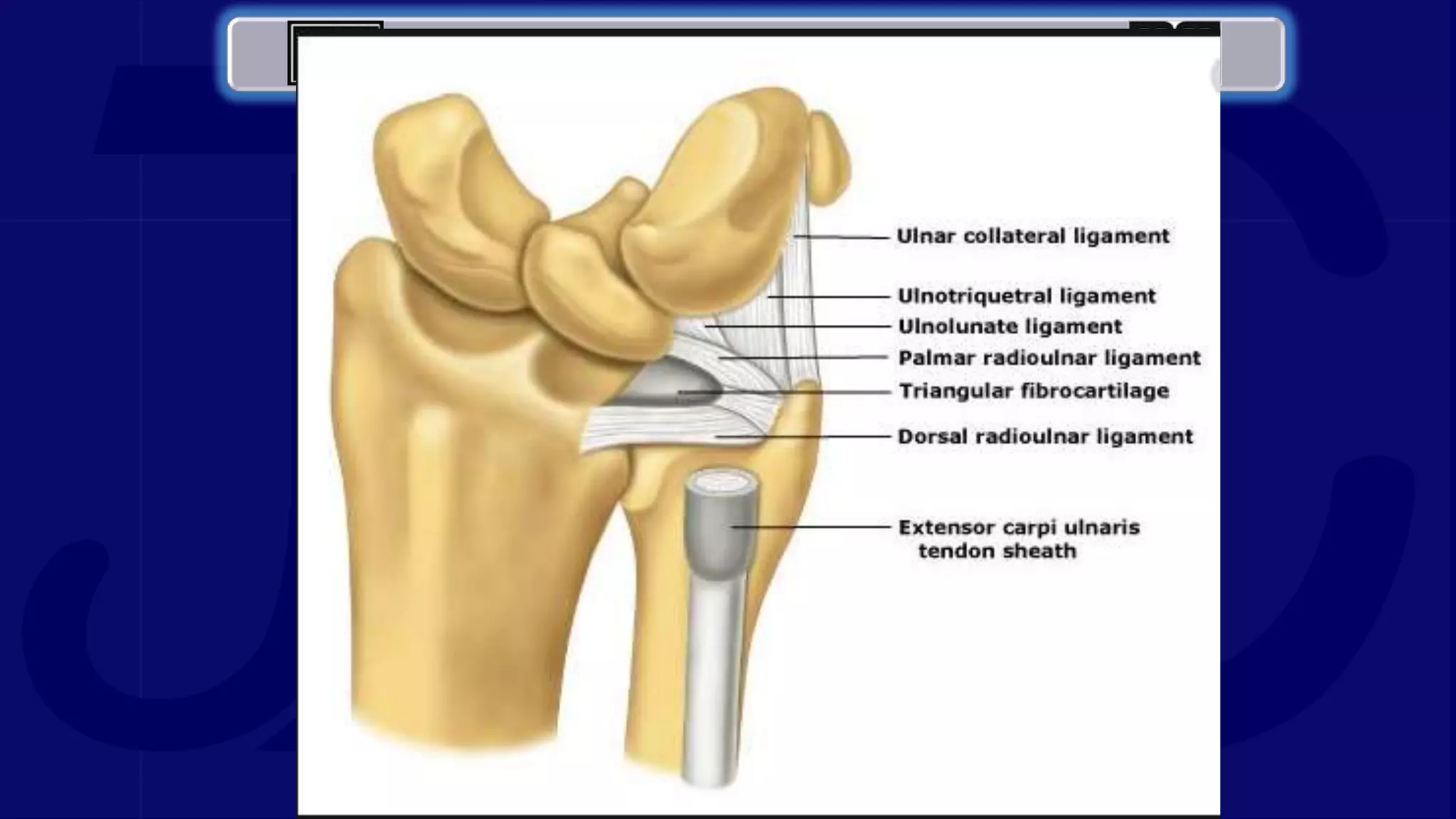
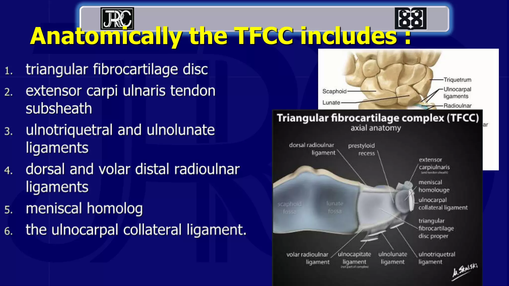
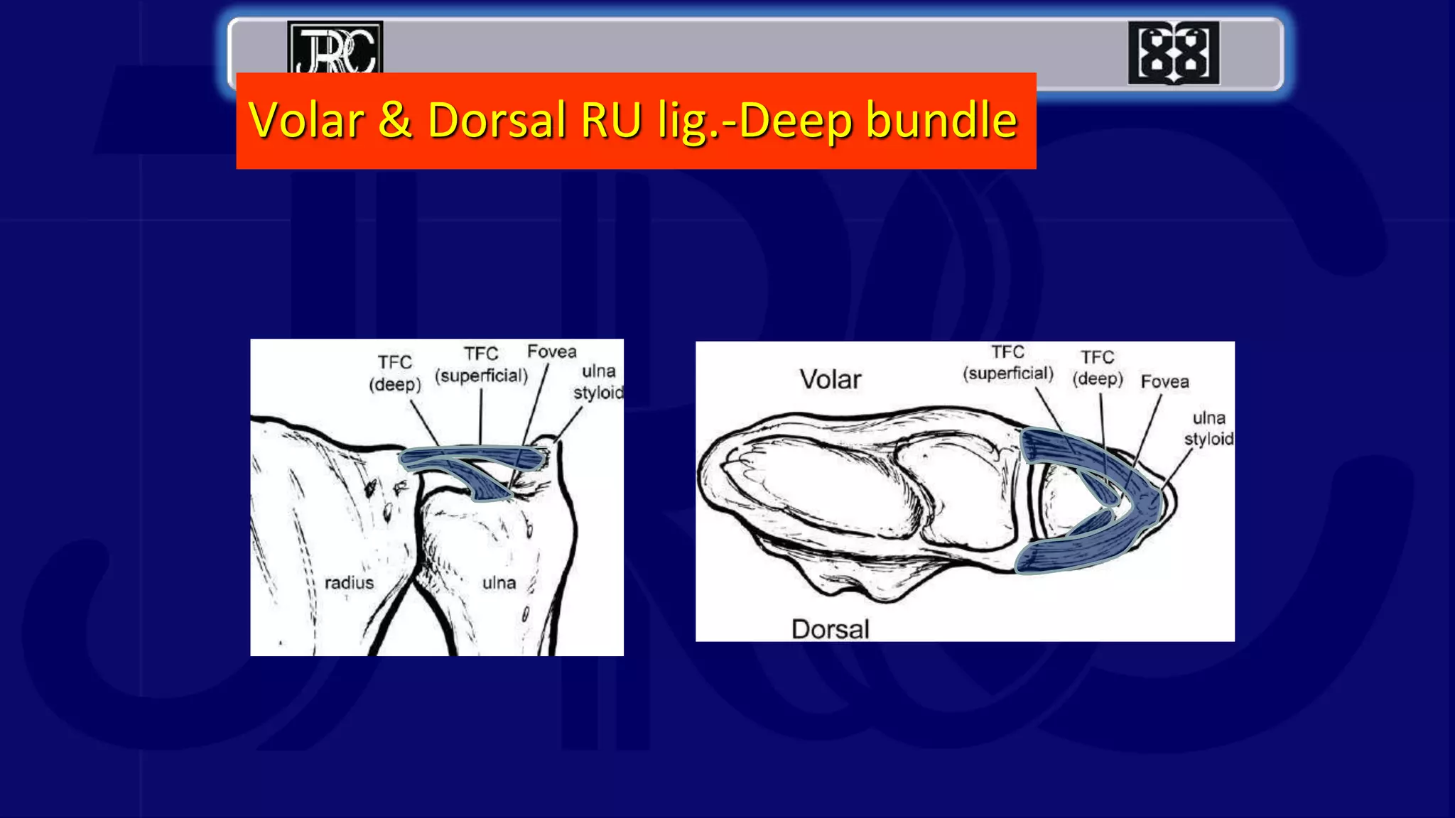
Hamidreza Zafari
Orthopedic Surgery Resident at Tehran University of Medical Scince
Imam khomeyni Hospital Center
TFCC Injury
Anatomically the TFCC includes :
Volar & Dorsal RU lig.-Deep bundle
 The triangular fibrocartilage complex (TFCC) is a load-
bearing structure between the lunate, triquetrum, and ulnar
head.
 The function of the TFCC is to act as a stabilizer for the
ulnar aspect of the wrist.

 Patients with TFCC injury will present with ulnar-sided wrist
pain that may present with clicking or point tenderness
between the pisiform and the ulnar head
 TFCC injury often occurs when there is a load compressed
on the TFCC while the wrist is in ulnar deviation.
 TFCC injury is also associated with positive ulnar variance;
this is when the articular surface of the ulna is more distal
than the articular surface of the radius.
 Positive ulnar variance is often due to prior surgery or prior
fracture.
 One study of 85 patients
with distal radius fractures
treated surgically found
that 53% of the patients
also had a TFCC lesion
diagnosed by arthroscopy.
History and Physical Exam:

 Several physical exam tests can suggest the
diagnosis of TFCC injury
 TFCC compression test:
forearm in the neutral
position with ulnar deviation
reproduces symptoms
 TFCC stress test:
applying a force across the
ulna with the wrist in ulnar
deviation reproduces
symptoms
 Press test:
Patient lifts themselves out of a
chair using the wrists in an
extended position. Pain indicates a
positive test.
 Supination test:
Patient grabs the underside of a table
with the forearms supinated; this
causes a load on the TFCC and dorsal
impingement, which will cause pain if
there is a peripheral, dorsal tear.
 Piano key test:
Place both hands on an exam
table and press the palms on the
table.
If the distal ulna is prominent on
the affected side, this suggests
distal radioulnar joint instability
which can have associations with
TFCC injury.

Evaluation :
 Initial workup typically starts with a radiograph to evaluate for
fracture and assess for ulnar variance.
 The next step will often be to get an MRI with or without an
arthrogram.
 If MR is unavailable or contraindicated, a CT can be an option,
although the sensitivity is less than MRI.

 Arthroscopy is the most
accurate means by which
to diagnose TFCC injury.
Treatment / Management
 Initial treatment includes rest, physical therapy, and
corticosteroid injections.
 Six months of conservative treatment is reasonable if
there is not DRUJ instability.
 There is limited evidence to support the use of bracing
as a treatment option for TFCC tears.
Palmar classification:
 1.traumatic injury
 2.degenerative injury
Palmer Classification of TFCC Lesions
•I Traumatic injury
– A: central perforation
– B: ulnar avulsion
– C: distal avulsion
– D: radial avulsion
•II Degenerative injury
1Palmer AK. Triangular fibrocartilage complex
lesions: a classification. JHS Am 1989
 2A: Degenerative changes of the triangular fibrocartilage
disc without evidence of perforation.
 2B: Grade 2A with the additional presence of
chondromalacia of the hyaline cartilage on the articular
surface.
 2C: Full thickness perforation of the triangular fibrocartilage
disc.
 2D: Any of the features in 2A through 2C plus lunotriquetral
ligament tear.
 2E: Grade 2D with the additional presence of ulnocarpal
arthritis
 Ulnar styloid impaction syndrome
involves repetitive friction between an
excessively long ulnar styloid and the
carpus, resulting in chondromalacia,
synovitis, and pain.
 The arthroscopic diagnosis, evaluation,
and management of this syndrome are
not well characterized.

 operative ulnar styloid excision, ultimately resolving chronic wrist
pain symptomology
 Type 2A, 2B, and 2C lesions can have conservative
therapy.
 If conservative management fails, a reasonable next step is
the Wafer procedure, which is resection of the distal aspect
of the ulnar head.
Arthroscopic Wafer
procedure
• Preferred when modest shortening needed
 Type 2D lesions can be
treated via ulnar shaft-
shortening with osteotomy
 Type 2E lesions can also be
treated by resection of the
ulnar head.
 Treating athletes can vary from treatment for
non-athletes.
A high school athlete who will not compete
beyond high school should begin with four weeks
of rest, ice, and anti-inflammatories.
 For elite athletes, if there is no distal radioulnar
joint instability, one week of rest with splinting
and re-examination after one week is reasonable.
 If there is a tear of the TFCC with instability of the distal
radioulnar joint, this is potentially career-threatening.
 If non-surgical treatment is elected, this could include long
arm immobilization for 3 weeks followed by short arm
immobilization for 3 weeks with a gradual return to play.
 Corticosteroid injections are
also an option, especially in
elite athletes who elect to
delay surgical intervention
in an attempt to finish the
season
Prognose :
 Prognosis for TFCC injury is generally favorable.
 Surgical management also has a good prognosis in children
as it has been found to have successful outcomes in
pediatric and adolescent high-level athletes who desire to
return to sport.
 They also found that degenerative tears and higher
positive ulnar variance generally had poorer outcomes
 Some poor prognostic factors include
negative DRUJ stress test
female gender
longer symptom duration.
Complication :
 Complications are mostly related to surgical
management.
 Post-operative complications include, but are not
limited to, infections, hypertrophic scarring, tendon
injury, nerve injury, reflex sympathetic dystrophy,
and joint stiffness with a limited range of motion
Postoperative and Rehabilitation Care :
 Recovery after surgery varies, but typically four to six
weeks should be expected for arthroscopy and
approximately three months for an open approach.
 Patients will undergo physical therapy after the procedure.
 If there is an osteotomy performed to shorten the ulna, patients
are immobilized for approximately 4 weeks before they begin
range of motion exercises.
 When grip strength is 80% of the expected normal, the athlete
can begin strengthening exercises and gradually return to play.
 If the surgery is performed on the throwing arm, an elite
athlete may be able to return to play in 8-12 weeks.
 If the surgical intervention is on the non-throwing arm, return
to play in 6-8 weeks is possible

TFCC (Triangular fibro cartilage complex) Injury

  • 1.
    Hamidreza Zafari Orthopedic SurgeryResident at Tehran University of Medical Scince Imam khomeyni Hospital Center
  • 2.
  • 4.
  • 5.
    Volar & DorsalRU lig.-Deep bundle
  • 6.
     The triangularfibrocartilage complex (TFCC) is a load- bearing structure between the lunate, triquetrum, and ulnar head.  The function of the TFCC is to act as a stabilizer for the ulnar aspect of the wrist.   Patients with TFCC injury will present with ulnar-sided wrist pain that may present with clicking or point tenderness between the pisiform and the ulnar head
  • 7.
     TFCC injuryoften occurs when there is a load compressed on the TFCC while the wrist is in ulnar deviation.  TFCC injury is also associated with positive ulnar variance; this is when the articular surface of the ulna is more distal than the articular surface of the radius.  Positive ulnar variance is often due to prior surgery or prior fracture.
  • 8.
     One studyof 85 patients with distal radius fractures treated surgically found that 53% of the patients also had a TFCC lesion diagnosed by arthroscopy.
  • 9.
  • 10.
  • 11.
     Several physicalexam tests can suggest the diagnosis of TFCC injury
  • 12.
     TFCC compressiontest: forearm in the neutral position with ulnar deviation reproduces symptoms  TFCC stress test: applying a force across the ulna with the wrist in ulnar deviation reproduces symptoms
  • 13.
     Press test: Patientlifts themselves out of a chair using the wrists in an extended position. Pain indicates a positive test.  Supination test: Patient grabs the underside of a table with the forearms supinated; this causes a load on the TFCC and dorsal impingement, which will cause pain if there is a peripheral, dorsal tear.
  • 14.
     Piano keytest: Place both hands on an exam table and press the palms on the table. If the distal ulna is prominent on the affected side, this suggests distal radioulnar joint instability which can have associations with TFCC injury. 
  • 15.
    Evaluation :  Initialworkup typically starts with a radiograph to evaluate for fracture and assess for ulnar variance.  The next step will often be to get an MRI with or without an arthrogram.  If MR is unavailable or contraindicated, a CT can be an option, although the sensitivity is less than MRI. 
  • 16.
     Arthroscopy isthe most accurate means by which to diagnose TFCC injury.
  • 17.
    Treatment / Management Initial treatment includes rest, physical therapy, and corticosteroid injections.  Six months of conservative treatment is reasonable if there is not DRUJ instability.  There is limited evidence to support the use of bracing as a treatment option for TFCC tears.
  • 18.
    Palmar classification:  1.traumaticinjury  2.degenerative injury
  • 19.
    Palmer Classification ofTFCC Lesions •I Traumatic injury – A: central perforation – B: ulnar avulsion – C: distal avulsion – D: radial avulsion •II Degenerative injury 1Palmer AK. Triangular fibrocartilage complex lesions: a classification. JHS Am 1989
  • 22.
     2A: Degenerativechanges of the triangular fibrocartilage disc without evidence of perforation.  2B: Grade 2A with the additional presence of chondromalacia of the hyaline cartilage on the articular surface.  2C: Full thickness perforation of the triangular fibrocartilage disc.  2D: Any of the features in 2A through 2C plus lunotriquetral ligament tear.  2E: Grade 2D with the additional presence of ulnocarpal arthritis
  • 23.
     Ulnar styloidimpaction syndrome involves repetitive friction between an excessively long ulnar styloid and the carpus, resulting in chondromalacia, synovitis, and pain.  The arthroscopic diagnosis, evaluation, and management of this syndrome are not well characterized.
  • 24.
      operative ulnarstyloid excision, ultimately resolving chronic wrist pain symptomology
  • 25.
     Type 2A,2B, and 2C lesions can have conservative therapy.  If conservative management fails, a reasonable next step is the Wafer procedure, which is resection of the distal aspect of the ulnar head.
  • 26.
    Arthroscopic Wafer procedure • Preferredwhen modest shortening needed
  • 27.
     Type 2Dlesions can be treated via ulnar shaft- shortening with osteotomy  Type 2E lesions can also be treated by resection of the ulnar head.
  • 28.
     Treating athletescan vary from treatment for non-athletes. A high school athlete who will not compete beyond high school should begin with four weeks of rest, ice, and anti-inflammatories.  For elite athletes, if there is no distal radioulnar joint instability, one week of rest with splinting and re-examination after one week is reasonable.
  • 29.
     If thereis a tear of the TFCC with instability of the distal radioulnar joint, this is potentially career-threatening.  If non-surgical treatment is elected, this could include long arm immobilization for 3 weeks followed by short arm immobilization for 3 weeks with a gradual return to play.
  • 30.
     Corticosteroid injectionsare also an option, especially in elite athletes who elect to delay surgical intervention in an attempt to finish the season
  • 31.
    Prognose :  Prognosisfor TFCC injury is generally favorable.  Surgical management also has a good prognosis in children as it has been found to have successful outcomes in pediatric and adolescent high-level athletes who desire to return to sport.
  • 32.
     They alsofound that degenerative tears and higher positive ulnar variance generally had poorer outcomes  Some poor prognostic factors include negative DRUJ stress test female gender longer symptom duration.
  • 33.
    Complication :  Complicationsare mostly related to surgical management.  Post-operative complications include, but are not limited to, infections, hypertrophic scarring, tendon injury, nerve injury, reflex sympathetic dystrophy, and joint stiffness with a limited range of motion
  • 34.
    Postoperative and RehabilitationCare :  Recovery after surgery varies, but typically four to six weeks should be expected for arthroscopy and approximately three months for an open approach.  Patients will undergo physical therapy after the procedure.
  • 35.
     If thereis an osteotomy performed to shorten the ulna, patients are immobilized for approximately 4 weeks before they begin range of motion exercises.  When grip strength is 80% of the expected normal, the athlete can begin strengthening exercises and gradually return to play.
  • 36.
     If thesurgery is performed on the throwing arm, an elite athlete may be able to return to play in 8-12 weeks.  If the surgical intervention is on the non-throwing arm, return to play in 6-8 weeks is possible