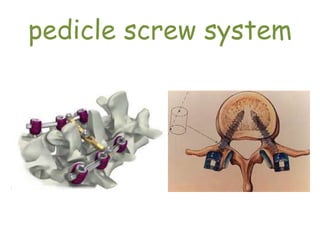
pedicle screw system
Introduction
• Pedicle screw fixation is most commonly used
technique in spinal stabilization
• screws traverse all three columns of the
vertebrae, so they can rigidly stabilize both
the ventral and dorsal aspects of the spine
Introduction
• Pedicle screw fixation does not require intact
dorsal elements. Thus, it can be used after a
laminectomy or traumatic disruption of
laminae, spinous processes and/or facets
Indications
1.Spinal instability:
post-laminectomy spondylolisthesis,
painful pseudoarthrosis
2. Potential instability:
spinal stenosis,
degenerative scoliosis
Indications
3. Unstable fractures.
4. Augmenting anterior strut grafting:
tumor,
infection
5. After correction of deformities
Contraindications
1. Recent infection.
2. Laminectomies that will not cause
instability
3. Fusions which are normally successful
without fixation.
Disadvantages
1. It requires extensive tissue dissection to
expose the entry points.
2. pedicle screw insertion can result in dural or
neural injury.
3. Postoperative imaging studies (especially
MRI) are, in part, obscured by the implants.
Disadvantages
4. Rigid fixation can accelerate adjacent
motion segment degeneration.
5. Steep learning curve.
6. Costly procedures.
Pedicle Anatomy
• Consists of a strong shell of cortical bone and
a core of cancellous bone
• Pedicle dimensions and angles change
progressively from the upper thoracic spine
distally
• Pedicles are widest at L5 and narrowest at T5
in the horizontal plane
Pedicle Anatomy
• The widest pedicles in the sagittal plane are at
T11 and the narrowest are at T1
• In the sagittal plane, the pedicles angle
caudad at L5 and cephalad at L3-T1
Pedicle Anatomy
T3
cc
c
T8 L4
d
dd
e
e
e
A B C
Pedicle Anatomy
FIGURE:
Pedicle dimensions of T3 (A), T8 (B), and L4 (C)
vertebrae. Vertical diameter (c) increases from
0.7 to 1.5 cm, horizontal diameter (d) increases
from 0.7 to 1.6 cm with minimum of 0.5 cm in T5.
Direction is almost sagittal from T4 to L4. Angle
(e) seldom extends beyond 10 degrees. More
proximally, direction is more oblique: T1 = 36
degrees, T2 = 34 degrees, T3 = 23 degrees. L5 is
oblique (30 degrees) but is large and easy to drill.
Pedicle Screw Entry Sites
• We use three techniques for localization of the
pedicle:
1) the intersection technique,
2) the pars interarticularis technique, and
3) the mammillary process technique
Pedicle Screw Entry Sites
• The intersection technique is perhaps the
most commonly used method of localizing the
pedicle
• Pedicle entrance point in thoracic spine at
intersection of lines drawn through middle of
inferior articular facets and middle of insertion
of transverse processes (1 mm below facet
joint).
Pedicle Screw Trajectory
(In practice)
1. Preoperative planning using plain radiographs
and CT scan is important in deciding the bone
quality, pedicle transverse diameter and screw
trajectory.
2. Sagittal pedicle angle increases in the thoracic
spine from an average of 0 degs at T1 to 10 degs
at T8 and then decreases to 0 degs at T12.
Pedicle Screw Trajectory
(In practice)
3. Usually the L4 sagittal pedicle angle is 0
degs and subsequent rostral and caudal levels
are associated with progressively greater
sagittal angles.
4. Lordotic curve of the lumbar spine produces
a rostral angulation for upper lumbar screws.
Pedicle Screw Trajectory
(In practice)
5. L5 pedicle screw is 5 degs to 10 degs
caudally inclined.
6. Coronal plane angulation (how medial?) at
T1 is 10 degs to 15 degs and at T12 is 5 degs.
7. At L1 the medial angulation of 5 degs to 10
degs is satisfactory.
Pedicle Screw Trajectory
(In practice)
* a wider angle in the coronal plane is
necessary to avoid lateral penetration of the
pedicle in the lower lumbar spine.
* the coronal plane angle increases
approximately 5 degs per level from L1 to the
sacrum
Checking trajectory by K- wire
Checking facet joint & intersection
point
intersection point
intersection point
intersection point
Screw insertion technique
After screw insertion
Decompression
Application of Rod
After application of Rod
Pedicle screw by professor shah alam

Pedicle screw by professor shah alam

  • 1.
  • 2.
    Introduction • Pedicle screwfixation is most commonly used technique in spinal stabilization • screws traverse all three columns of the vertebrae, so they can rigidly stabilize both the ventral and dorsal aspects of the spine
  • 3.
    Introduction • Pedicle screwfixation does not require intact dorsal elements. Thus, it can be used after a laminectomy or traumatic disruption of laminae, spinous processes and/or facets
  • 4.
    Indications 1.Spinal instability: post-laminectomy spondylolisthesis, painfulpseudoarthrosis 2. Potential instability: spinal stenosis, degenerative scoliosis
  • 5.
    Indications 3. Unstable fractures. 4.Augmenting anterior strut grafting: tumor, infection 5. After correction of deformities
  • 6.
    Contraindications 1. Recent infection. 2.Laminectomies that will not cause instability 3. Fusions which are normally successful without fixation.
  • 7.
    Disadvantages 1. It requiresextensive tissue dissection to expose the entry points. 2. pedicle screw insertion can result in dural or neural injury. 3. Postoperative imaging studies (especially MRI) are, in part, obscured by the implants.
  • 8.
    Disadvantages 4. Rigid fixationcan accelerate adjacent motion segment degeneration. 5. Steep learning curve. 6. Costly procedures.
  • 9.
    Pedicle Anatomy • Consistsof a strong shell of cortical bone and a core of cancellous bone • Pedicle dimensions and angles change progressively from the upper thoracic spine distally • Pedicles are widest at L5 and narrowest at T5 in the horizontal plane
  • 10.
    Pedicle Anatomy • Thewidest pedicles in the sagittal plane are at T11 and the narrowest are at T1 • In the sagittal plane, the pedicles angle caudad at L5 and cephalad at L3-T1
  • 11.
  • 12.
    Pedicle Anatomy FIGURE: Pedicle dimensionsof T3 (A), T8 (B), and L4 (C) vertebrae. Vertical diameter (c) increases from 0.7 to 1.5 cm, horizontal diameter (d) increases from 0.7 to 1.6 cm with minimum of 0.5 cm in T5. Direction is almost sagittal from T4 to L4. Angle (e) seldom extends beyond 10 degrees. More proximally, direction is more oblique: T1 = 36 degrees, T2 = 34 degrees, T3 = 23 degrees. L5 is oblique (30 degrees) but is large and easy to drill.
  • 13.
    Pedicle Screw EntrySites • We use three techniques for localization of the pedicle: 1) the intersection technique, 2) the pars interarticularis technique, and 3) the mammillary process technique
  • 14.
    Pedicle Screw EntrySites • The intersection technique is perhaps the most commonly used method of localizing the pedicle • Pedicle entrance point in thoracic spine at intersection of lines drawn through middle of inferior articular facets and middle of insertion of transverse processes (1 mm below facet joint).
  • 15.
    Pedicle Screw Trajectory (Inpractice) 1. Preoperative planning using plain radiographs and CT scan is important in deciding the bone quality, pedicle transverse diameter and screw trajectory. 2. Sagittal pedicle angle increases in the thoracic spine from an average of 0 degs at T1 to 10 degs at T8 and then decreases to 0 degs at T12.
  • 16.
    Pedicle Screw Trajectory (Inpractice) 3. Usually the L4 sagittal pedicle angle is 0 degs and subsequent rostral and caudal levels are associated with progressively greater sagittal angles. 4. Lordotic curve of the lumbar spine produces a rostral angulation for upper lumbar screws.
  • 17.
    Pedicle Screw Trajectory (Inpractice) 5. L5 pedicle screw is 5 degs to 10 degs caudally inclined. 6. Coronal plane angulation (how medial?) at T1 is 10 degs to 15 degs and at T12 is 5 degs. 7. At L1 the medial angulation of 5 degs to 10 degs is satisfactory.
  • 18.
    Pedicle Screw Trajectory (Inpractice) * a wider angle in the coronal plane is necessary to avoid lateral penetration of the pedicle in the lower lumbar spine. * the coronal plane angle increases approximately 5 degs per level from L1 to the sacrum
  • 19.
  • 20.
    Checking facet joint& intersection point
  • 21.
  • 22.
  • 23.
  • 24.
  • 25.
  • 26.
  • 27.
  • 28.