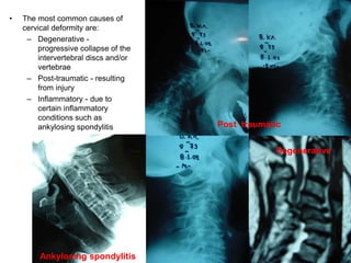
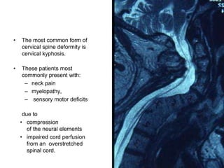
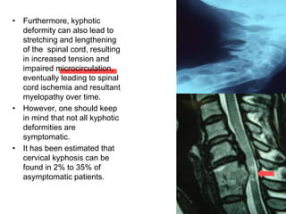
Cervical Spine Deformity
George Sapkas
Professor of Orthopaedics
• Cervical spine deformities can
have a significant negative
impact on the quality of life by
causing :
– pain,
– myelopathy,
– radiculopathy,
– sensory motor deficits,
– inability to maintain
horizontal gaze
in severe cases.
• Deformity of the cervical spine
typically results in an abnormal
position of the head relative to the
chest and shoulders.
• While this is usually cosmetically
bothersome to a person, cervical
deformity also can result in :
– difficulty swallowing
– breathing,
– can severely disrupt the
person’s ability to perform
normal tasks such as:
• driving,
• eating
• and reading.
• The most common causes of
cervical deformity are:
– Degenerative -
progressive collapse of the
intervertebral discs and/or
vertebrae
– Post-traumatic - resulting
from injury
– Inflammatory - due to
certain inflammatory
conditions such as
ankylosing spondylitis
Ankylosing spondylitis
Post traumatic
Degenerative
• The most common form of
cervical spine deformity is
cervical kyphosis.
• These patients most
commonly present with:
– neck pain
– myelopathy,
– sensory motor deficits
due to
• compression
of the neural elements
• impaired cord perfusion
from an overstretched
spinal cord.
• Cervical deformities
are categorized in two
primary ways:
1. Fixed — meaning that
the deformity is rigid
regardless of patient
position
2. Reducible — meaning
that the deformity is
flexible.
• Depending on whether it
is fixed or reducible, the
deformity may require
surgery from :
– the front of the neck,
– the back of the neck,
– sometimes multiple
stages from both the
front and back.
• The aim of this presentation
is to provide an overview
of cervical spine deformity
including
– the biomechanics,
– radiographic parameters,
– classification
– surgical procedures
Biomechanics
of
the Cervical Spine
Lee A. Tan, et al, Cervical Spine Deformity part 1-2-3, Neurosurgery 2017
• The cervical spine is a weight-
bearing mechanical structure with
6 degrees of freedom of
movement.
• The principle motions of the
cervical spine include:
– flexion/extension,
– axial rotation
– lateral bending,
– along with a small amount of
coupled anterior/posterior
translational movements
along the Cartesian
coordinates
• The cervical spine is able to
move within the neutral zone with
relatively little force, therefore
requires very little energy
expenditure from the paraspinal
muscles.
An illustration demonstrating the 6 degrees
of freedom in the cervical spine.
• Additional movement beyond
the neutral zone, requires
more effort to overcome the
elastic force from the soft
tissues;
therefore, this zone is called
the elastic zone.
• Adding the movement
realized in both
the neural and elastic zones
provides the total range of
motion (ROM) at a given
segment.
• An abnormal increase in
neutral zone or ROM may
indicate ligamentous injury or
spinal instability.
• The global physiological ROM
in the cervical spine is
approximately :
– 90◦ of flexion,
– 70◦ of extension,
– 20◦ to 45◦ of lateral
bending,
– up to 90◦ of rotation on
each side.
• When cervical kyphotic
deformity is present, the
head Center of Mass
moves anteriorly and the
movement arm increases
relative to the IAR, thus
creating a larger bending
moment.
• The resultant larger
bending moment requires
greater paraspinal muscle
contraction to keep the
head erect, which in turn
can cause muscle fatigue
and pain.
• In addition, kyphotic
cervical deformity
shifts the axial load
anteriorly, thus can
potentially accelerate
cervical disc
degeneration.
• Decreased disc height
from degenerative
changes can cause
more cervical
kyphosis, thus
creating the notion
“kyphosis begets
kyphosis.”
• Furthermore, kyphotic
deformity can also lead to
stretching and lengthening
of the spinal cord, resulting
in increased tension and
impaired microcirculation,
eventually leading to spinal
cord ischemia and resultant
myelopathy over time.
• However, one should keep
in mind that not all kyphotic
deformities are
symptomatic.
• It has been estimated that
cervical kyphosis can be
found in 2% to 35% of
asymptomatic patients.
• The fundamental functions of
the cervical spine include:
– transmitting axial load from
the cranium,
– maintaining horizontal
gaze,
– allowing normal head and
neck movement,
– protecting important
neurovascular structures
such as
• spinal cord,
• nerve roots,
• vertebral arteries
• If the kyphotic deformity is severe
:
– chin-on-chest deformity,
– dropped head syndrome,
– etc.,
• patients can have
significant difficulty with:
– swallowing
– maintaining horizontal
gaze.
Radiographic Parameters
• There are several radiographic
parameters commonly used to
assess the cervical spine,
including:
– cervical lordosis (CL),
– C2- 7 sagittal vertical axis
(C2-7 SVA),
– chin-brow vertical angle
(CBVA),
– T1 slope (T1S), t
– Thoracic inlet angle (TIA),
– and neck tilt.
A drawing demonstrating TIA, T1S, and neck tilt.
Cervical Lordosis
• There is no universally accepted
definition currently for “normal”
CL.
• By convention, a lordotic
alignment is usually reported as a
negative angle, whereas a
kyphotic alignment is generally
reported as a positive angle.
• The most common methods for
measuring CL include :
– the modified Cobb method
(mCM),
– Jackson physiological stress
lines (JPS),
– Harrison’s posterior tangent
method (HPT),
– the Ishihara index.
A, mCM, B, JPS lines, C, HPT method D, the Ishihara index.
mCM: modified Cobb
method;
JPS, Jackson
physiological
stress
HPT, Harrison’s
posterior
tangent
• Hardacker et al reported an
average C1 to C7 lordosis
of –39.4◦ ±9.5◦ after studying
100 asymptomatic volunteers.
• The majority of CL (77%)
occurred at the C1–2 level, with
the subaxial cervical segments
accounting for the remaining 23%
of CL.
• Lyer et al studied 120
asymptomatic adults and found a
mean C2-7 lordosis to be –12.2◦
(measured with HPT method).
Hardacker JW, et al. Spine 1997
Lyer S. et al Spine. 2016.
• This result is similar to the mean
C2-7 lordosis of –9.9◦
• It is also important to note that CL
can be influenced by
– posture
– thoracic kyphosis.
• An average increase of CL
by 3.45◦ from standing to sitting.
• In addition, CL tends
– to increase with age as a
compensatory mechanism
for the increased thoracic
kyphosis
– reduced lumbar lordosis to
maintain the horizontal Gaze.
Hey HW, Spine J. 2016
C2-7 SVA
• Regional sagittal alignment of
the cervical spine is usually
measured by C2-7 SVA, which
has been shown to correlate
(albeit weakly) with health-
related quality of life.
• The C2-7 SVA is obtained by
measuring the distance
between the C2 plumb line
and the vertical line drawn
from the posterior superior end
plate of C7.
• Park et al found a mean C2-7
SVA of 4.74 mm in 80
asymptomatic patients.
Park JH, et al J Korean Neurosurg Soc.
Lyer S. et al Spine. 2016.
A lateral x-ray showing natural spinal
alignment with
proportional CL, thoracic kyphosis and
lumbar lordosis, along with the center of
gravity line passing through the femoral
heads demonstrating good sagittal balance.
A lateral cervical x-ray
showing C2-7 SVA.
Clinical photo demonstrating a
patient with chin-on-chest
deformity with CBVA measurement
• However, the measurements
were obtained from cervical
computed tomography (CT)
images, thus the results are
almost certainly erroneous
due to the supine position.
• Lyer et al reported a mean
C2-7 SVA of 21.3 mm in 120
asymptomatic patients from
upright radiographs obtained
from EOS imaging system.
• Tang et al retrospectively
reviewed 113 patients
receiving multilevel posterior
cervical fusions.
Park JH, et al J Korean Neurosurg Soc.
Lyer S. et al Spine. 2016.
Tang JA, et al Neurosurgery. 2012
Cervical Deformity Classification
• Given the paucity of
high-level evidence data
and relative rarity compared
to other more common
degenerative conditions of
cervical spine, there has not
been a universally accepted
classification system for
cervical deformity.
• More importantly, it is of
dubious benefit to classify
cervical deformities other
than with terms already in
use, such as flexible, rigid,
kyphotic, scoliotic, etc.
• Unlike scoliosis,
where
classifications are
used to determine
fusion levels and
research requires
uniform
descriptors,
in the cervical
spine, the levels of
deformity are rather
obvious.
• In 2015, Ames et al
proposed a classification
system for cervical spine
deformity including
– a deformity descriptor
– plus 5 modifiers.
Ames CP, et al. J Neurosurg Spine. 2015
• The 5 deformity descriptors
include:
– C(cervical),
– CT (cervicothoracic),
– T (thoracic),
– S (coronal),
– CVJ (craniovertebral
junction),
which are selected based
on the apex of the
cervical deformity.
Ames CP, et al. J Neurosurg Spine. 2015
• The 5 modifiers included :
– C2-7 SVA
(sagittal vertical axis),
– CBVA
(chin-brow vertical angle),
– T1–C2-7 lordosis,
• modified Japanese Orthopedic
Association (JOA) score,
• SRS-Schwab classification
for thoraco-lumbar deformity.
Ames CP, et al. J Neurosurg Spine. 2015
Cervical Deformity Classification
System Proposed
Ames CP, et al. J Neurosurg Spine. 2015
Surgical Procedures
• The general goals of cervical
spine deformity surgery
include:
– correction of deformity,
– restoration of the
horizontal gaze,
– decompression of the
neural elements as
necessary,
– solid arthrodesis to
maintain the surgical
correction
– and spinal alignment,
– and avoidance of
complications.
Specific surgical techniques
include:
Anterior procedures
• cervical discectomy and
fusion,
• cervical corpectomy,
• osteotomy,
An illustration demonstrating sequential reduction of cervical kyphosis using the
anterior cervical screws and plate system. (Reprinted with
permission from Traynelis VC,7 2010, Journal of Neurosurgery: Spine)
A drawing demonstrating placing the pins perpendicular to the anterior plane of the
cervicalspine to facilitate correction of kyphotic deformity. (Reprinted with permission
from Traynelis VC,7 2010, Journal of Neurosurgery: Spine)
Anterior Osteotomy
A drawing showing the buttress plate being
used to prevent graft
extrusion during the posterior operation
when the patient is in the prone
position.
Anterior Osteotomy
• Posterior procedures
Cranio – Cervical – Thorasic Spondylodesia
Posterior procedures
• Ponte type
osteotomy
• Smith-Petersen
osteotomy,
• Pedicle subtraction
osteotomy (PSO),
35
degrees
wedge to
be
resected
Ponte type osteotomy
Smith-Petersen osteotomy
Pedicle subtraction osteotomy
– Combination
techniques.
CONCLUSIONS
• Cervical spine
deformity can
significantly impair
a patient’s quality
of life.
• Various surgical
strategies and
techniques exist
to treat this
challenging
condition.
• Regardless of the specific
surgical approach used,
– a solid understanding of
spine biomechanics,
– a thorough preoperative
neurological examination,
– a detailed review of
preoperative images,
careful surgical planning,
– meticulous surgical
techniques
are essential to ensure the
best clinical outcome in
cervical deformity
correction.
Cervical Spine Deformity 2019

Cervical Spine Deformity 2019

  • 1.
    Cervical Spine Deformity GeorgeSapkas Professor of Orthopaedics
  • 2.
    • Cervical spinedeformities can have a significant negative impact on the quality of life by causing : – pain, – myelopathy, – radiculopathy, – sensory motor deficits, – inability to maintain horizontal gaze in severe cases.
  • 3.
    • Deformity ofthe cervical spine typically results in an abnormal position of the head relative to the chest and shoulders. • While this is usually cosmetically bothersome to a person, cervical deformity also can result in : – difficulty swallowing – breathing, – can severely disrupt the person’s ability to perform normal tasks such as: • driving, • eating • and reading.
  • 4.
    • The mostcommon causes of cervical deformity are: – Degenerative - progressive collapse of the intervertebral discs and/or vertebrae – Post-traumatic - resulting from injury – Inflammatory - due to certain inflammatory conditions such as ankylosing spondylitis Ankylosing spondylitis Post traumatic Degenerative
  • 5.
    • The mostcommon form of cervical spine deformity is cervical kyphosis. • These patients most commonly present with: – neck pain – myelopathy, – sensory motor deficits due to • compression of the neural elements • impaired cord perfusion from an overstretched spinal cord.
  • 6.
    • Cervical deformities arecategorized in two primary ways: 1. Fixed — meaning that the deformity is rigid regardless of patient position 2. Reducible — meaning that the deformity is flexible.
  • 7.
    • Depending onwhether it is fixed or reducible, the deformity may require surgery from : – the front of the neck, – the back of the neck, – sometimes multiple stages from both the front and back.
  • 8.
    • The aimof this presentation is to provide an overview of cervical spine deformity including – the biomechanics, – radiographic parameters, – classification – surgical procedures
  • 9.
    Biomechanics of the Cervical Spine LeeA. Tan, et al, Cervical Spine Deformity part 1-2-3, Neurosurgery 2017
  • 10.
    • The cervicalspine is a weight- bearing mechanical structure with 6 degrees of freedom of movement. • The principle motions of the cervical spine include: – flexion/extension, – axial rotation – lateral bending, – along with a small amount of coupled anterior/posterior translational movements along the Cartesian coordinates • The cervical spine is able to move within the neutral zone with relatively little force, therefore requires very little energy expenditure from the paraspinal muscles. An illustration demonstrating the 6 degrees of freedom in the cervical spine.
  • 11.
    • Additional movementbeyond the neutral zone, requires more effort to overcome the elastic force from the soft tissues; therefore, this zone is called the elastic zone. • Adding the movement realized in both the neural and elastic zones provides the total range of motion (ROM) at a given segment. • An abnormal increase in neutral zone or ROM may indicate ligamentous injury or spinal instability.
  • 12.
    • The globalphysiological ROM in the cervical spine is approximately : – 90◦ of flexion, – 70◦ of extension, – 20◦ to 45◦ of lateral bending, – up to 90◦ of rotation on each side.
  • 13.
    • When cervicalkyphotic deformity is present, the head Center of Mass moves anteriorly and the movement arm increases relative to the IAR, thus creating a larger bending moment. • The resultant larger bending moment requires greater paraspinal muscle contraction to keep the head erect, which in turn can cause muscle fatigue and pain.
  • 14.
    • In addition,kyphotic cervical deformity shifts the axial load anteriorly, thus can potentially accelerate cervical disc degeneration. • Decreased disc height from degenerative changes can cause more cervical kyphosis, thus creating the notion “kyphosis begets kyphosis.”
  • 15.
    • Furthermore, kyphotic deformitycan also lead to stretching and lengthening of the spinal cord, resulting in increased tension and impaired microcirculation, eventually leading to spinal cord ischemia and resultant myelopathy over time. • However, one should keep in mind that not all kyphotic deformities are symptomatic. • It has been estimated that cervical kyphosis can be found in 2% to 35% of asymptomatic patients.
  • 16.
    • The fundamentalfunctions of the cervical spine include: – transmitting axial load from the cranium, – maintaining horizontal gaze, – allowing normal head and neck movement, – protecting important neurovascular structures such as • spinal cord, • nerve roots, • vertebral arteries
  • 17.
    • If thekyphotic deformity is severe : – chin-on-chest deformity, – dropped head syndrome, – etc., • patients can have significant difficulty with: – swallowing – maintaining horizontal gaze.
  • 18.
    Radiographic Parameters • Thereare several radiographic parameters commonly used to assess the cervical spine, including: – cervical lordosis (CL), – C2- 7 sagittal vertical axis (C2-7 SVA), – chin-brow vertical angle (CBVA), – T1 slope (T1S), t – Thoracic inlet angle (TIA), – and neck tilt. A drawing demonstrating TIA, T1S, and neck tilt.
  • 19.
    Cervical Lordosis • Thereis no universally accepted definition currently for “normal” CL. • By convention, a lordotic alignment is usually reported as a negative angle, whereas a kyphotic alignment is generally reported as a positive angle. • The most common methods for measuring CL include : – the modified Cobb method (mCM), – Jackson physiological stress lines (JPS), – Harrison’s posterior tangent method (HPT), – the Ishihara index.
  • 20.
    A, mCM, B,JPS lines, C, HPT method D, the Ishihara index. mCM: modified Cobb method; JPS, Jackson physiological stress HPT, Harrison’s posterior tangent
  • 21.
    • Hardacker etal reported an average C1 to C7 lordosis of –39.4◦ ±9.5◦ after studying 100 asymptomatic volunteers. • The majority of CL (77%) occurred at the C1–2 level, with the subaxial cervical segments accounting for the remaining 23% of CL. • Lyer et al studied 120 asymptomatic adults and found a mean C2-7 lordosis to be –12.2◦ (measured with HPT method). Hardacker JW, et al. Spine 1997 Lyer S. et al Spine. 2016.
  • 22.
    • This resultis similar to the mean C2-7 lordosis of –9.9◦ • It is also important to note that CL can be influenced by – posture – thoracic kyphosis. • An average increase of CL by 3.45◦ from standing to sitting. • In addition, CL tends – to increase with age as a compensatory mechanism for the increased thoracic kyphosis – reduced lumbar lordosis to maintain the horizontal Gaze. Hey HW, Spine J. 2016
  • 23.
    C2-7 SVA • Regionalsagittal alignment of the cervical spine is usually measured by C2-7 SVA, which has been shown to correlate (albeit weakly) with health- related quality of life. • The C2-7 SVA is obtained by measuring the distance between the C2 plumb line and the vertical line drawn from the posterior superior end plate of C7. • Park et al found a mean C2-7 SVA of 4.74 mm in 80 asymptomatic patients. Park JH, et al J Korean Neurosurg Soc. Lyer S. et al Spine. 2016. A lateral x-ray showing natural spinal alignment with proportional CL, thoracic kyphosis and lumbar lordosis, along with the center of gravity line passing through the femoral heads demonstrating good sagittal balance.
  • 24.
    A lateral cervicalx-ray showing C2-7 SVA. Clinical photo demonstrating a patient with chin-on-chest deformity with CBVA measurement • However, the measurements were obtained from cervical computed tomography (CT) images, thus the results are almost certainly erroneous due to the supine position. • Lyer et al reported a mean C2-7 SVA of 21.3 mm in 120 asymptomatic patients from upright radiographs obtained from EOS imaging system. • Tang et al retrospectively reviewed 113 patients receiving multilevel posterior cervical fusions. Park JH, et al J Korean Neurosurg Soc. Lyer S. et al Spine. 2016. Tang JA, et al Neurosurgery. 2012
  • 25.
    Cervical Deformity Classification •Given the paucity of high-level evidence data and relative rarity compared to other more common degenerative conditions of cervical spine, there has not been a universally accepted classification system for cervical deformity. • More importantly, it is of dubious benefit to classify cervical deformities other than with terms already in use, such as flexible, rigid, kyphotic, scoliotic, etc.
  • 26.
    • Unlike scoliosis, where classificationsare used to determine fusion levels and research requires uniform descriptors, in the cervical spine, the levels of deformity are rather obvious.
  • 27.
    • In 2015,Ames et al proposed a classification system for cervical spine deformity including – a deformity descriptor – plus 5 modifiers. Ames CP, et al. J Neurosurg Spine. 2015
  • 28.
    • The 5deformity descriptors include: – C(cervical), – CT (cervicothoracic), – T (thoracic), – S (coronal), – CVJ (craniovertebral junction), which are selected based on the apex of the cervical deformity. Ames CP, et al. J Neurosurg Spine. 2015
  • 29.
    • The 5modifiers included : – C2-7 SVA (sagittal vertical axis), – CBVA (chin-brow vertical angle), – T1–C2-7 lordosis, • modified Japanese Orthopedic Association (JOA) score, • SRS-Schwab classification for thoraco-lumbar deformity. Ames CP, et al. J Neurosurg Spine. 2015
  • 30.
    Cervical Deformity Classification SystemProposed Ames CP, et al. J Neurosurg Spine. 2015
  • 31.
  • 32.
    • The generalgoals of cervical spine deformity surgery include: – correction of deformity, – restoration of the horizontal gaze, – decompression of the neural elements as necessary, – solid arthrodesis to maintain the surgical correction – and spinal alignment, – and avoidance of complications.
  • 33.
    Specific surgical techniques include: Anteriorprocedures • cervical discectomy and fusion, • cervical corpectomy, • osteotomy,
  • 34.
    An illustration demonstratingsequential reduction of cervical kyphosis using the anterior cervical screws and plate system. (Reprinted with permission from Traynelis VC,7 2010, Journal of Neurosurgery: Spine)
  • 35.
    A drawing demonstratingplacing the pins perpendicular to the anterior plane of the cervicalspine to facilitate correction of kyphotic deformity. (Reprinted with permission from Traynelis VC,7 2010, Journal of Neurosurgery: Spine) Anterior Osteotomy
  • 36.
    A drawing showingthe buttress plate being used to prevent graft extrusion during the posterior operation when the patient is in the prone position. Anterior Osteotomy
  • 37.
    • Posterior procedures Cranio– Cervical – Thorasic Spondylodesia
  • 38.
    Posterior procedures • Pontetype osteotomy • Smith-Petersen osteotomy, • Pedicle subtraction osteotomy (PSO), 35 degrees wedge to be resected Ponte type osteotomy Smith-Petersen osteotomy Pedicle subtraction osteotomy
  • 39.
  • 40.
    CONCLUSIONS • Cervical spine deformitycan significantly impair a patient’s quality of life. • Various surgical strategies and techniques exist to treat this challenging condition.
  • 41.
    • Regardless ofthe specific surgical approach used, – a solid understanding of spine biomechanics, – a thorough preoperative neurological examination, – a detailed review of preoperative images, careful surgical planning, – meticulous surgical techniques are essential to ensure the best clinical outcome in cervical deformity correction.