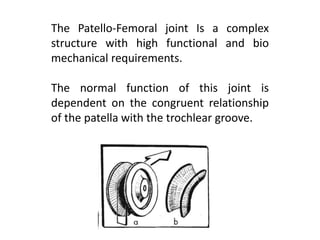
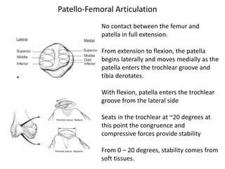
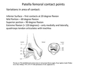
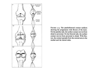
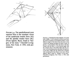
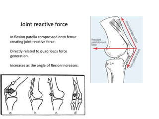
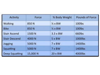
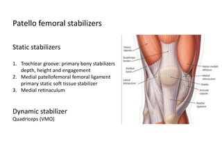
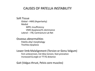
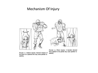
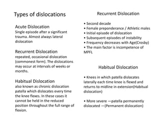
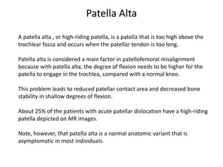
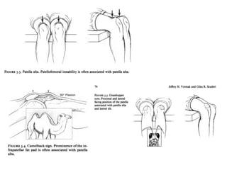
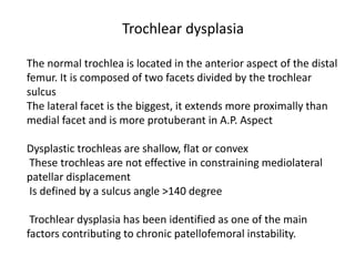
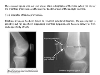
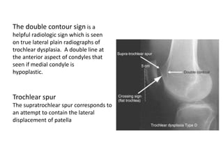
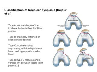
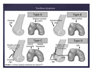
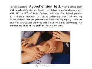
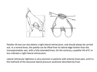
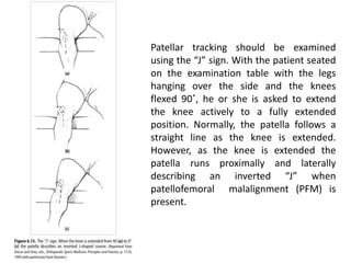
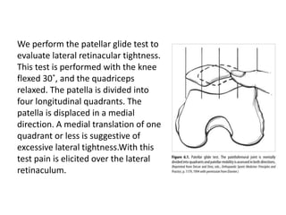
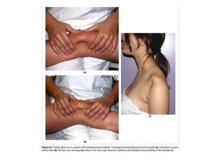
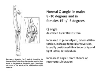
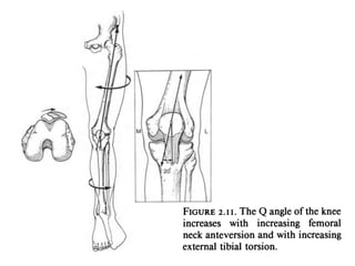
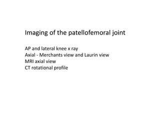
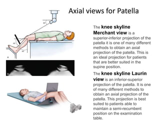
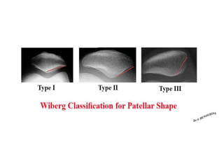
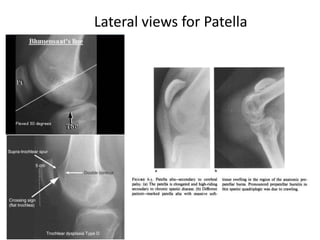
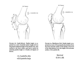
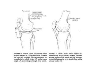
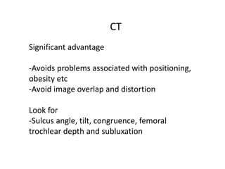
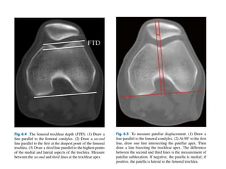
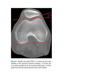
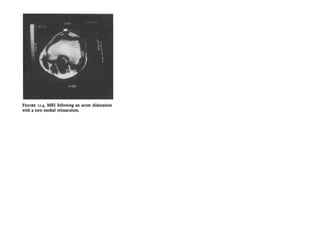
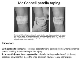
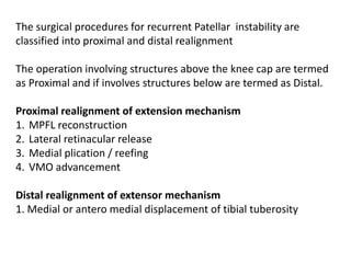
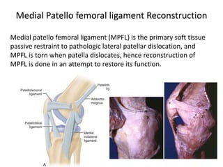
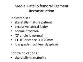
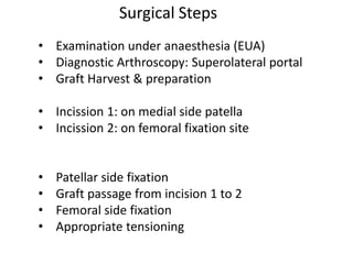
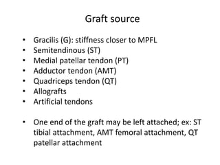
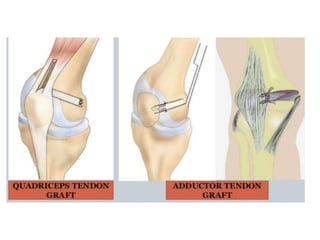
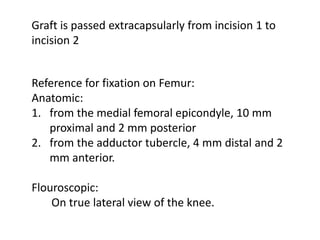
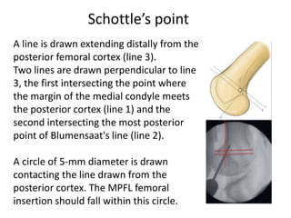
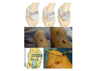
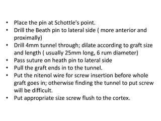
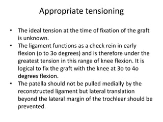
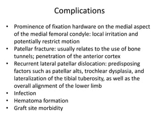
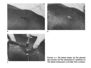
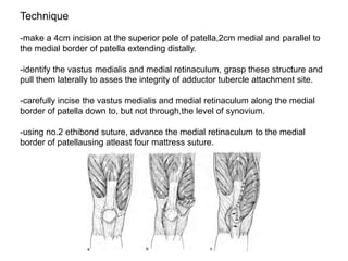
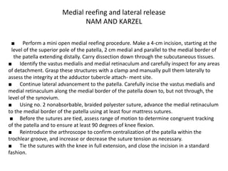
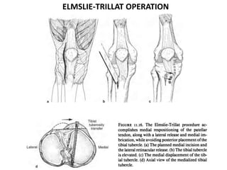
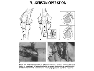
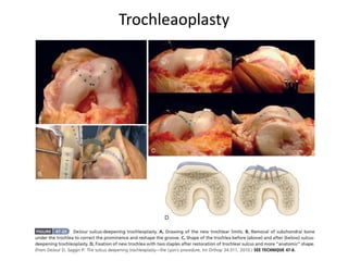
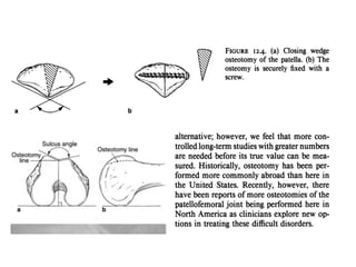
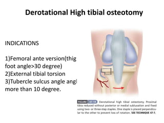
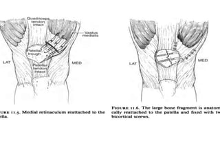
Patellar Dislocations
By Dr Kota Gandhi
I yr PG Orthopaedics
Kamineni Institute Of Medical Sciences
The patella is a flat, triangular bone, situated on the front of the knee-joint. It is usually
regarded as a sesamoid bone, developed in the tendon of the Quadriceps femoris.
Anatomy
Anatomy
The upper three quarters of the patella articulates with femur
and subdivided into medial and a lateral facet by vertical ledge
which varies in shape.
Wiberg classification is a system for describing the
shape of the patella based mainly on asymmetry between
the patellar medial and lateral facet on axial views of the
patella.
Type I: the facets are concave, symmetrical, and of equal
size.
Type II: the medial facet is smaller than the lateral facet
and flat or only slightly convex. The lateral facet is
concave.
• Rectus femoris tendon : 8 -10 cm in length, triangular in shape with
insertion 3-5 cm in width at superior pole of patella.
• VMO tendon : Inserts obliquely at superomedial border of patella only
a few mm in length; primary stabiliser of patella medially against VL
• Vastus Lateralis : Inserts obliquely At superior lateral aspect of Patella,
2.8 cm in length
• Lateral expansion of Vastus Lateralis With a superficial and deep layer
Forms the Lateral retinaculum; Deep layer is the lateral patellofemoral
ligament : this is a static guide for the patella; this may decrease medial
excursion and increase the lateral tracking.
• Medial side also has a patellofemoral ligament but it is much weaker
than lateral side.
Attachments around Patella
The Patello-Femoral joint Is a complex
structure with high functional and bio
mechanical requirements.
The normal function of this joint is
dependent on the congruent relationship
of the patella with the trochlear groove.
No contact between the femur and
patella in full extension.
From extension to flexion, the patella
begins laterally and moves medially as the
patella enters the trochlear groove and
tibia derotates.
With flexion, patella enters the trochlear
groove from the lateral side
Seats in the trochlear at ~20 degrees at
this point the congruence and
compressive forces provide stability
From 0 – 20 degrees, stability comes from
soft tissues.
Patello-Femoral Articulation
Variations in area of contact:
Inferior Surface – first contacts at 20 degree flexion
Mid Portion – 60 degree flexion
Superior portion – 90 degree flexion
Extreme flexion (> 120 degrees) – only medially and laterally,
quadriceps tendon articulates with trochlea
Patella femoral contact points
In flexion patella compressed onto femur
creating joint reactive force.
Directly related to quadriceps force
generation.
Increases as the angle of flexion increases.
Joint reactive force
Static stabilizers
1. Trochlear groove: primary bony stabilizers
depth, height and engagement
2. Medial patellofemoral femoral ligament
primary static soft tissue stabilizer
3. Medial retinaculum
Dynamic stabilizer
Quadriceps (VMO)
Patello femoral stabilizers
Soft Tissue
Global --HMS (Hyperlaxity)
Medial
MPFL Insufficiency
VMO dysplasia/VL dominance
Lateral -- ITB, Contracture Lat Ret
Osseous abnormalities
Patella alta/ morphology
Trochlea dysplasia
Lower limb Malalignment (Torsion or Genu Valgum)
Fem anteversion, Ext tibia torsion, foot pronation
Increased Q angle or TT:TG distance
Gait (Valgus thrust, Pelvis core muscles)
CAUSES OF PATELLA INSTABILITY
Mechanism Of Injury
Acute Dislocation
Single episode after a significant
trauma. Almost always lateral
dislocation
Recurrent Dislocation
repeated, occasional dislocation
(commonest form). The dislocations
may occur at intervals of weeks or
months.
Habitual Dislocation
also known as chronic dislocation
patella which dislocates every time
the knee flexes. In these cases it
cannot be held in the reduced
position throughout the full range of
flexion.
Types of dislocations Recurrent Dislocation
• Second decade
• Female preponderance / Athletic males
• Initial episode of dislocation
• Subsequent episodes of instability
• Frequency decreases with Age(Crosby)
• The main factor is incompetence of
MPFL
Habitual Dislocation
• Knees in which patella dislocates
laterally each time knee is flexed and
returns to midline in extension(Habitual
dislocation)
• More severe —patella permanently
dislocated —(Permanent dislocation)
• Young
• Female
• Family history
• Bilateral
• Atraumatic disorders
• Anatomic abnormalities
Patella alta
Trochlear hypoplasia
TT-TG distance
‘Q’ angle
Quadriceps dysfunction
Hyper mobility
Predisposing factors responsible for recurrence
Evaluation
We evaluate the following features
1. Integrity of medial patello femoral ligament
2. Height of patella on physical and radiographic
examination
3. Length of patellar tendon
4. Position of patella in relationship to trochlea
A patella alta , or high-riding patella, is a patella that is too high above the
trochlear fossa and occurs when the patellar tendon is too long.
Patella alta is considered a main factor in patellofemoral misalignment
because with patella alta, the degree of flexion needs to be higher for the
patella to engage in the trochlea, compared with a normal knee.
This problem leads to reduced patellar contact area and decreased bone
stability in shallow degrees of flexion.
About 25% of the patients with acute patellar dislocation have a high-riding
patella depicted on MR images.
Note, however, that patella alta is a normal anatomic variant that is
asymptomatic in most individuals.
Patella Alta
The normal trochlea is located in the anterior aspect of the distal
femur. It is composed of two facets divided by the trochlear
sulcus
The lateral facet is the biggest, it extends more proximally than
medial facet and is more protuberant in A.P. Aspect
Dysplastic trochleas are shallow, flat or convex
These trochleas are not effective in constraining mediolateral
patellar displacement
Is defined by a sulcus angle >140 degree
Trochlear dysplasia has been identified as one of the main
factors contributing to chronic patellofemoral instability.
Trochlear dysplasia
The crossing sign is seen on true lateral plain radiographs of the knee when the line of
the trochlear groove crosses the anterior border of one of the condyle trochlea.
It is a predictor of trochlear dysplasia.
Trochlear dysplasia has been linked to recurrent patellar dislocation. The crossing sign is
sensitive but not specific in diagnosing trochlear dysplasia, and has a sensitivity of 94%
and a specificity of 56%
The double contour sign is a
helpful radiologic sign which is seen
on true lateral plain radiographs of
trochlear dysplasia. A double line at
the anterior aspect of condyles that
seen if medial condyle is
hypoplastic.
Trochlear spur
The supratrochlear spur corresponds to
an attempt to contain the lateral
displacement of patella
Type A: normal shape of the
trochlea, but a shallow trochlear
groove
Type B: markedly flattened or
even convex trochlea
Type C: trochlear facet
asymmetry, with too high lateral
facet, and hypo plastic medial
facet
Type D: type C features and a
vertical link between facets ('cliff
pattern') 3
Classification of trochlear dysplasia (Dejour
et al)
Fairbanks patellar Apprehension test, when positive (pain
and muscle defensive contraction on lateral patellar displacement
with 20˚ to 30˚ of knee flexion), indicates that lateral patellar
instability is an important part of the patient’s problem. This test may
be so positive that the patient withdraws the leg rapidly when the
examiner approaches the knee with his or her hand, preventing thus
any contact, or he or she grabs the examiner’s arm.
Patellar tilt test can also detect a tight lateral retinaculum, and should always be carried
out. In a normal knee, the patella can be lifted from its lateral edge farther than the
transepicondylar axis, with a fully extended knee. On the contrary, a patellar tilt of 0˚ or
less indicates a tight lateral retinaculum.
Lateral retinacular tightness is very common in patients with anterior knee pain, and it is
the hallmark of the excessive lateral pressure syndrome described by Ficat.
Patellar tracking should be examined
using the “J” sign. With the patient seated
on the examination table with the legs
hanging over the side and the knees
flexed 90˚, he or she is asked to extend
the knee actively to a fully extended
position. Normally, the patella follows a
straight line as the knee is extended.
However, as the knee is extended the
patella runs proximally and laterally
describing an inverted “J” when
patellofemoral malalignment (PFM) is
present.
We perform the patellar glide test to
evaluate lateral retinacular tightness.
This test is performed with the knee
flexed 30˚, and the quadriceps
relaxed. The patella is divided into
four longitudinal quadrants. The
patella is displaced in a medial
direction. A medial translation of one
quadrant or less is suggestive of
excessive lateral tightness.With this
test pain is elicited over the lateral
retinaculum.
Q angle
described by Sir Brasttstrom
Increased in genu valgum, external tibial
torsion, increase femoral anteversion,
laterally positioned tibial tuberosity and
tight lateral retinaculum.
Increase Q angle : more chance of
recurrent subluxation
Normal Q angle in males
8 -10 degrees and in
females 15 +/- 5 degrees
Imaging of the patellofemoral joint
AP and lateral knee x ray
Axial - Merchants view and Laurin view
MRI axial view
CT rotational profile
The knee skyline
Merchant view is a
superior-inferior projection of the
patella it is one of many different
methods to obtain an axial
projection of the patella. This is
an ideal projection for patients
that are better suited in the
supine position.
The knee skyline Laurin
view is an inferior-superior
projection of the patella. It is one
of many different methods to
obtain an axial projection of the
patella. This projection is best
suited to patients able to
maintain a semi-recumbent
position on the examination
table.
Axial views for Patella
Lateral views for Patella
Normal
0.54-1.06
>1.2 patella alta
<0.8 patella baja
CT
Significant advantage
-Avoids problems associated with positioning,
obesity etc
-Avoid image overlap and distortion
Look for
-Sulcus angle, tilt, congruence, femoral
trochlear depth and subluxation
Congruence Angle
CT classification of malalignment
Type 1 - lateral subluxation without tilt
Type 2 - lateral subluxation with tilt
Type 3 - lateral tilt without subluxation
Type 4 - radiographically normal alignment
MRI Scan
MRI can be used to diagnose prior patellar dislocations on
the basis of typical injury patterns.
In general, deformity or deems of the inferno medial
patella and the lateral condyle, in conjunction with MPFL
disruption and patellar lateralisation, is diagnostic for
recent patellar dislocation
More than two-thirds of the patients will show osteo-
chondral lesions of the medial patella.
Management
• Non Operative management To be attempted in all
patients.
• Goals —Normal flexibility,Balanced quadriceps
strength,Stretching of tight lateral structures
• Push back w/o difficulty .
• Jt aspiration and immobilized in full extension for 3
weeks. >Splint;
• If no sign of soft tissue lesion
• Retained for 2-3 weeks
• Quadriceps strengthening exercise ; 2-3 months.
1.Quadriceps strengthening
2.Core stability
3.Mc Connell taping
4.Insoles
Treatment of Patella Instability
Always conservative first
Quadriceps Training
1.Most Essential component
2.Strengthening of quadriceps especially
VMO
3.Isometric and progressive resistance
exercises with knee in extension
4.With increasing strength short arc
exercises in last 30°.
Indications
With certain knee injuries – such as patellofemoral pain syndrome where abnormal
patella tracking is contributing to the injury.
To prevent injury or injury aggravation – Patella taping maybe beneficial during
sports or activities that place the knee at-risk of injury or injury aggravation
Mc Connell patella taping
Barefoot running
Barefoot running may reduce patellofemoral joint
stress as a result of reduced joint reaction forces.
Barefoot runners are more likely to use it forefoot
versus a heel strike Pattern in the initial loading
response, which has been shown to increase ankle
eccentric work and simultaneously decrease the
loading on the knee joint
Surgical treatment
Surgery in acute patella dislocation is indicated in
1. Osteochondral fracture
2. Loose body formation or joint incongruity
3. Incompetency of MPFL
Removal of loose bodies and MPFL repair required in these conditions.
Complications
• Recurrent dislocation
• Anterior knee pain
• Knee swelling
• Recurrent haemarthrosis
Recurrent Patellofemoral dislocation management
If dislocation of the patella continues despite appropriate
nonoperative treatment, surgery is indicated. Otherwise,
the patient may become apprehensive and afraid to use
the knee, and with continued recurrence the joint may
be severely damaged.
More than 100 surgical procedures have been described
for the treatment of patellofemoral instability. The key to
successful surgical intervention is correctly identifying
and treating the pathologic anatomy producing the
instability.
The surgical procedures for recurrent Patellar instability are
classified into proximal and distal realignment
The operation involving structures above the knee cap are termed
as Proximal and if involves structures below are termed as Distal.
Proximal realignment of extension mechanism
1. MPFL reconstruction
2. Lateral retinacular release
3. Medial plication / reefing
4. VMO advancement
Distal realignment of extensor mechanism
1. Medial or antero medial displacement of tibial tuberosity
Medial patello femoral ligament (MPFL) is the primary soft tissue
passive restraint to pathologic lateral patellar dislocation, and
MPFL is torn when patella dislocates, hence reconstruction of
MPFL is done in an attempt to restore its function.
Medial Patello femoral ligament Reconstruction
Indicated in :
• skeletally mature patient
• excessive lateral laxity
• normal trochlea
• ‘Q' angle is normal
• TT-TG distance is < 20mm
• low grade trochlear dysplasia
Contraindications :
• skeletally immaturity
Medial Patello femoral ligament
Reconstruction
• Examination under anaesthesia (EUA)
• Diagnostic Arthroscopy: Superolateral portal
• Graft Harvest & preparation
• Incission 1: on medial side patella
• Incission 2: on femoral fixation site
• Patellar side fixation
• Graft passage from incision 1 to 2
• Femoral side fixation
• Appropriate tensioning
Surgical Steps
• Gracilis (G): stiffness closer to MPFL
• Semitendinous (ST)
• Medial patellar tendon (PT)
• Adductor tendon (AMT)
• Quadriceps tendon (QT)
• Allografts
• Artificial tendons
• One end of the graft may be left attached; ex: ST
tibial attachment, AMT femoral attachment, QT
patellar attachment
Graft source
Graft is passed extracapsularly from incision 1 to
incision 2
Reference for fixation on Femur:
Anatomic:
1. from the medial femoral epicondyle, 10 mm
proximal and 2 mm posterior
2. from the adductor tubercle, 4 mm distal and 2
mm anterior.
Flouroscopic:
On true lateral view of the knee.
A line is drawn extending distally from the
posterior femoral cortex (line 3).
Two lines are drawn perpendicular to line
3, the first intersecting the point where
the margin of the medial condyle meets
the posterior cortex (line 1) and the
second intersecting the most posterior
point of Blumensaat's line (line 2).
A circle of 5-mm diameter is drawn
contacting the line drawn from the
posterior cortex. The MPFL femoral
insertion should fall within this circle.
Schottle’s point
• Place the pin at Schottle's point.
• Drill the Beath pin to lateral side ( more anterior and
proximally)
• Drill 4mm tunnel through; dilate according to graft size
and length ( usually 25mm long, 6 rum diameter)
• Pass suture on heath pin to lateral side
• Pull the graft ends in to the tunnel.
• Put the nitenol wire for screw insertion before whole
graft goes in; otherwise finding the tunnel to put screw
will be difficult.
• Put appropriate size screw flush to the cortex.
Appropriate tensioning
• The ideal tension at the time of fixation of the graft
is unknown.
• The ligament functions as a check rein in early
flexion (o to 3o degrees) and is therefore under the
greatest tension in this range of knee flexion. It is
logical to fix the graft with the knee at 3o to 4o
degrees flexion.
• The patella should not be pulled medially by the
reconstructed ligament but lateral translation
beyond the lateral margin of the trochlear should be
prevented.
• Prominence of fixation hardware on the medial aspect
of the medial femoral condyle: local irritation and
potentially restrict motion
• Patellar fracture: usually relates to the use of bone
tunnels; penetration of the anterior cortex
• Recurrent lateral patellar dislocation: predisposing
factors such as patellar alts, trochlear dysplasia, and
lateralization of the tibial tuberosity, as well as the
overall alignment of the lower limb
• Infection
• Hematoma formation
• Graft site morbidity
Complications
Indication
1)Tight lateral structure prevent patellar centring
2)Lateral patellar pressure syndrome
3)Can be done in skeletally immature patients
Release to include
1)Lateral retinaculum from distal third of vastus
lateralis
2)Lateral patellofemoral ligament
3)Lateral patellotibial ligament
Lateral Release
Can be done open or arthroscopy procedure ( now a days arthroscopic
release preferred )
Complications
1)Extending the release too far can cause medial subluxation of the
patella; infact medial patella subluxation or dislocation is almost always
iatrogenic, secondary to an overzealous lateral release.
2)injury to superolateral geniculate vessel to prevent this make a
superior anterolateral 2cm incision starting just lateral to the proximal
pole of patella.
Results varied, good results in short term(metcalf,Simpson),poorer in
long term(Christensen)
Anatomical and biomechanical studies have indicated that the
MPFL and the VMO are the primary restraints to lateral patella
translation, particularly early in flexion before full trochlear
engagement.
There are 3 types of primary procedures for medial repair the
techniques include
(1)Plication of the medial patellar retinaculum.
(2)Anatomic repair of the MPFL, and
(3)Anatomical repair surgery of the VMO.
Medial Repair
Technique
-make a 4cm incision at the superior pole of patella,2cm medial and parallel to
the medial border of patella extending distally.
-identify the vastus medialis and medial retinaculum, grasp these structure and
pull them laterally to asses the integrity of adductor tubercle attachment site.
-carefully incise the vastus medialis and medial retinaculum along the medial
border of patella down to, but not through,the level of synovium.
-using no.2 ethibond suture, advance the medial retinaculum to the medial
border of patellausing atleast four mattress suture.
Medial reefing and lateral release
NAM AND KARZEL
■ Perform a mini open medial reefing procedure. Make a 4-cm incision, starting at the
level of the superior pole of the patella, 2 cm medial and parallel to the medial border of
the patella extending distally. Carry dissection down through the subcutaneous tissues.
■ Identify the vastus medialis and medial retinaculum and carefully inspect for any areas
of detachment. Grasp these structures with a clamp and manually pull them laterally to
assess the integrity at the adductor tubercle attach- ment site.
■ Continue lateral advancement to the patella. Carefully incise the vastus medialis and
medial retinaculum along the medial border of the patella down to, but not through, the
level of the synovium.
■ Using no. 2 nonabsorbable, braided polyester suture, advance the medial retinaculum
to the medial border of the patella using at least four mattress sutures.
■ Before the sutures are tied, assess range of motion to determine congruent tracking
of the patella and to ensure at least 90 degrees of knee flexion.
■ Reintroduce the arthroscope to confirm centralization of the patella within the
trochlear groove, and increase or decrease the suture tension as necessary.
■ Tie the sutures with the knee in full extension, and close the incision in a standard
fashion.
Medial reefing and lateral release
NAM AND KARZEL
Distal Realignment Surgeries
Aims to diminish the Q angle or TT-TG distance with
anteromedialisation of tibial tuberosity and unloads
patello femoral articulation .
Indications
1. Q angle or /1" TT-TG distance > 20mm
2. Patellar alta
3. Normal patellar glide
4. Medial facet arthritis
Contraindications
1.Skeletally immature patients
2. incompetent MPFL
3. Diffuse patellar arthritis
ELMSLIE-TRILLAT OPERATION
FULKERSON OPERATION
Surgical indications
• High grade trochlear dysplasia with patellar instability in
the absence of patellofemoral osteoarthritis
• Type of dysplasia should be identified when deciding the
procedure
• Associated abnormalities including TT-TG distance,
patellar alta, patellar tilt should be identified and
rectified
• MPFL reconstruction is always done
Contra indications
• Skeletally immature patients
• Associated osteoarthritis
Management of Trochlear dysplasia
Type A dysplasia : medial patellofemoral ligament
reconstruction
Type B and D dysplasia : sulcus deepening
trochleoplasty with MPFL reconstruction
Type C dvsplasia : lateral facet elevation trochleoplasty
with MPFL reconstruction
Management of Trochlear dysplasia
Trochleaoplasty
Derotational High tibial osteotomy
INDICATIONS
1)Femoral ante version(thigh
foot angle>30 degree)
2)External tibial torsion
3)Tubercle sulcus angle angle
more than 10 degree.
Thank You!

Patella dislocations

  • 1.
    Patellar Dislocations By DrKota Gandhi I yr PG Orthopaedics Kamineni Institute Of Medical Sciences
  • 2.
    The patella isa flat, triangular bone, situated on the front of the knee-joint. It is usually regarded as a sesamoid bone, developed in the tendon of the Quadriceps femoris. Anatomy
  • 3.
    Anatomy The upper threequarters of the patella articulates with femur and subdivided into medial and a lateral facet by vertical ledge which varies in shape. Wiberg classification is a system for describing the shape of the patella based mainly on asymmetry between the patellar medial and lateral facet on axial views of the patella. Type I: the facets are concave, symmetrical, and of equal size. Type II: the medial facet is smaller than the lateral facet and flat or only slightly convex. The lateral facet is concave.
  • 6.
    • Rectus femoristendon : 8 -10 cm in length, triangular in shape with insertion 3-5 cm in width at superior pole of patella. • VMO tendon : Inserts obliquely at superomedial border of patella only a few mm in length; primary stabiliser of patella medially against VL • Vastus Lateralis : Inserts obliquely At superior lateral aspect of Patella, 2.8 cm in length • Lateral expansion of Vastus Lateralis With a superficial and deep layer Forms the Lateral retinaculum; Deep layer is the lateral patellofemoral ligament : this is a static guide for the patella; this may decrease medial excursion and increase the lateral tracking. • Medial side also has a patellofemoral ligament but it is much weaker than lateral side. Attachments around Patella
  • 8.
    The Patello-Femoral jointIs a complex structure with high functional and bio mechanical requirements. The normal function of this joint is dependent on the congruent relationship of the patella with the trochlear groove.
  • 9.
    No contact betweenthe femur and patella in full extension. From extension to flexion, the patella begins laterally and moves medially as the patella enters the trochlear groove and tibia derotates. With flexion, patella enters the trochlear groove from the lateral side Seats in the trochlear at ~20 degrees at this point the congruence and compressive forces provide stability From 0 – 20 degrees, stability comes from soft tissues. Patello-Femoral Articulation
  • 10.
    Variations in areaof contact: Inferior Surface – first contacts at 20 degree flexion Mid Portion – 60 degree flexion Superior portion – 90 degree flexion Extreme flexion (> 120 degrees) – only medially and laterally, quadriceps tendon articulates with trochlea Patella femoral contact points
  • 13.
    In flexion patellacompressed onto femur creating joint reactive force. Directly related to quadriceps force generation. Increases as the angle of flexion increases. Joint reactive force
  • 15.
    Static stabilizers 1. Trochleargroove: primary bony stabilizers depth, height and engagement 2. Medial patellofemoral femoral ligament primary static soft tissue stabilizer 3. Medial retinaculum Dynamic stabilizer Quadriceps (VMO) Patello femoral stabilizers
  • 16.
    Soft Tissue Global --HMS(Hyperlaxity) Medial MPFL Insufficiency VMO dysplasia/VL dominance Lateral -- ITB, Contracture Lat Ret Osseous abnormalities Patella alta/ morphology Trochlea dysplasia Lower limb Malalignment (Torsion or Genu Valgum) Fem anteversion, Ext tibia torsion, foot pronation Increased Q angle or TT:TG distance Gait (Valgus thrust, Pelvis core muscles) CAUSES OF PATELLA INSTABILITY
  • 17.
  • 18.
    Acute Dislocation Single episodeafter a significant trauma. Almost always lateral dislocation Recurrent Dislocation repeated, occasional dislocation (commonest form). The dislocations may occur at intervals of weeks or months. Habitual Dislocation also known as chronic dislocation patella which dislocates every time the knee flexes. In these cases it cannot be held in the reduced position throughout the full range of flexion. Types of dislocations Recurrent Dislocation • Second decade • Female preponderance / Athletic males • Initial episode of dislocation • Subsequent episodes of instability • Frequency decreases with Age(Crosby) • The main factor is incompetence of MPFL Habitual Dislocation • Knees in which patella dislocates laterally each time knee is flexed and returns to midline in extension(Habitual dislocation) • More severe —patella permanently dislocated —(Permanent dislocation)
  • 19.
    • Young • Female •Family history • Bilateral • Atraumatic disorders • Anatomic abnormalities Patella alta Trochlear hypoplasia TT-TG distance ‘Q’ angle Quadriceps dysfunction Hyper mobility Predisposing factors responsible for recurrence
  • 20.
    Evaluation We evaluate thefollowing features 1. Integrity of medial patello femoral ligament 2. Height of patella on physical and radiographic examination 3. Length of patellar tendon 4. Position of patella in relationship to trochlea
  • 21.
    A patella alta, or high-riding patella, is a patella that is too high above the trochlear fossa and occurs when the patellar tendon is too long. Patella alta is considered a main factor in patellofemoral misalignment because with patella alta, the degree of flexion needs to be higher for the patella to engage in the trochlea, compared with a normal knee. This problem leads to reduced patellar contact area and decreased bone stability in shallow degrees of flexion. About 25% of the patients with acute patellar dislocation have a high-riding patella depicted on MR images. Note, however, that patella alta is a normal anatomic variant that is asymptomatic in most individuals. Patella Alta
  • 24.
    The normal trochleais located in the anterior aspect of the distal femur. It is composed of two facets divided by the trochlear sulcus The lateral facet is the biggest, it extends more proximally than medial facet and is more protuberant in A.P. Aspect Dysplastic trochleas are shallow, flat or convex These trochleas are not effective in constraining mediolateral patellar displacement Is defined by a sulcus angle >140 degree Trochlear dysplasia has been identified as one of the main factors contributing to chronic patellofemoral instability. Trochlear dysplasia
  • 25.
    The crossing signis seen on true lateral plain radiographs of the knee when the line of the trochlear groove crosses the anterior border of one of the condyle trochlea. It is a predictor of trochlear dysplasia. Trochlear dysplasia has been linked to recurrent patellar dislocation. The crossing sign is sensitive but not specific in diagnosing trochlear dysplasia, and has a sensitivity of 94% and a specificity of 56%
  • 26.
    The double contoursign is a helpful radiologic sign which is seen on true lateral plain radiographs of trochlear dysplasia. A double line at the anterior aspect of condyles that seen if medial condyle is hypoplastic. Trochlear spur The supratrochlear spur corresponds to an attempt to contain the lateral displacement of patella
  • 27.
    Type A: normalshape of the trochlea, but a shallow trochlear groove Type B: markedly flattened or even convex trochlea Type C: trochlear facet asymmetry, with too high lateral facet, and hypo plastic medial facet Type D: type C features and a vertical link between facets ('cliff pattern') 3 Classification of trochlear dysplasia (Dejour et al)
  • 29.
    Fairbanks patellar Apprehensiontest, when positive (pain and muscle defensive contraction on lateral patellar displacement with 20˚ to 30˚ of knee flexion), indicates that lateral patellar instability is an important part of the patient’s problem. This test may be so positive that the patient withdraws the leg rapidly when the examiner approaches the knee with his or her hand, preventing thus any contact, or he or she grabs the examiner’s arm.
  • 30.
    Patellar tilt testcan also detect a tight lateral retinaculum, and should always be carried out. In a normal knee, the patella can be lifted from its lateral edge farther than the transepicondylar axis, with a fully extended knee. On the contrary, a patellar tilt of 0˚ or less indicates a tight lateral retinaculum. Lateral retinacular tightness is very common in patients with anterior knee pain, and it is the hallmark of the excessive lateral pressure syndrome described by Ficat.
  • 31.
    Patellar tracking shouldbe examined using the “J” sign. With the patient seated on the examination table with the legs hanging over the side and the knees flexed 90˚, he or she is asked to extend the knee actively to a fully extended position. Normally, the patella follows a straight line as the knee is extended. However, as the knee is extended the patella runs proximally and laterally describing an inverted “J” when patellofemoral malalignment (PFM) is present.
  • 32.
    We perform thepatellar glide test to evaluate lateral retinacular tightness. This test is performed with the knee flexed 30˚, and the quadriceps relaxed. The patella is divided into four longitudinal quadrants. The patella is displaced in a medial direction. A medial translation of one quadrant or less is suggestive of excessive lateral tightness.With this test pain is elicited over the lateral retinaculum.
  • 34.
    Q angle described bySir Brasttstrom Increased in genu valgum, external tibial torsion, increase femoral anteversion, laterally positioned tibial tuberosity and tight lateral retinaculum. Increase Q angle : more chance of recurrent subluxation Normal Q angle in males 8 -10 degrees and in females 15 +/- 5 degrees
  • 37.
    Imaging of thepatellofemoral joint AP and lateral knee x ray Axial - Merchants view and Laurin view MRI axial view CT rotational profile
  • 38.
    The knee skyline Merchantview is a superior-inferior projection of the patella it is one of many different methods to obtain an axial projection of the patella. This is an ideal projection for patients that are better suited in the supine position. The knee skyline Laurin view is an inferior-superior projection of the patella. It is one of many different methods to obtain an axial projection of the patella. This projection is best suited to patients able to maintain a semi-recumbent position on the examination table. Axial views for Patella
  • 40.
  • 41.
  • 43.
    CT Significant advantage -Avoids problemsassociated with positioning, obesity etc -Avoid image overlap and distortion Look for -Sulcus angle, tilt, congruence, femoral trochlear depth and subluxation
  • 46.
  • 47.
    CT classification ofmalalignment Type 1 - lateral subluxation without tilt Type 2 - lateral subluxation with tilt Type 3 - lateral tilt without subluxation Type 4 - radiographically normal alignment
  • 49.
    MRI Scan MRI canbe used to diagnose prior patellar dislocations on the basis of typical injury patterns. In general, deformity or deems of the inferno medial patella and the lateral condyle, in conjunction with MPFL disruption and patellar lateralisation, is diagnostic for recent patellar dislocation More than two-thirds of the patients will show osteo- chondral lesions of the medial patella.
  • 51.
    Management • Non Operativemanagement To be attempted in all patients. • Goals —Normal flexibility,Balanced quadriceps strength,Stretching of tight lateral structures • Push back w/o difficulty . • Jt aspiration and immobilized in full extension for 3 weeks. >Splint; • If no sign of soft tissue lesion • Retained for 2-3 weeks • Quadriceps strengthening exercise ; 2-3 months.
  • 52.
    1.Quadriceps strengthening 2.Core stability 3.McConnell taping 4.Insoles Treatment of Patella Instability Always conservative first
  • 53.
    Quadriceps Training 1.Most Essentialcomponent 2.Strengthening of quadriceps especially VMO 3.Isometric and progressive resistance exercises with knee in extension 4.With increasing strength short arc exercises in last 30°.
  • 54.
    Indications With certain kneeinjuries – such as patellofemoral pain syndrome where abnormal patella tracking is contributing to the injury. To prevent injury or injury aggravation – Patella taping maybe beneficial during sports or activities that place the knee at-risk of injury or injury aggravation Mc Connell patella taping
  • 55.
    Barefoot running Barefoot runningmay reduce patellofemoral joint stress as a result of reduced joint reaction forces. Barefoot runners are more likely to use it forefoot versus a heel strike Pattern in the initial loading response, which has been shown to increase ankle eccentric work and simultaneously decrease the loading on the knee joint
  • 56.
    Surgical treatment Surgery inacute patella dislocation is indicated in 1. Osteochondral fracture 2. Loose body formation or joint incongruity 3. Incompetency of MPFL Removal of loose bodies and MPFL repair required in these conditions. Complications • Recurrent dislocation • Anterior knee pain • Knee swelling • Recurrent haemarthrosis
  • 57.
    Recurrent Patellofemoral dislocationmanagement If dislocation of the patella continues despite appropriate nonoperative treatment, surgery is indicated. Otherwise, the patient may become apprehensive and afraid to use the knee, and with continued recurrence the joint may be severely damaged. More than 100 surgical procedures have been described for the treatment of patellofemoral instability. The key to successful surgical intervention is correctly identifying and treating the pathologic anatomy producing the instability.
  • 58.
    The surgical proceduresfor recurrent Patellar instability are classified into proximal and distal realignment The operation involving structures above the knee cap are termed as Proximal and if involves structures below are termed as Distal. Proximal realignment of extension mechanism 1. MPFL reconstruction 2. Lateral retinacular release 3. Medial plication / reefing 4. VMO advancement Distal realignment of extensor mechanism 1. Medial or antero medial displacement of tibial tuberosity
  • 59.
    Medial patello femoralligament (MPFL) is the primary soft tissue passive restraint to pathologic lateral patellar dislocation, and MPFL is torn when patella dislocates, hence reconstruction of MPFL is done in an attempt to restore its function. Medial Patello femoral ligament Reconstruction
  • 60.
    Indicated in : •skeletally mature patient • excessive lateral laxity • normal trochlea • ‘Q' angle is normal • TT-TG distance is < 20mm • low grade trochlear dysplasia Contraindications : • skeletally immaturity Medial Patello femoral ligament Reconstruction
  • 61.
    • Examination underanaesthesia (EUA) • Diagnostic Arthroscopy: Superolateral portal • Graft Harvest & preparation • Incission 1: on medial side patella • Incission 2: on femoral fixation site • Patellar side fixation • Graft passage from incision 1 to 2 • Femoral side fixation • Appropriate tensioning Surgical Steps
  • 62.
    • Gracilis (G):stiffness closer to MPFL • Semitendinous (ST) • Medial patellar tendon (PT) • Adductor tendon (AMT) • Quadriceps tendon (QT) • Allografts • Artificial tendons • One end of the graft may be left attached; ex: ST tibial attachment, AMT femoral attachment, QT patellar attachment Graft source
  • 64.
    Graft is passedextracapsularly from incision 1 to incision 2 Reference for fixation on Femur: Anatomic: 1. from the medial femoral epicondyle, 10 mm proximal and 2 mm posterior 2. from the adductor tubercle, 4 mm distal and 2 mm anterior. Flouroscopic: On true lateral view of the knee.
  • 65.
    A line isdrawn extending distally from the posterior femoral cortex (line 3). Two lines are drawn perpendicular to line 3, the first intersecting the point where the margin of the medial condyle meets the posterior cortex (line 1) and the second intersecting the most posterior point of Blumensaat's line (line 2). A circle of 5-mm diameter is drawn contacting the line drawn from the posterior cortex. The MPFL femoral insertion should fall within this circle. Schottle’s point
  • 67.
    • Place thepin at Schottle's point. • Drill the Beath pin to lateral side ( more anterior and proximally) • Drill 4mm tunnel through; dilate according to graft size and length ( usually 25mm long, 6 rum diameter) • Pass suture on heath pin to lateral side • Pull the graft ends in to the tunnel. • Put the nitenol wire for screw insertion before whole graft goes in; otherwise finding the tunnel to put screw will be difficult. • Put appropriate size screw flush to the cortex.
  • 68.
    Appropriate tensioning • Theideal tension at the time of fixation of the graft is unknown. • The ligament functions as a check rein in early flexion (o to 3o degrees) and is therefore under the greatest tension in this range of knee flexion. It is logical to fix the graft with the knee at 3o to 4o degrees flexion. • The patella should not be pulled medially by the reconstructed ligament but lateral translation beyond the lateral margin of the trochlear should be prevented.
  • 69.
    • Prominence offixation hardware on the medial aspect of the medial femoral condyle: local irritation and potentially restrict motion • Patellar fracture: usually relates to the use of bone tunnels; penetration of the anterior cortex • Recurrent lateral patellar dislocation: predisposing factors such as patellar alts, trochlear dysplasia, and lateralization of the tibial tuberosity, as well as the overall alignment of the lower limb • Infection • Hematoma formation • Graft site morbidity Complications
  • 70.
    Indication 1)Tight lateral structureprevent patellar centring 2)Lateral patellar pressure syndrome 3)Can be done in skeletally immature patients Release to include 1)Lateral retinaculum from distal third of vastus lateralis 2)Lateral patellofemoral ligament 3)Lateral patellotibial ligament Lateral Release
  • 72.
    Can be doneopen or arthroscopy procedure ( now a days arthroscopic release preferred ) Complications 1)Extending the release too far can cause medial subluxation of the patella; infact medial patella subluxation or dislocation is almost always iatrogenic, secondary to an overzealous lateral release. 2)injury to superolateral geniculate vessel to prevent this make a superior anterolateral 2cm incision starting just lateral to the proximal pole of patella. Results varied, good results in short term(metcalf,Simpson),poorer in long term(Christensen)
  • 73.
    Anatomical and biomechanicalstudies have indicated that the MPFL and the VMO are the primary restraints to lateral patella translation, particularly early in flexion before full trochlear engagement. There are 3 types of primary procedures for medial repair the techniques include (1)Plication of the medial patellar retinaculum. (2)Anatomic repair of the MPFL, and (3)Anatomical repair surgery of the VMO. Medial Repair
  • 74.
    Technique -make a 4cmincision at the superior pole of patella,2cm medial and parallel to the medial border of patella extending distally. -identify the vastus medialis and medial retinaculum, grasp these structure and pull them laterally to asses the integrity of adductor tubercle attachment site. -carefully incise the vastus medialis and medial retinaculum along the medial border of patella down to, but not through,the level of synovium. -using no.2 ethibond suture, advance the medial retinaculum to the medial border of patellausing atleast four mattress suture.
  • 75.
    Medial reefing andlateral release NAM AND KARZEL ■ Perform a mini open medial reefing procedure. Make a 4-cm incision, starting at the level of the superior pole of the patella, 2 cm medial and parallel to the medial border of the patella extending distally. Carry dissection down through the subcutaneous tissues. ■ Identify the vastus medialis and medial retinaculum and carefully inspect for any areas of detachment. Grasp these structures with a clamp and manually pull them laterally to assess the integrity at the adductor tubercle attach- ment site. ■ Continue lateral advancement to the patella. Carefully incise the vastus medialis and medial retinaculum along the medial border of the patella down to, but not through, the level of the synovium. ■ Using no. 2 nonabsorbable, braided polyester suture, advance the medial retinaculum to the medial border of the patella using at least four mattress sutures. ■ Before the sutures are tied, assess range of motion to determine congruent tracking of the patella and to ensure at least 90 degrees of knee flexion. ■ Reintroduce the arthroscope to confirm centralization of the patella within the trochlear groove, and increase or decrease the suture tension as necessary. ■ Tie the sutures with the knee in full extension, and close the incision in a standard fashion.
  • 76.
    Medial reefing andlateral release NAM AND KARZEL
  • 77.
    Distal Realignment Surgeries Aimsto diminish the Q angle or TT-TG distance with anteromedialisation of tibial tuberosity and unloads patello femoral articulation . Indications 1. Q angle or /1" TT-TG distance > 20mm 2. Patellar alta 3. Normal patellar glide 4. Medial facet arthritis Contraindications 1.Skeletally immature patients 2. incompetent MPFL 3. Diffuse patellar arthritis
  • 78.
  • 79.
  • 80.
    Surgical indications • Highgrade trochlear dysplasia with patellar instability in the absence of patellofemoral osteoarthritis • Type of dysplasia should be identified when deciding the procedure • Associated abnormalities including TT-TG distance, patellar alta, patellar tilt should be identified and rectified • MPFL reconstruction is always done Contra indications • Skeletally immature patients • Associated osteoarthritis Management of Trochlear dysplasia
  • 81.
    Type A dysplasia: medial patellofemoral ligament reconstruction Type B and D dysplasia : sulcus deepening trochleoplasty with MPFL reconstruction Type C dvsplasia : lateral facet elevation trochleoplasty with MPFL reconstruction Management of Trochlear dysplasia
  • 82.
  • 84.
    Derotational High tibialosteotomy INDICATIONS 1)Femoral ante version(thigh foot angle>30 degree) 2)External tibial torsion 3)Tubercle sulcus angle angle more than 10 degree.
  • 93.