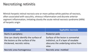
Spirochaetal Uveitis
DR SHRUTI LADDHA
SYPHILIS
• Sexually transmitted disease
• Enter through small abrasions of skin and mucous membrane
• Primary syphilis- painless non suppurative chancre, painless lymphadenopathy.
• Secondary syphilis- if no t/t given in primary syphilis, treponema disseminate ,- flu like
symptoms-headache, fever, myalgia, sore throat, painless maculopapular rash.
Rash disappear spontaneously in most cases
40%- shows CNS involvement.
optic neuritis, optic perineuritis, cranial nerve palsies.
anterior uveitis -10%
• Latent stage- early latent-first year following infection
late latent- later years
Treponema is present in liver and spleen
Patient is non infectious
Reactivation of dormant treponema may occur
May occur several year to several decades of infection
• Tertiary syphilis-
may be bening tertiary, cardiovascular or neurosyphilis.
Bening tertiary syphilis – gumma of skin, mucous membrane, uveal tract.
Cardiovascular syphilis - obliterative endarteritis of the vasa vasorum of the aorta
and causes aortitis, aortic aneurysms, and aortic valvular insufficiency
Neurosyphilis – meningovascular and parenchymal
• Congenital syphilis
IUFD.
Mucocutaneous manifestation-facial and tooth deformity.
Hutchinsons triad- interstitial keratitis, peg shaped upper inscisors, SNHL.
Ghost vessels
Peg shaped incisors
• Ophthalmic involvement can be in secondary or tertiary syphilis.
• Chronic gummatous or granulomatous inflammation of the ocular structures is typical of
late stage disease.
• Whereas more aggressive inflammation (iridocyclitis with vascularized nodules or
roseolae and necrotizing retinitis) is associated with early disease.
• Anterior uveitis -poor response to topical steroid treatment and a history of a skin rash in
the recent past should alert the clinician to the possibility of syphilitic anterior uveitis
POSTERIOR SEGMENT
Treponema thrive in all layers of eyes
• Focal/multifocal chorioretinitis
• Acute posterior placoid chorioretinitis
• Necrotizing retinitis
• Retinal vasculitis
• Intermediate uveitis
• Panuveitis
• Optic nerve involvement
CHOROIDITIS
• Deep chorioretinitis is most common manifestation.
• Focal syphilitic chorioretinitis presents as a deep, yellow gray lesion often with a
shallow serous retinal detachment and inflammatory cells in the vitreous.
• Multifocal lesions from one half to one disk diameter can coalesce to become
confluent
ACUTE SYPHILITIC POSTERIOR PLACOID
CHORIORETINITIS (ASPPC)
• Usually bilateral.
• With large, solitary, placoid, pale-yellowish subretinal lesions .
• Vitritis.
• Lesions show evidence of central fading and a pattern of coarsely
stippled hyperpigmentation of the pigment epithelium
• It is thought to be
• Due to retinal pigment epithelial infection and occurs more
• Commonly in the immunocompromised.
ARN Syphylitic necrosis
Starts in periphery Posterior pole
One can clearly identify the surface of
the lesions as the surface of the
thickened, necrotic retina.
Surface of the lesion is somewhat
indistinct, as if a layer of exudate
obscures the underlying retina from
view
Necrotic area-homogenous Necrotic area- mottled
Necrotizing retinitis
Mimick herpetic retinal necrosis-one or more yellow-white patches of necrosis,
often associated with vasculitis, vitreous inflammation and discrete anterior
segment inflammation, imitating closely the acute retinal necrosis syndrome (ARN)
of herpetic origin
Foci of syphilitic retinal necrosis in a patient at
presentation
after initiation of penicillin therapy
Dense compact necrosis observed in a patients
with viral ARN
Foci of syphilitic retinal necrosis in a patient at
presentation
after initiation of penicillin therapy
Healed syphilitic retinitis presenting as pseudo-retinitis
pigmentosa
OPTIC NERVE INFLAMMATION
• Acute meningitis occurs in 1 to 2 percent of patients with secondary syphilis and this can
cause increased intracranial pressure and papilledema.
• In papillitis there is a swollen disk with intraretinal exudates and perivasculitis around it.
• Neuroretinitis - When the inflammatory changes extend into the peripapillary retina
resulting in hard exudates.
• Optic perineuritis is a distinct entity due to an inflammation of the meningeal sheaths of
the optic nerve and causes mild swelling of the optic disk, without affecting its function.
This condition should be suspected in patients with normal visual acuity and colour vision
who seem to have papilledema but in whom lumbar puncture reveals normal cerebrospinal
fluid pressure and the presence of inflammatory cells or increased protein
Chronic bilateral papilledema, more pronounced in the left eye, in a white patient
with asymptomatic neurosyphilis
Syphilitic perioptic neuritis in a young woman with normal visual acuity and
normal intracranial pressure on lumbar puncture and on imaging
DIAGNOSIS OF OCULAR SYPHLIS
Testing for syphilis is indicated if
• the history or the presentation are suggestive
• the inflammation has unusual characteristics or it fails to respond to the usual
treatment (often steroids)
• the patient belongs to a high risk group for sexually transmitted diseases
Serologic testing that includes non-treponemal tests and treponemal tests is
considered the standard detection method
Non treponemal tests - VDRL, RPR
• Non-treponemal antibody titers decline as a result of treatment.
• A fourfold reduction in antibody titer of the same non-treponemal test is considered a
significant response to treatment.
• Lack of expected reduction in titer or an increase in titer suggests treatment failure or
reinfection.
• Non-treponemal tests may give false positive results in conditions other than syphilis (viral
infection, pregnancy, post-immunization).
• Moreover, they may be negative in as many as 30 percent of patients during the late latent or
tertiary stages.
• Direct treponemal tests detect antibodies specific to T. pallidum.
• This test stays reactive for life and indicates that infection has occurred but does not
distinguish active versus latent or treated infection.
• Thus, a positive direct test will indicate whether the patient has been exposed to
syphilis in the past and a positive indirect test such as the RPR or VDRL will indicate
active untreated infection.
TREATMENT
• Antibiotics
• Cortisosteroids
OCULAR LEPTOSPIROSIS-some differentiating
features
• May present 2 days to 4 yrs after systemic infection(mostly 6 months)
• Acute onset
• Non granulomatous in 90% cases
• hypopyon
• Post synechiae- easily break
• Cataract- common complication, progresses rapidly ,sometimes get self absorbed
• Membranous vitritis-These vitreous veil like membranous opacities are either
attached to the disc or they freely float in the vitreous
• sometimes string of pearls
• Disc edema
• Vasculitis-veins more than arteries
• Occlusion and neovascularisation less common
• Resolves quickly with restoration of vision in most cases
• Lab- primary-IgM ELISIA
Confirmatory-MAT (Microaglutination test)
PCR
• For mild to moderate cases-Doxycycline may be given in doses of 100 mg twice
daily for one week.
• Leptospires are sensitive to most antimicrobial agents, including penicillin,
amoxicillin, doxycycline and ceftriaxone
• Corticosteroids

Spirocheteal uveitis

  • 1.
  • 2.
    SYPHILIS • Sexually transmitteddisease • Enter through small abrasions of skin and mucous membrane • Primary syphilis- painless non suppurative chancre, painless lymphadenopathy. • Secondary syphilis- if no t/t given in primary syphilis, treponema disseminate ,- flu like symptoms-headache, fever, myalgia, sore throat, painless maculopapular rash. Rash disappear spontaneously in most cases 40%- shows CNS involvement. optic neuritis, optic perineuritis, cranial nerve palsies. anterior uveitis -10%
  • 3.
    • Latent stage-early latent-first year following infection late latent- later years Treponema is present in liver and spleen Patient is non infectious Reactivation of dormant treponema may occur May occur several year to several decades of infection • Tertiary syphilis- may be bening tertiary, cardiovascular or neurosyphilis. Bening tertiary syphilis – gumma of skin, mucous membrane, uveal tract. Cardiovascular syphilis - obliterative endarteritis of the vasa vasorum of the aorta and causes aortitis, aortic aneurysms, and aortic valvular insufficiency Neurosyphilis – meningovascular and parenchymal
  • 4.
    • Congenital syphilis IUFD. Mucocutaneousmanifestation-facial and tooth deformity. Hutchinsons triad- interstitial keratitis, peg shaped upper inscisors, SNHL. Ghost vessels Peg shaped incisors
  • 5.
    • Ophthalmic involvementcan be in secondary or tertiary syphilis. • Chronic gummatous or granulomatous inflammation of the ocular structures is typical of late stage disease. • Whereas more aggressive inflammation (iridocyclitis with vascularized nodules or roseolae and necrotizing retinitis) is associated with early disease. • Anterior uveitis -poor response to topical steroid treatment and a history of a skin rash in the recent past should alert the clinician to the possibility of syphilitic anterior uveitis
  • 6.
    POSTERIOR SEGMENT Treponema thrivein all layers of eyes • Focal/multifocal chorioretinitis • Acute posterior placoid chorioretinitis • Necrotizing retinitis • Retinal vasculitis • Intermediate uveitis • Panuveitis • Optic nerve involvement
  • 7.
    CHOROIDITIS • Deep chorioretinitisis most common manifestation. • Focal syphilitic chorioretinitis presents as a deep, yellow gray lesion often with a shallow serous retinal detachment and inflammatory cells in the vitreous. • Multifocal lesions from one half to one disk diameter can coalesce to become confluent
  • 8.
    ACUTE SYPHILITIC POSTERIORPLACOID CHORIORETINITIS (ASPPC) • Usually bilateral. • With large, solitary, placoid, pale-yellowish subretinal lesions . • Vitritis. • Lesions show evidence of central fading and a pattern of coarsely stippled hyperpigmentation of the pigment epithelium • It is thought to be • Due to retinal pigment epithelial infection and occurs more • Commonly in the immunocompromised.
  • 9.
    ARN Syphylitic necrosis Startsin periphery Posterior pole One can clearly identify the surface of the lesions as the surface of the thickened, necrotic retina. Surface of the lesion is somewhat indistinct, as if a layer of exudate obscures the underlying retina from view Necrotic area-homogenous Necrotic area- mottled Necrotizing retinitis Mimick herpetic retinal necrosis-one or more yellow-white patches of necrosis, often associated with vasculitis, vitreous inflammation and discrete anterior segment inflammation, imitating closely the acute retinal necrosis syndrome (ARN) of herpetic origin
  • 10.
    Foci of syphiliticretinal necrosis in a patient at presentation after initiation of penicillin therapy Dense compact necrosis observed in a patients with viral ARN
  • 11.
    Foci of syphiliticretinal necrosis in a patient at presentation after initiation of penicillin therapy
  • 12.
    Healed syphilitic retinitispresenting as pseudo-retinitis pigmentosa
  • 13.
    OPTIC NERVE INFLAMMATION •Acute meningitis occurs in 1 to 2 percent of patients with secondary syphilis and this can cause increased intracranial pressure and papilledema. • In papillitis there is a swollen disk with intraretinal exudates and perivasculitis around it. • Neuroretinitis - When the inflammatory changes extend into the peripapillary retina resulting in hard exudates. • Optic perineuritis is a distinct entity due to an inflammation of the meningeal sheaths of the optic nerve and causes mild swelling of the optic disk, without affecting its function. This condition should be suspected in patients with normal visual acuity and colour vision who seem to have papilledema but in whom lumbar puncture reveals normal cerebrospinal fluid pressure and the presence of inflammatory cells or increased protein
  • 14.
    Chronic bilateral papilledema,more pronounced in the left eye, in a white patient with asymptomatic neurosyphilis
  • 15.
    Syphilitic perioptic neuritisin a young woman with normal visual acuity and normal intracranial pressure on lumbar puncture and on imaging
  • 16.
    DIAGNOSIS OF OCULARSYPHLIS Testing for syphilis is indicated if • the history or the presentation are suggestive • the inflammation has unusual characteristics or it fails to respond to the usual treatment (often steroids) • the patient belongs to a high risk group for sexually transmitted diseases Serologic testing that includes non-treponemal tests and treponemal tests is considered the standard detection method
  • 17.
    Non treponemal tests- VDRL, RPR • Non-treponemal antibody titers decline as a result of treatment. • A fourfold reduction in antibody titer of the same non-treponemal test is considered a significant response to treatment. • Lack of expected reduction in titer or an increase in titer suggests treatment failure or reinfection. • Non-treponemal tests may give false positive results in conditions other than syphilis (viral infection, pregnancy, post-immunization). • Moreover, they may be negative in as many as 30 percent of patients during the late latent or tertiary stages.
  • 18.
    • Direct treponemaltests detect antibodies specific to T. pallidum. • This test stays reactive for life and indicates that infection has occurred but does not distinguish active versus latent or treated infection. • Thus, a positive direct test will indicate whether the patient has been exposed to syphilis in the past and a positive indirect test such as the RPR or VDRL will indicate active untreated infection.
  • 20.
  • 21.
    OCULAR LEPTOSPIROSIS-some differentiating features •May present 2 days to 4 yrs after systemic infection(mostly 6 months) • Acute onset • Non granulomatous in 90% cases • hypopyon • Post synechiae- easily break • Cataract- common complication, progresses rapidly ,sometimes get self absorbed • Membranous vitritis-These vitreous veil like membranous opacities are either attached to the disc or they freely float in the vitreous • sometimes string of pearls • Disc edema
  • 22.
    • Vasculitis-veins morethan arteries • Occlusion and neovascularisation less common • Resolves quickly with restoration of vision in most cases • Lab- primary-IgM ELISIA Confirmatory-MAT (Microaglutination test) PCR • For mild to moderate cases-Doxycycline may be given in doses of 100 mg twice daily for one week. • Leptospires are sensitive to most antimicrobial agents, including penicillin, amoxicillin, doxycycline and ceftriaxone • Corticosteroids