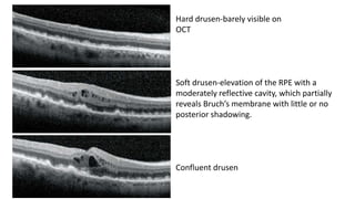
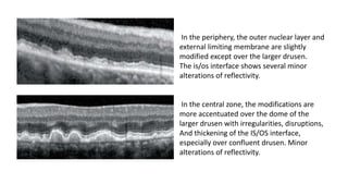
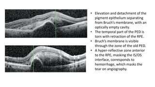
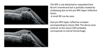
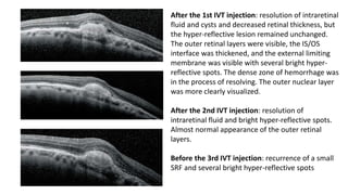
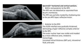
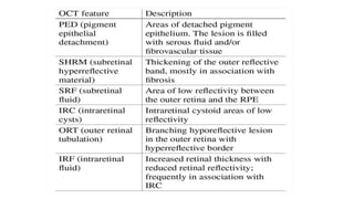
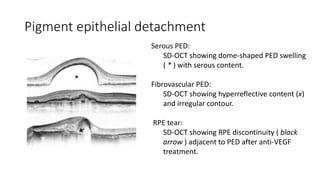
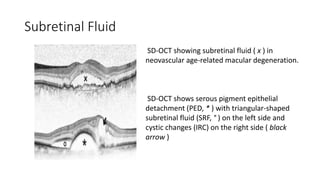
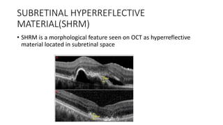
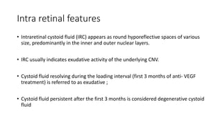
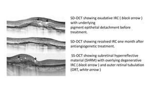
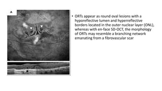
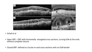
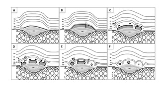
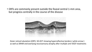
OCT IN WET AMD
DR SHRUTI LADDHA
Hard drusen-barely visible on
OCT
Soft drusen-elevation of the RPE with a
moderately reflective cavity, which partially
reveals Bruch’s membrane with little or no
posterior shadowing.
Confluent drusen
In the periphery, the outer nuclear layer and
external limiting membrane are slightly
modified except over the larger drusen.
The is/os interface shows several minor
alterations of reflectivity.
In the central zone, the modifications are
more accentuated over the dome of the
larger drusen with irregularities, disruptions,
And thickening of the IS/OS interface,
especially over confluent drusen. Minor
alterations of reflectivity.
• As drusen increase in size, they raise the RPE band, IS/ OS interface, and external
limiting membrane, which then can appear irregular, thinned, and disrupted. The
outer nuclear layer can also be modified, thinned, and invaded by a homogeneous
or jagged moderately reflective zone .
• These alterations reflect damage to inner and outer segments and photoreceptors
as a whole over the largest drusen. They therefore constitute valuable signs of the
impact of drusen and possible photoreceptor damage.
• Numerous soft drusen some of which
have resulted in atrophy.
• Loss of drusen in the atrophic area,
thinning of the rpe, loss of the external
limiting membrane and is/os interface
and alteration of the outer nuclear
layer.
• The inner layers of the retina remain
almost normal.
Small, regular dome-shaped juxtafoveal drusenoid PED has a less reflective cavity,
Allowing good visualization of bruch’s membrane.
There is adjacent alterations of the IS/OS interface and the denser zone that invades the
outer nuclear layer
Occult CNV can induce various types of retinal pigment epithelium detachment (PED):
• Predominantly Fibrovascular PED
The most frequent from of occult CNV (65%) is a fibrovascualar PED, in which the
exudative reaction is usually moderate
• CNV Accompanied by a Serous PED
This fluid accumulation is due to an intense exudative reaction. This form (12% to
15% of cases) consists of formation of a bullous PED adjacent to an occult CNV,
which is often located in a notch of the PED .
Occult CNV can subsequently invade the entire PED cavity.
TYPE 1 CNVM
Alteration of the outer nuclear layer just over
the PED with increased retinal thickness.
Normal visual acuity: 6/6
.
At 12th month: marked accentuation of
exudation with intraretinal fluid and especially
SRF VA: 6/9.
At 30th month: slight extension of the lesion
and marked deterioration of the SRF and the
PED. VA: 6/12 →Decision to initiate intravitreal
anti-VEGF therapy.
Presence of a fairly prominent, rounded,
regular, juxtafoveal PED associated with SRF
(surrounding the PED) and slightly increased
retinal thickness.
FA
Typical early occult CNV on FFA
• Moderate, regular flat with multiple domes of elevation, giving a
wavy appearance and Suggesting PED.
• The PED cavity is hypo-reflective and almost optically empty.
• Intraretinal fluid infiltration, causing slightly increased retinal
thickness and subretinal fluid with a sub foveal SRF.
Moderately large occult CNV.
Loss of the IS/OS interface over the PED; a zone of hyper-reflectivity and
several sub foveal bright hyper-reflective spots.
• Elevation and detachment of the
pigment epithelium separating
from Bruch’s membrane, with an
optically empty cavity.
• The temporal part of the PED is
torn with retraction of the RPE.
• Bruch’s membrane is visible
through the zone of the old PED.
• A hyper-reflective zone anterior
to the RPE, masking the IS/OS
interface, corresponds to
hemorrhage, which masks the
tear on angiography.
TYPE 2 CNVM
The RPE is not detached or separated from
Bruch’s membrane but is partially masked by
shadowing due to the pre-RPE hyper-reflective
lesion.
A small IRF can be seen.
Vast pre-RPE hyper-reflective complex
corresponding to classic CNV. The dense zone
anterior to the classic CNV probably
corresponds to retinal hemorrhage.
After the 1st IVT injection: resolution of intraretinal
fluid and cysts and decreased retinal thickness, but
the hyper-reflective lesion remained unchanged.
The outer retinal layers were visible, the IS/OS
interface was thickened, and the external limiting
membrane was visible with several bright hyper-
reflective spots. The dense zone of hemorrhage was
in the process of resolving. The outer nuclear layer
was more clearly visualized.
After the 2nd IVT injection: resolution of
intraretinal fluid and bright hyper-reflective spots.
Almost normal appearance of the outer retinal
layers.
Before the 3rd IVT injection: recurrence of a small
SRF and several bright hyper-reflective spots
Spectralis* horizontal and vertical sections.
• Within and posterior to the RPE:
The RPE was not detached or separated from
Bruch’s membrane.
The RPE was partially masked by shadowing due
to the pre-RPE hyper-reflective lesion.
• Anterior to the RPE:
The IS/OS interface was gradually thickened,
delineating a hyper-reflective complex anterior
to the RPE.
The outer nuclear layer was visible and invaded
by a more reflective zone, related to
hemorrhage.
Increased retinal thickness (687 μm), intraretinal
fluid, and cysts
Spectralis horizontal and vertical sections.
• Within and posterior to the RPE:
The RPE was partially masked.
The hyper-reflective lesion was denser and
more fibrotic.
• Anterior to the RPE:
No exudative signs.
The outer retinal layers were well-visualized
and well-organized.
The IS/OS interface remained thickened. The
subfoveal outer nuclear layer was well-
visualized. Thickness (Spectralis*):351 μm.
The fovea remained elevated by the hyper-
reflective lesion, suggesting progressive fibrosis
of the classic CNV.
SUMMARY
Pigment epithelial detachment
Serous PED:
SD-OCT showing dome-shaped PED swelling
( * ) with serous content.
Fibrovascular PED:
SD-OCT showing hyperreflective content (x)
and irregular contour.
RPE tear:
SD-OCT showing RPE discontinuity ( black
arrow ) adjacent to PED after anti-VEGF
treatment.
• RPE tears may result during the course of the disease and after anti-VEGF
treatment.
• Eyes with fibrovascular PEDs, where the CNV is attached to the RPE, are
especially prone to RPE tears.
• SD-OCT showed that RPE tears usually occur after the first anti-VEGF treatment,
as contraction of the CNV induces tension on bare RPE areas, leading to adjacent
tears if the RPE is not stabilized by CNV or attached to Bruch’s membrane
• Post-tear OCT images reveal a discontinuous RPE layer
• The subretinal compartment is located between the neurosensory retina and the
RPE.
• Subretinal fluid can be easily detected on OCT as a hyporeflective space under
the neurosensory retina
• The optical density ratio of SRF in SD-OCT may help in distinguishing CNV from
other entities associated with CNV and fluid exudation such as central serous
chorioretinopathy
• Optical density was significantly higher in subretinal fluid associated with CNV
Subretinal Fluid
SD-OCT showing subretinal fluid ( x ) in
neovascular age-related macular degeneration.
SD-OCT shows serous pigment epithelial
detachment (PED, * ) with triangular-shaped
subretinal fluid (SRF, ° ) on the left side and
cystic changes (IRC) on the right side ( black
arrow )
SUBRETINAL HYPERREFLECTIVE
MATERIAL(SHRM)
• SHRM is a morphological feature seen on OCT as hyperreflective
material located in subretinal space
• SHRM (both greater height and width)- worse outcome
• After anti-VEGF initiated ,within first 4 weeks SHRM decrease rapidly
,thereafter slowly
• SHRM is composed of fluid ,fibrin blood ,scar ,CNV
• Initiation of anti VEGF decrease endothelium permeability-decrease
vascular leakage, decrease fluid component
• Persistent SRHM-poor visual acuity
• Thick SRHM – toxic effect to photoreceptors, decrease normal
photoreceptor function
• Persistent SRHM- scar risk factor
Intra retinal features
• Intraretinal cystoid fluid (IRC) appears as round hyporeflective spaces of various
size, predominantly in the inner and outer nuclear layers.
• IRC usually indicates exudative activity of the underlying CNV.
• Cystoid fluid resolving during the loading interval (first 3 months of anti- VEGF
treatment) is referred to as exudative ;
• Cystoid fluid persistent after the first 3 months is considered degenerative cystoid
fluid
SD-OCT showing exudative IRC ( black arrow )
with underlying
pigment epithelial detachment before
treatment.
SD-OCT showing resolved IRC one month after
antiangiogenetic treatment.
SS-OCT showing subretinal hyperreflective
material (SHRM) with overlying degenerative
IRC ( black arrow ) and outer retinal tubulation
(ORT, white arrow )
OUTER RETINAL
TUBULATIONS
• Curcio et al
• Structural rearrangement of photoreceptors in response to retinal
injury
• Develop in the presence of significant photoreceptor disruption by
invagination of remaining cells and scrolling of the external limiting
membrane (ELM)
• ORTs appear as round oval lesions with a
hyporeflective lumen and hyperreflective
borders located in the outer nuclear layer (ONL),
whereas with en-face SD-OCT, the morphology
of ORTs may resemble a branching network
emanating from a fibrovascular scar
• Schall et al
• Open ORT – ORT with horizontally elongated cross sections, curving ELM at the ends
without complete closure
• Closed ORT- defined as circular or oval cross-sections with an ELM border
PATHOPHYSIOLOGY
• Progressive damage to photoreceptors may activate the Müller cells and promote
the expression of glial fibrillary acidic protein, facilitating ORT formation and
bifurcation.
• This fact may explain the significant association of ORTs with the presence of GA
in the present report, suggesting that a dysmorphic or absent RPE facilitates ORT
formation.
• ORTs are commonly present outside the foveal central 1-mm area,
but progress centrally in the course of the disease
Outer retinal tubulation (ORT). SD-OCT showing hyperreflective borders ( white arrow )
as well as SRHM and overlying neurosensory atrophy after multiple anti-VEGF treatments
• Should be differentiated from intraretinal or subretinal fluid cysts located at the
outer retinal layers.
• Intraretinal fluid cysts in cystoid macular edema (CME) have the arrangement as a
petaloid manner, while ORTs are randomly arranged at the macula.
• Pseudocysts are usually distinguished from ORTs because they are located in the
inner nuclear layer.
• Retinal tubulations are always located at the level of the ONL

OCT IN WET AMD

  • 1.
    OCT IN WETAMD DR SHRUTI LADDHA
  • 2.
    Hard drusen-barely visibleon OCT Soft drusen-elevation of the RPE with a moderately reflective cavity, which partially reveals Bruch’s membrane with little or no posterior shadowing. Confluent drusen
  • 3.
    In the periphery,the outer nuclear layer and external limiting membrane are slightly modified except over the larger drusen. The is/os interface shows several minor alterations of reflectivity. In the central zone, the modifications are more accentuated over the dome of the larger drusen with irregularities, disruptions, And thickening of the IS/OS interface, especially over confluent drusen. Minor alterations of reflectivity.
  • 4.
    • As drusenincrease in size, they raise the RPE band, IS/ OS interface, and external limiting membrane, which then can appear irregular, thinned, and disrupted. The outer nuclear layer can also be modified, thinned, and invaded by a homogeneous or jagged moderately reflective zone . • These alterations reflect damage to inner and outer segments and photoreceptors as a whole over the largest drusen. They therefore constitute valuable signs of the impact of drusen and possible photoreceptor damage.
  • 5.
    • Numerous softdrusen some of which have resulted in atrophy. • Loss of drusen in the atrophic area, thinning of the rpe, loss of the external limiting membrane and is/os interface and alteration of the outer nuclear layer. • The inner layers of the retina remain almost normal.
  • 6.
    Small, regular dome-shapedjuxtafoveal drusenoid PED has a less reflective cavity, Allowing good visualization of bruch’s membrane. There is adjacent alterations of the IS/OS interface and the denser zone that invades the outer nuclear layer
  • 7.
    Occult CNV caninduce various types of retinal pigment epithelium detachment (PED): • Predominantly Fibrovascular PED The most frequent from of occult CNV (65%) is a fibrovascualar PED, in which the exudative reaction is usually moderate • CNV Accompanied by a Serous PED This fluid accumulation is due to an intense exudative reaction. This form (12% to 15% of cases) consists of formation of a bullous PED adjacent to an occult CNV, which is often located in a notch of the PED . Occult CNV can subsequently invade the entire PED cavity.
  • 8.
  • 9.
    Alteration of theouter nuclear layer just over the PED with increased retinal thickness. Normal visual acuity: 6/6 . At 12th month: marked accentuation of exudation with intraretinal fluid and especially SRF VA: 6/9. At 30th month: slight extension of the lesion and marked deterioration of the SRF and the PED. VA: 6/12 →Decision to initiate intravitreal anti-VEGF therapy.
  • 10.
    Presence of afairly prominent, rounded, regular, juxtafoveal PED associated with SRF (surrounding the PED) and slightly increased retinal thickness. FA
  • 11.
    Typical early occultCNV on FFA • Moderate, regular flat with multiple domes of elevation, giving a wavy appearance and Suggesting PED. • The PED cavity is hypo-reflective and almost optically empty. • Intraretinal fluid infiltration, causing slightly increased retinal thickness and subretinal fluid with a sub foveal SRF.
  • 12.
    Moderately large occultCNV. Loss of the IS/OS interface over the PED; a zone of hyper-reflectivity and several sub foveal bright hyper-reflective spots.
  • 13.
    • Elevation anddetachment of the pigment epithelium separating from Bruch’s membrane, with an optically empty cavity. • The temporal part of the PED is torn with retraction of the RPE. • Bruch’s membrane is visible through the zone of the old PED. • A hyper-reflective zone anterior to the RPE, masking the IS/OS interface, corresponds to hemorrhage, which masks the tear on angiography.
  • 14.
  • 15.
    The RPE isnot detached or separated from Bruch’s membrane but is partially masked by shadowing due to the pre-RPE hyper-reflective lesion. A small IRF can be seen. Vast pre-RPE hyper-reflective complex corresponding to classic CNV. The dense zone anterior to the classic CNV probably corresponds to retinal hemorrhage.
  • 16.
    After the 1stIVT injection: resolution of intraretinal fluid and cysts and decreased retinal thickness, but the hyper-reflective lesion remained unchanged. The outer retinal layers were visible, the IS/OS interface was thickened, and the external limiting membrane was visible with several bright hyper- reflective spots. The dense zone of hemorrhage was in the process of resolving. The outer nuclear layer was more clearly visualized. After the 2nd IVT injection: resolution of intraretinal fluid and bright hyper-reflective spots. Almost normal appearance of the outer retinal layers. Before the 3rd IVT injection: recurrence of a small SRF and several bright hyper-reflective spots
  • 17.
    Spectralis* horizontal andvertical sections. • Within and posterior to the RPE: The RPE was not detached or separated from Bruch’s membrane. The RPE was partially masked by shadowing due to the pre-RPE hyper-reflective lesion. • Anterior to the RPE: The IS/OS interface was gradually thickened, delineating a hyper-reflective complex anterior to the RPE. The outer nuclear layer was visible and invaded by a more reflective zone, related to hemorrhage. Increased retinal thickness (687 μm), intraretinal fluid, and cysts
  • 18.
    Spectralis horizontal andvertical sections. • Within and posterior to the RPE: The RPE was partially masked. The hyper-reflective lesion was denser and more fibrotic. • Anterior to the RPE: No exudative signs. The outer retinal layers were well-visualized and well-organized. The IS/OS interface remained thickened. The subfoveal outer nuclear layer was well- visualized. Thickness (Spectralis*):351 μm. The fovea remained elevated by the hyper- reflective lesion, suggesting progressive fibrosis of the classic CNV.
  • 19.
  • 21.
    Pigment epithelial detachment SerousPED: SD-OCT showing dome-shaped PED swelling ( * ) with serous content. Fibrovascular PED: SD-OCT showing hyperreflective content (x) and irregular contour. RPE tear: SD-OCT showing RPE discontinuity ( black arrow ) adjacent to PED after anti-VEGF treatment.
  • 22.
    • RPE tearsmay result during the course of the disease and after anti-VEGF treatment. • Eyes with fibrovascular PEDs, where the CNV is attached to the RPE, are especially prone to RPE tears. • SD-OCT showed that RPE tears usually occur after the first anti-VEGF treatment, as contraction of the CNV induces tension on bare RPE areas, leading to adjacent tears if the RPE is not stabilized by CNV or attached to Bruch’s membrane • Post-tear OCT images reveal a discontinuous RPE layer
  • 23.
    • The subretinalcompartment is located between the neurosensory retina and the RPE. • Subretinal fluid can be easily detected on OCT as a hyporeflective space under the neurosensory retina • The optical density ratio of SRF in SD-OCT may help in distinguishing CNV from other entities associated with CNV and fluid exudation such as central serous chorioretinopathy • Optical density was significantly higher in subretinal fluid associated with CNV
  • 24.
    Subretinal Fluid SD-OCT showingsubretinal fluid ( x ) in neovascular age-related macular degeneration. SD-OCT shows serous pigment epithelial detachment (PED, * ) with triangular-shaped subretinal fluid (SRF, ° ) on the left side and cystic changes (IRC) on the right side ( black arrow )
  • 25.
    SUBRETINAL HYPERREFLECTIVE MATERIAL(SHRM) • SHRMis a morphological feature seen on OCT as hyperreflective material located in subretinal space
  • 26.
    • SHRM (bothgreater height and width)- worse outcome • After anti-VEGF initiated ,within first 4 weeks SHRM decrease rapidly ,thereafter slowly • SHRM is composed of fluid ,fibrin blood ,scar ,CNV • Initiation of anti VEGF decrease endothelium permeability-decrease vascular leakage, decrease fluid component
  • 27.
    • Persistent SRHM-poorvisual acuity • Thick SRHM – toxic effect to photoreceptors, decrease normal photoreceptor function • Persistent SRHM- scar risk factor
  • 28.
    Intra retinal features •Intraretinal cystoid fluid (IRC) appears as round hyporeflective spaces of various size, predominantly in the inner and outer nuclear layers. • IRC usually indicates exudative activity of the underlying CNV. • Cystoid fluid resolving during the loading interval (first 3 months of anti- VEGF treatment) is referred to as exudative ; • Cystoid fluid persistent after the first 3 months is considered degenerative cystoid fluid
  • 29.
    SD-OCT showing exudativeIRC ( black arrow ) with underlying pigment epithelial detachment before treatment. SD-OCT showing resolved IRC one month after antiangiogenetic treatment. SS-OCT showing subretinal hyperreflective material (SHRM) with overlying degenerative IRC ( black arrow ) and outer retinal tubulation (ORT, white arrow )
  • 30.
  • 31.
    • Curcio etal • Structural rearrangement of photoreceptors in response to retinal injury • Develop in the presence of significant photoreceptor disruption by invagination of remaining cells and scrolling of the external limiting membrane (ELM)
  • 32.
    • ORTs appearas round oval lesions with a hyporeflective lumen and hyperreflective borders located in the outer nuclear layer (ONL), whereas with en-face SD-OCT, the morphology of ORTs may resemble a branching network emanating from a fibrovascular scar
  • 33.
    • Schall etal • Open ORT – ORT with horizontally elongated cross sections, curving ELM at the ends without complete closure • Closed ORT- defined as circular or oval cross-sections with an ELM border
  • 34.
    PATHOPHYSIOLOGY • Progressive damageto photoreceptors may activate the Müller cells and promote the expression of glial fibrillary acidic protein, facilitating ORT formation and bifurcation. • This fact may explain the significant association of ORTs with the presence of GA in the present report, suggesting that a dysmorphic or absent RPE facilitates ORT formation.
  • 36.
    • ORTs arecommonly present outside the foveal central 1-mm area, but progress centrally in the course of the disease Outer retinal tubulation (ORT). SD-OCT showing hyperreflective borders ( white arrow ) as well as SRHM and overlying neurosensory atrophy after multiple anti-VEGF treatments
  • 37.
    • Should bedifferentiated from intraretinal or subretinal fluid cysts located at the outer retinal layers. • Intraretinal fluid cysts in cystoid macular edema (CME) have the arrangement as a petaloid manner, while ORTs are randomly arranged at the macula. • Pseudocysts are usually distinguished from ORTs because they are located in the inner nuclear layer. • Retinal tubulations are always located at the level of the ONL