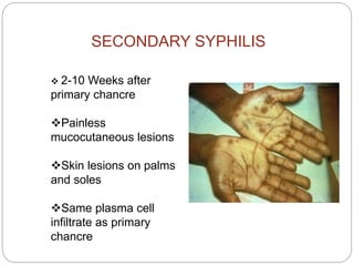
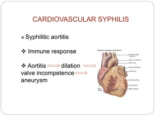
"He who knows syphilis,
knows medicine"
Sir William Osler
SYPHILIS
NIBIN M
MBBS Student
Calicut Medical College
INTRODUCTION
 Syphilis is a chronic sexually
transmitted disease with varied
clinical and pathological
manifestations
Causative org: Treponema
pallidum
 Transmission: Sexual and
vertical
SPIROCHETES
Long, slender, helically tightly coiled bacteria
Gram-negative
Corkscrew motility
Can be free living or parasitic
Best-known are those which cause disease: Syphilis
and Lyme’s disease
TRYPONEMA PALLIDUM
Microaerophilic spirochete
Not seen in gram stain
By silver stains and
immunofluorescence or dark-
field microscopy
PATHOGENESIS
 Penetration
• Via skin and mucuos membrane
through abrasions during sexual contact
•Also transplacentally
Dissemination
• Lymphatic and haematological
PATHOLOGY
 Proliferative endarteritis
 Ischaemia produced by vascular lesions
 Immune response
 Chancres and rashes – T-cells, plasma cells and
macrophages
 TprK – structural diversity by gene recombination
PRIMARY SYPHILIS
 3 weeks after infection
 CHANCRE : Single firm non-tender, raised, red
lesion •Progresses from macule to papule to ulcer
• Plasma cells , scattered macrophages,
lymphocytes
• Proliferative endarteritis
• Endothelial cell activation to proliferation to
fibrosis
Regional lymphadenopathy: classically rubbery,
painless, bilateral
 Serologic tests for syphilis may not be positive during
early primary syphilis
SECONDARY SYPHILIS
 2-10 Weeks after
primary chancre
Painless
mucocutaneous lesions
Skin lesions on palms
and soles
Same plasma cell
infiltrate as primary
chancre
SECONDARY SYPHILIS
 Condylomata lata – broad based elevated
plaques
- in moist areas of skin
Other manifestations of
secondary syphilis
- lymphadenopathy,Rashes,
-Malaise, fever,weight loss
- Alopecia
- Neurosyphilis
 Serological tests – highest
especially in red lesions in vagina
or mouth
Latent Syphilis
 Host suppresses the infection enough so that
no lesions are clinically apparent
 Only evidence is positive serologic test for
syphilis
 May occur between primary and secondary
stages, between secondary relapses, and
after secondary stage
 Categories:
 Early latent: <1 year duration
 Late latent: 1 year duration
TERTIARY SYPHILIS
CARDIOVASCULAR SYPHILIS
NEUROSYPHILIS
BENIGN TERTIARY SYPHILIS
CARDIOVASCULAR SYPHILIS
 Syphilitic aortitis
 Immune response
 Aortitis dilation
valve incompetence
aneurysm
CARDIOVASCULAR SYPHILIS –
MORPHOLOGIC CHANGES
 Aortitis – endarteritis of vaso vasorum of proximal
aorta
 Occlusion of vaso vasorum- scaring of media of
proximal aortic wall
- loss of elasticity
Narrowing of coronary artery ostia – subintimal
scarring
NEUROSYPHILIS
SYMPTOMATIC
• 10% untreated individuals
•Meningovascular, paretic, tabes dorsalis
•Taboparesis
•AIDS patients - meningovascular
Meningovascular neurosyphilis
• Chronic meningitis
• obliterative endarteritis
Paretic neurosyphilis
• progressive mental deficits leading to
dementia
• parenchymal damage in cerebral cortex
Tabes dorsalis
• Damage to sensory nerves – loss of
position&pain sense
• loss of axons and myelin in dorsal roots
ASYMPTOMATIC NEUROSYPHILIS
• One third of neurosyphilic cases
CSF shows:
•Lymphocytic pleocytosis
•Elevated protein and usually normal
glucose concentrations
•VDRL test is usually reactive.
• It can mimic tuberculous or fungal
meningitis or aseptic meningitis of
various causes.
• Often involves the base of the brain
and may result in unilateral or bilateral
cranial nerve palsies.
• Without treatment, syphilitic meningitis
usually resolves, like the other
manifestations
BENIGN TERTIARY SYPHILIS
 Gummas in bone, skin , and mucous membrane
 Gummas – nodular lesions
- delayed hypersensitivity
 Bone – pain, tenderness, swelling, pathologic
fractures
 Skin & mucuos membrane – Nodular lesions
- ulcerative lesions
Syphilitic gummas
 White, gray, and rubbery
 single or multiple, vary in size
 In liver – scarring causes hepar lobatum
 Histologically, centre- coagulated necrotic material
margins - macrophages, fibroblasts,
- large no. of MNL
• Treponemes are scanty
Syphilis Diagnosis And
Treatment
Aspects of Syphilis Diagnosis
1. Clinical history
2. Physical examination
3. Laboratory diagnosis
Clinical History
Assess:
 History of syphilis
 Known contact to an early case of syphilis
 Typical signs or symptoms of syphilis in the past
12 months
 Most recent serologic test for syphilis
Physical Examination
 Oral cavity
 Lymph nodes
 Skin
 Palms and soles
 Genitalia and perianal area
 Neurologic examination
Laboratory Diagnosis
 Identification of Treponema pallidum in lesions
 Darkfield microscopy
 Direct fluorescent antibody - T. pallidum (DFA-TP)
 Serologic tests
 Nontreponemal tests
 Treponemal tests
Nontreponemal Serologic Tests
 Principles
 Measure antibody directed against a cardiolipin-lecithin-
cholesterol antigen
 Not specific for T. pallidum
 Titers usually correlate with disease activity and results are
reported quantitatively
 May be reactive for life
 Nontreponemal tests include VDRL, RPR
Diagnosis
Treponemal Serologic Tests
 Principles
 Measure antibody directed against T. pallidum antigens
 Qualitative
 Usually reactive for life
 Treponemal tests include , FTA-ABS, EIA
Usefullness
 Primary syphilis – both moderatively sensitive
 secondary – very sensitive
 Tertiary and latent – Treponemal sensitive
- non treponemal less
 To monitor therapy – non –treponemal
 Good for screening ,confirmatory test req
 False positive results – Pregnancy, autoimmune
diseases, infections
TREATMENT
 PENICILLIN
 Intramuscularly or
intravenously
 Treatment at any stage
PREVENTION
 Abstinance is the most effective way to
prevent the contraction of the disease
 practice safe sex
 be tested regularly for syphilis if married or
sexually active
 avoid direct contact with blood, sores or bodily
fluid
learn about safe sex and injection practices
 get tested for syphilis if pregnant


Syphilis

  • 1.
    "He who knowssyphilis, knows medicine" Sir William Osler SYPHILIS NIBIN M MBBS Student Calicut Medical College
  • 2.
    INTRODUCTION  Syphilis isa chronic sexually transmitted disease with varied clinical and pathological manifestations Causative org: Treponema pallidum  Transmission: Sexual and vertical
  • 3.
    SPIROCHETES Long, slender, helicallytightly coiled bacteria Gram-negative Corkscrew motility Can be free living or parasitic Best-known are those which cause disease: Syphilis and Lyme’s disease
  • 4.
    TRYPONEMA PALLIDUM Microaerophilic spirochete Notseen in gram stain By silver stains and immunofluorescence or dark- field microscopy
  • 5.
    PATHOGENESIS  Penetration • Viaskin and mucuos membrane through abrasions during sexual contact •Also transplacentally Dissemination • Lymphatic and haematological
  • 6.
    PATHOLOGY  Proliferative endarteritis Ischaemia produced by vascular lesions  Immune response  Chancres and rashes – T-cells, plasma cells and macrophages  TprK – structural diversity by gene recombination
  • 8.
    PRIMARY SYPHILIS  3weeks after infection  CHANCRE : Single firm non-tender, raised, red lesion •Progresses from macule to papule to ulcer • Plasma cells , scattered macrophages, lymphocytes • Proliferative endarteritis • Endothelial cell activation to proliferation to fibrosis Regional lymphadenopathy: classically rubbery, painless, bilateral  Serologic tests for syphilis may not be positive during early primary syphilis
  • 10.
    SECONDARY SYPHILIS  2-10Weeks after primary chancre Painless mucocutaneous lesions Skin lesions on palms and soles Same plasma cell infiltrate as primary chancre
  • 11.
    SECONDARY SYPHILIS  Condylomatalata – broad based elevated plaques - in moist areas of skin
  • 12.
    Other manifestations of secondarysyphilis - lymphadenopathy,Rashes, -Malaise, fever,weight loss - Alopecia - Neurosyphilis  Serological tests – highest especially in red lesions in vagina or mouth
  • 13.
    Latent Syphilis  Hostsuppresses the infection enough so that no lesions are clinically apparent  Only evidence is positive serologic test for syphilis  May occur between primary and secondary stages, between secondary relapses, and after secondary stage  Categories:  Early latent: <1 year duration  Late latent: 1 year duration
  • 14.
  • 15.
    CARDIOVASCULAR SYPHILIS  Syphiliticaortitis  Immune response  Aortitis dilation valve incompetence aneurysm
  • 16.
    CARDIOVASCULAR SYPHILIS – MORPHOLOGICCHANGES  Aortitis – endarteritis of vaso vasorum of proximal aorta  Occlusion of vaso vasorum- scaring of media of proximal aortic wall - loss of elasticity Narrowing of coronary artery ostia – subintimal scarring
  • 17.
    NEUROSYPHILIS SYMPTOMATIC • 10% untreatedindividuals •Meningovascular, paretic, tabes dorsalis •Taboparesis •AIDS patients - meningovascular
  • 18.
    Meningovascular neurosyphilis • Chronicmeningitis • obliterative endarteritis Paretic neurosyphilis • progressive mental deficits leading to dementia • parenchymal damage in cerebral cortex Tabes dorsalis • Damage to sensory nerves – loss of position&pain sense • loss of axons and myelin in dorsal roots
  • 19.
    ASYMPTOMATIC NEUROSYPHILIS • Onethird of neurosyphilic cases CSF shows: •Lymphocytic pleocytosis •Elevated protein and usually normal glucose concentrations •VDRL test is usually reactive. • It can mimic tuberculous or fungal meningitis or aseptic meningitis of various causes. • Often involves the base of the brain and may result in unilateral or bilateral cranial nerve palsies. • Without treatment, syphilitic meningitis usually resolves, like the other manifestations
  • 20.
    BENIGN TERTIARY SYPHILIS Gummas in bone, skin , and mucous membrane  Gummas – nodular lesions - delayed hypersensitivity  Bone – pain, tenderness, swelling, pathologic fractures  Skin & mucuos membrane – Nodular lesions - ulcerative lesions
  • 22.
    Syphilitic gummas  White,gray, and rubbery  single or multiple, vary in size  In liver – scarring causes hepar lobatum  Histologically, centre- coagulated necrotic material margins - macrophages, fibroblasts, - large no. of MNL • Treponemes are scanty
  • 23.
  • 24.
    Aspects of SyphilisDiagnosis 1. Clinical history 2. Physical examination 3. Laboratory diagnosis
  • 25.
    Clinical History Assess:  Historyof syphilis  Known contact to an early case of syphilis  Typical signs or symptoms of syphilis in the past 12 months  Most recent serologic test for syphilis
  • 26.
    Physical Examination  Oralcavity  Lymph nodes  Skin  Palms and soles  Genitalia and perianal area  Neurologic examination
  • 27.
    Laboratory Diagnosis  Identificationof Treponema pallidum in lesions  Darkfield microscopy  Direct fluorescent antibody - T. pallidum (DFA-TP)  Serologic tests  Nontreponemal tests  Treponemal tests
  • 28.
    Nontreponemal Serologic Tests Principles  Measure antibody directed against a cardiolipin-lecithin- cholesterol antigen  Not specific for T. pallidum  Titers usually correlate with disease activity and results are reported quantitatively  May be reactive for life  Nontreponemal tests include VDRL, RPR Diagnosis
  • 29.
    Treponemal Serologic Tests Principles  Measure antibody directed against T. pallidum antigens  Qualitative  Usually reactive for life  Treponemal tests include , FTA-ABS, EIA
  • 30.
    Usefullness  Primary syphilis– both moderatively sensitive  secondary – very sensitive  Tertiary and latent – Treponemal sensitive - non treponemal less  To monitor therapy – non –treponemal  Good for screening ,confirmatory test req  False positive results – Pregnancy, autoimmune diseases, infections
  • 31.
    TREATMENT  PENICILLIN  Intramuscularlyor intravenously  Treatment at any stage
  • 32.
    PREVENTION  Abstinance isthe most effective way to prevent the contraction of the disease  practice safe sex  be tested regularly for syphilis if married or sexually active  avoid direct contact with blood, sores or bodily fluid learn about safe sex and injection practices  get tested for syphilis if pregnant 