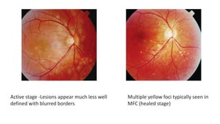
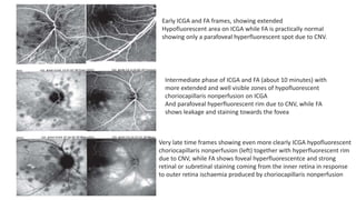
MULTIFOCAL CHOROIDITIS
DR SHRUTI LADDHA
• MC-Female
• Young to middle age
• small randomly distributed chorioretinal scars
• Responds to corticosteroids
• Recurence common
• CNV develops in 1/3rd of cases
• Lesions tends to leave scars
CLINICAL FEATURES
• Photopsia and scotoma
• Photopsia is present also when there is no clinical evidence of reactivation of the
disease
• May be bilateral –asymmetrical or may be unilateral
• Anterior granulomatous or sometimes non granulomatous inflammation
• FUNDUS-atrophic yellow-white foci with pigment spots that sometimes can
become adjacent to each other and form a ribbon of pearls.
• posterior pole or the periphery or both.
• In the active phases of disease new lesions are not always visible and can be very
discreet on FA, whereas ICGA is the most sensitive method to detect new lesions
Active stage -Lesions appear much less well
defined with blurred borders
Multiple yellow foci typically seen in
MFC (healed stage)
ICGA
• Scars-hypofluroscent
• Active –new hypofluroscent patches, sometimes peripapillary hypofluroscence-
enlargement of blind spot-regress if therapy started early
Left picture shows quiet stage. Middle picture shows reactivation of MFC 8 months later with a parafoveal CNV.
Far right image shows healed stage of choriocapillaritis and cicatricial membrane (arrows) 6 months later after
systemic corticosteroid therapy
FFA
• Scars –late staining or masking effect due to pigmentation
• Active- silent on FFA or late hyperfluroscence
• If CNV present-classical signs of leakage
Left image shows late hyperfluorescence indicating both CNV (parafoveal) and reactivation of MFC. Image
on the right taken 6 months after introduction of systemic corticosteroid therapy shows complete healing with only
scars (late hyperfluorescence) with no staining and no CNV activity
Early ICGA and FA frames, showing extended
Hypofluorescent area on ICGA while FA is practically normal
showing only a parafoveal hyperfluorescent spot due to CNV.
Intermediate phase of ICGA and FA (about 10 minutes) with
more extended and well visible zones of hypofluorescent
choriocapillaris nonperfusion on ICGA
And parafoveal hyperfluorescent rim due to CNV, while FA
shows leakage and staining towards the fovea
Very late time frames showing even more clearly ICGA hypofluorescent
choriocapillaris nonperfusion (left) together with hyperfluorescent rim
due to CNV, while FA shows foveal hyperfluorescentce and strong
retinal or subretinal staining coming from the inner retina in response
to outer retina ischaemia produced by choriocapillaris nonperfusion
OCT
All the 3 areas of foci show profound re-modelling
of outer retina with a bump (nodular lesion at and
above the level of RPE) for all 3 foci. The overlying
inner retinal is also disorganised and there is
pooling of fluid under the retinal around the 3 foci
FAF
• Increased autofluorescence in those areas that have silent (meaning without FA
signs), ICGA hypofluorescent lesions
• Hypoautofluorescence in the cicatricial areas.
• After corticosteroid therapy hyperautofluorescence disappears in parallel with
resolution of ICGA hypofluorescence.
• The areas showing hyperautofluorescence go beyond the ICGA hypofluorescent
areas indicating that dysfunction of cells and inflammatory involvement go even
beyond the areas detected by ICGA.
Bright hyperautofluorescence is present all over the fundus (left image) that
decreases sustantially after corticosteroid treatment (right)
VISUAL FIELD
• Small scotomas corresponding to chorioretinal scars
• Active phase-scotomas are larger and correspond to choriocapillaris non perfusion
shown on ICGA.
• Visual field recovery is well correlated with the regression of ICGA hypofluorescent
areas.
ERG
• MERG- diffusely depressed
• MERG recovered partially only in 78% of patients.
• This might be the explanation why patients continue to be symptomatic and
have photopsia despite apparent quiescence of the disease
TREATMENT
• Oral steroid
• Sub tenons corticosteroid-CME
• CNVM – generally regress in response to steroid ,if not then additional anti-VEGF
PUNCTATE INNER CHOROID
• Subset of multifocal choroiditis.
• Similar characteristics except smaller lesion.
• More common in young, myopic females.
• CNVM
• T/t – steroids ,PST
SUBRETINAL FIBROSIS
• Some patient may show to subretinal fibrosis
• Suggest subclinical MFC
• It might also reflect the propensity of the disease to develop subretinal
neovascularization that might result in spontaneous fibrosis in some cases.
PRESUMED OCULAR HISTOPLASMOSIS
SYNDROME (POHS)
• Histoplasma capsulatum-fungus
• Missipi, Ohio, Italy, Central America, Turkey, Israel and Australia.
• Pulmonary inhalation
• In case of a positive histoplasma skin test and the presence of multifocal
choroiditis the diagnosis of POHS has to be considered
• In non-endemic areas this terminology should not be applied and the diagnosis of
POHS should be questioned unless there is a positive skin test
The criteria that differentiate POHS from
multifocal choroiditis are
• the absence of anterior chamber reaction,
• punched-out multifocal lesions also in the
periphery (histospots)
• peripapillary scaring
• asymptomatic course unless neovascular
membranes develop.
• ICGA-does not show hypofluorescent
areas seen in MFC in case of active disease
but shows pinpoint hyperfluorescent ICGA
spots not detected by funduscopy or
fluorescein angiography.
Serpiginous Choroiditis
• Bilateral, chronic, progressive, recurrent inflammation of the choriocapillaris,
choroid and retinal pigment epithelium, of unknown aetiology.
• Disease was called geographic choroidopathy and placoid chorioretinitis
• Rare cause of posterior uveitis (usually< 5%)
• Affect mostly healthy young to middle-aged adults
• M>F
CLINICAL FEATURES
• Unilateral
• Decrease in central vision,
• Metamorphopsia,
• scotoma.
• No inflammation is usually seen in the
anterior segment or anterior vitreous.
• Peripapillary serpentine lesions in the
fundus are characteristic
Classic peripapillary case of serpiginous
choroiditis with focal choriocapillaris and
RPE atrophy, RPE clumping at the border of
the lesions and fibrosis
PERIPAPILLARY GEOGRAPHIC SC
• 80%
• begins with patches of grayish or creamy yellow sub-retinal infiltrates originating
in the peripapillary area and progressing usually toward the periphery like a
serpentine.
• The overlying retina is usually edematous with rare cases of serous retinal
detachments.
• Resolves in 6-8 weeks leaving focal choriocapillaris and RPE atrophy.
• Recurrences usually occur at the edges of previous atrophic scars
• Late stage-chorioretinal atrophy, subretinal fibrosis, and extensive RPE pigment
clumping.
MACULAR SC
• Atypical
• Begins at macula
• Worst prognosis –due to early macular
involvement and high risk of CNVM
AMPIGINOUS CHOROIDITIS
• Also described as “relentless placoid chorioretinitis”
• Evolution of acute posterior multifocal placoid pigment epitheliopathy (APMPPE)
can mimic the clinical course of SC.
• New small, isolated round white plaque-like lesions similar to those seen in
APMPPE.
• These isolated lesions coalesced leaving the typical serpiginous atrophic lesions
FFA
• The active lesions, at the borders of the old atrophic lesions, block fluorescein early and show
diffuse late staining and leakage of dye.
• In the atrophic areas, there is early hypofluorescence secondary to atrophy of choriocapillaris and
progressive hyperfluorescence at the margins of the lesions
• phlebitis
Active inferior lesions are dark at early phases of fluorescein and become hyperfluorescent at late phase of
fluorescein angiogram. It is associated with a perivascular inflammatory leakage of the infero-temporal vein.
A diffuse late staining of the underlying sclera can also be observed in the superior inactive lesions
ICGA
• Best to evaluate extend and activity of disease and its follow-up
• Hypofluorescent areas beginning from the early to the late phase which
supposed to correspond to choriocapillaris non-perfusion or delayed filling and
corresponding to a primary inflammatory choriocapillaropathy.
• Undetected lesions with the fluorescein angiography can be apparent on ICGA.
OCT
• CME
• Retinal spot atrophy-diffuse thinning of retina
• Loss of photoreceptors and RPE
VISUAL FIELDS
• Scotoma corresponding to geographic lesions
PATHOGENESIS
• Inflammation
• HLA B7 or C3 decreased blood level
• May be infectious-TB,HZV
• Vasculopathy either primary or secondary to systemic disease and characteristic
angiographic findings of choriocapillaris non-perfusion,
COMPLICATIONS
• CNVM
• Branch retinal vein occlusion,
• Periphlebitis
• Pigment epithelium detachment
• Serous retinal detachment
• Cystoid macular oedema
• Optic disk neovascularisation,
• Serous detachments
• Subretinal fibrosis.
TREATMENT
• Fundus photo and angiography may be necessary to decide the treatment
• Steroids and immunosuppressive therapy

Multifocal choroiditis

  • 1.
  • 2.
    • MC-Female • Youngto middle age • small randomly distributed chorioretinal scars • Responds to corticosteroids • Recurence common • CNV develops in 1/3rd of cases • Lesions tends to leave scars
  • 3.
    CLINICAL FEATURES • Photopsiaand scotoma • Photopsia is present also when there is no clinical evidence of reactivation of the disease • May be bilateral –asymmetrical or may be unilateral • Anterior granulomatous or sometimes non granulomatous inflammation • FUNDUS-atrophic yellow-white foci with pigment spots that sometimes can become adjacent to each other and form a ribbon of pearls. • posterior pole or the periphery or both. • In the active phases of disease new lesions are not always visible and can be very discreet on FA, whereas ICGA is the most sensitive method to detect new lesions
  • 4.
    Active stage -Lesionsappear much less well defined with blurred borders Multiple yellow foci typically seen in MFC (healed stage)
  • 5.
    ICGA • Scars-hypofluroscent • Active–new hypofluroscent patches, sometimes peripapillary hypofluroscence- enlargement of blind spot-regress if therapy started early Left picture shows quiet stage. Middle picture shows reactivation of MFC 8 months later with a parafoveal CNV. Far right image shows healed stage of choriocapillaritis and cicatricial membrane (arrows) 6 months later after systemic corticosteroid therapy
  • 6.
    FFA • Scars –latestaining or masking effect due to pigmentation • Active- silent on FFA or late hyperfluroscence • If CNV present-classical signs of leakage Left image shows late hyperfluorescence indicating both CNV (parafoveal) and reactivation of MFC. Image on the right taken 6 months after introduction of systemic corticosteroid therapy shows complete healing with only scars (late hyperfluorescence) with no staining and no CNV activity
  • 7.
    Early ICGA andFA frames, showing extended Hypofluorescent area on ICGA while FA is practically normal showing only a parafoveal hyperfluorescent spot due to CNV. Intermediate phase of ICGA and FA (about 10 minutes) with more extended and well visible zones of hypofluorescent choriocapillaris nonperfusion on ICGA And parafoveal hyperfluorescent rim due to CNV, while FA shows leakage and staining towards the fovea Very late time frames showing even more clearly ICGA hypofluorescent choriocapillaris nonperfusion (left) together with hyperfluorescent rim due to CNV, while FA shows foveal hyperfluorescentce and strong retinal or subretinal staining coming from the inner retina in response to outer retina ischaemia produced by choriocapillaris nonperfusion
  • 8.
    OCT All the 3areas of foci show profound re-modelling of outer retina with a bump (nodular lesion at and above the level of RPE) for all 3 foci. The overlying inner retinal is also disorganised and there is pooling of fluid under the retinal around the 3 foci
  • 9.
    FAF • Increased autofluorescencein those areas that have silent (meaning without FA signs), ICGA hypofluorescent lesions • Hypoautofluorescence in the cicatricial areas. • After corticosteroid therapy hyperautofluorescence disappears in parallel with resolution of ICGA hypofluorescence. • The areas showing hyperautofluorescence go beyond the ICGA hypofluorescent areas indicating that dysfunction of cells and inflammatory involvement go even beyond the areas detected by ICGA.
  • 10.
    Bright hyperautofluorescence ispresent all over the fundus (left image) that decreases sustantially after corticosteroid treatment (right)
  • 11.
    VISUAL FIELD • Smallscotomas corresponding to chorioretinal scars • Active phase-scotomas are larger and correspond to choriocapillaris non perfusion shown on ICGA. • Visual field recovery is well correlated with the regression of ICGA hypofluorescent areas.
  • 12.
    ERG • MERG- diffuselydepressed • MERG recovered partially only in 78% of patients. • This might be the explanation why patients continue to be symptomatic and have photopsia despite apparent quiescence of the disease
  • 13.
    TREATMENT • Oral steroid •Sub tenons corticosteroid-CME • CNVM – generally regress in response to steroid ,if not then additional anti-VEGF
  • 14.
    PUNCTATE INNER CHOROID •Subset of multifocal choroiditis. • Similar characteristics except smaller lesion. • More common in young, myopic females. • CNVM • T/t – steroids ,PST
  • 15.
    SUBRETINAL FIBROSIS • Somepatient may show to subretinal fibrosis • Suggest subclinical MFC • It might also reflect the propensity of the disease to develop subretinal neovascularization that might result in spontaneous fibrosis in some cases.
  • 16.
    PRESUMED OCULAR HISTOPLASMOSIS SYNDROME(POHS) • Histoplasma capsulatum-fungus • Missipi, Ohio, Italy, Central America, Turkey, Israel and Australia. • Pulmonary inhalation • In case of a positive histoplasma skin test and the presence of multifocal choroiditis the diagnosis of POHS has to be considered • In non-endemic areas this terminology should not be applied and the diagnosis of POHS should be questioned unless there is a positive skin test
  • 17.
    The criteria thatdifferentiate POHS from multifocal choroiditis are • the absence of anterior chamber reaction, • punched-out multifocal lesions also in the periphery (histospots) • peripapillary scaring • asymptomatic course unless neovascular membranes develop. • ICGA-does not show hypofluorescent areas seen in MFC in case of active disease but shows pinpoint hyperfluorescent ICGA spots not detected by funduscopy or fluorescein angiography.
  • 18.
    Serpiginous Choroiditis • Bilateral,chronic, progressive, recurrent inflammation of the choriocapillaris, choroid and retinal pigment epithelium, of unknown aetiology. • Disease was called geographic choroidopathy and placoid chorioretinitis • Rare cause of posterior uveitis (usually< 5%) • Affect mostly healthy young to middle-aged adults • M>F
  • 19.
    CLINICAL FEATURES • Unilateral •Decrease in central vision, • Metamorphopsia, • scotoma. • No inflammation is usually seen in the anterior segment or anterior vitreous. • Peripapillary serpentine lesions in the fundus are characteristic Classic peripapillary case of serpiginous choroiditis with focal choriocapillaris and RPE atrophy, RPE clumping at the border of the lesions and fibrosis
  • 20.
    PERIPAPILLARY GEOGRAPHIC SC •80% • begins with patches of grayish or creamy yellow sub-retinal infiltrates originating in the peripapillary area and progressing usually toward the periphery like a serpentine. • The overlying retina is usually edematous with rare cases of serous retinal detachments. • Resolves in 6-8 weeks leaving focal choriocapillaris and RPE atrophy. • Recurrences usually occur at the edges of previous atrophic scars • Late stage-chorioretinal atrophy, subretinal fibrosis, and extensive RPE pigment clumping.
  • 21.
    MACULAR SC • Atypical •Begins at macula • Worst prognosis –due to early macular involvement and high risk of CNVM
  • 22.
    AMPIGINOUS CHOROIDITIS • Alsodescribed as “relentless placoid chorioretinitis” • Evolution of acute posterior multifocal placoid pigment epitheliopathy (APMPPE) can mimic the clinical course of SC. • New small, isolated round white plaque-like lesions similar to those seen in APMPPE. • These isolated lesions coalesced leaving the typical serpiginous atrophic lesions
  • 23.
    FFA • The activelesions, at the borders of the old atrophic lesions, block fluorescein early and show diffuse late staining and leakage of dye. • In the atrophic areas, there is early hypofluorescence secondary to atrophy of choriocapillaris and progressive hyperfluorescence at the margins of the lesions • phlebitis Active inferior lesions are dark at early phases of fluorescein and become hyperfluorescent at late phase of fluorescein angiogram. It is associated with a perivascular inflammatory leakage of the infero-temporal vein. A diffuse late staining of the underlying sclera can also be observed in the superior inactive lesions
  • 24.
    ICGA • Best toevaluate extend and activity of disease and its follow-up • Hypofluorescent areas beginning from the early to the late phase which supposed to correspond to choriocapillaris non-perfusion or delayed filling and corresponding to a primary inflammatory choriocapillaropathy. • Undetected lesions with the fluorescein angiography can be apparent on ICGA.
  • 25.
    OCT • CME • Retinalspot atrophy-diffuse thinning of retina • Loss of photoreceptors and RPE
  • 26.
    VISUAL FIELDS • Scotomacorresponding to geographic lesions
  • 27.
    PATHOGENESIS • Inflammation • HLAB7 or C3 decreased blood level • May be infectious-TB,HZV • Vasculopathy either primary or secondary to systemic disease and characteristic angiographic findings of choriocapillaris non-perfusion,
  • 28.
    COMPLICATIONS • CNVM • Branchretinal vein occlusion, • Periphlebitis • Pigment epithelium detachment • Serous retinal detachment • Cystoid macular oedema • Optic disk neovascularisation, • Serous detachments • Subretinal fibrosis.
  • 29.
    TREATMENT • Fundus photoand angiography may be necessary to decide the treatment • Steroids and immunosuppressive therapy