A spinal cord injury (SCI) is damage to the spinal cord that causes temporary or permanent changes in its function. Symptoms may include loss of muscle function, sensation, or autonomic function in the parts of the body served by the spinal cord below the level of the injury.
Central nervous system defects include disorders caused by an imbalance of cerebrospinal fluid (as in hydrocephalus) and a range of disorders resulting from malformations of the neural tube during embryonic development (often called “neural tube defects”). These defects vary from mild to severely disabling.
Spina bifida is a birth defect where there is an incomplete closing of the backbone and membranes around the spinal cord. It is a developmental congenital anomaly
A stroke occurs when the blood supply to part of your brain is interrupted or reduced, depriving brain tissue of oxygen and nutrients. Within minutes, brain cells begin to die.
Encephalitis is a rare yet serious disease that can be life-threatening.
Encephalitis is an inflammation of the brain tissue.
The most common cause is viral infections.
In rare cases it can be caused by bacteria or even fungi.
Encephalitis is an inflammation of the brain tissue.
Primary encephalitis- It occurs when a virus directly infects the brain and spinal cord.
Secondary encephalitis- It occurs when an infection starts elsewhere in the body and then travels to your brain.
Older adults
Children under the age of 1 year
People with weak immune systems
Primary (infectious) encephalitis
Common viruses, including HSV (herpes simplex virus) and EBV (Epstein-Barr virus)
Childhood viruses, including measles and mumps
Arboviruses (spread by mosquitoes, ticks, and other insects), including Japanese encephalitis, West Nile encephalitis, and tick-borne encephalitis
Secondary encephalitis: could be caused by a complication of a viral infection.
The hip joint is a pivotal joint of the lower extremity, and its functional demands require great stability coupled with a wide range of motion that allows poly axial motion, including flexion, extension, abduction, adduction, internal and external rotation and circumduction.
Hydrocephalus
introduction
Hydrocephalus, also known years ago as “water on the brain”, is a condition where the circulation system of the body’s cerebrospinal fluid (CSF) is not functioning properly. The CSF accumulates in the brain and causes intracranial pressure. A shunt is usually placed to equalize the flow of CSF, which requires surgery. The diagnosis and surgery can be very frightening for the parents as well as the child
definition
Hydrocephalus is a condition characterized by an excess of cerebrospinal fluid (CSF) within the ventricular and subarachnoid spaces of the cranial cavity
INCIDENCE
It is found in 1-3 of every 1000 born children in world wide
Classification
Non communicating. In the non communicating type of congenital hydrocephalus, an obstruction occurs in the free circulation of CSF.
Communicating. In the communicating type of hydrocephalus, no obstruction of the free flow of the CSF exists between the ventricles and the spinal theca; rather, the condition is caused by defective absorption of CSF, thus causing increased pressure on the brain or spinal cord.
CAUSES
Obstruction. The most common problem is a partial obstruction of the normal flow of CSF, either from one ventricle to another or from the ventricles to other spaces around the brain.
Poor absorption. Less common is a problem with the mechanisms that enable the blood vessels to absorb CSF; this is often related to inflammation of brain tissues from disease or injury.
Overproduction. Rarely, the mechanisms for producing CSF create more than normal and more quickly than it can be absorbed.
PATHOPHYSIOLOGY
CLINICAL MANIFESTATION
Poor feeding. The infant with hydrocephalus has trouble in feeding due to the difficulty of his condition.
Large head. An excessively large head at birth is suggestive of hydrocephalus.
Bulging of the anterior fontanelles. The anterior fontanelle becomes tense and bulging, the skull enlarges in all diameters, and the scalp becomes shiny and its veins dilate.
Setting sun sign. If pressure continues to increase without intervention, the eyes appear to be pushed downward slightly with the sclera visible above the iris- the so-called setting sun sign.
High-pitched cry. The intracranial pressure may increase and the infant’s cry could become high-pitched.
Irritability. Irritability is also caused by an increase in the intracranial pressure.
Projectile vomiting. An increase in the intracranial pressure can cause projectile vomiting
,
,
,
,
,
,
,
,
,
,
,
,
,
,
,
,
,
,
,
,
,
,
,
,
,
,
,
,
,
,
,
,
,
,
,
,
,
,
,
,
,
,
,,
,
,
,
,
,
,
,
,
,
,
,
,
,
,
,
,
,
,
conclusions
Health Assessment of the Newborn
The newborn requires thorough skilled observation to ensure a satisfactory adjustment to extra uterine life.
Health assessment of newborn after delivery can be divided into:
1. Initial Assessment
2. Transitional Assessment
3. Assessment of gestational age
4. Behavioural asessment
5. Systemic physical examination
Initial Assessment:
Initial assessment is done by using the APGAR scoring system.
APGAR score: It is method use to assess the newborn’s immediate adjustment to extra uterine life.
• The score based on five signs
1. Appearance (colour)
2. Pulse (Heart rate)
3. Grimace (Reflex irritability )
4. Activity (Muscle tone)
5. Respiratory rate
• Each item is given a score 0, 1, or 2
• 0-3 severe distress
• 4-6 moderate difficulty
• 7-10 no difficulty adjusting to life
• Evaluations of all five categories are made on 1-5 min after birth.
APGAR score:
Sign 0 1 2
Appearance (colour) Blue or pale Body pink, Extrimities Blue Completely Pink
Pulse (Heart rate) Absent Slow (<100 /> 100/m
Grimace (Reflex irritability ) No response Grimace Cough Or Sneeze
Activity(Muscle tone Limp Some flexion Active movement
Respiratory rate Absent Slow, Irregular Good, Crying
Other initial assessment are-
• Stabilization
• Measuring weight.
Transitional Assessment during the period of reactivity
First period of reactivity (6- 8 hours after birth):
During the first 30 minutes the newborn is very alert, cries vigorously, may suck a first greedily, and appears very interested in the environment. Physiologically the respiratory rate can be as high as 80 breaths/ min, crackles may be heard, heart rate may reach 180 beats/min, bowel sound are active, mucus secretions are increased and temperature may decrease slightly.
Second period of reactivity:
Began when the newborn awake from the deep sleep, it lasts about 2-5 hours. The newborn is alert and responsive, heart and respiratory rate are increased, gastric and respiratory secretions are increased, and passage of meconium commonly occurs.
Following this stage is a period of stabilization of physiologic systems & vacillating patern of sleep & activity.
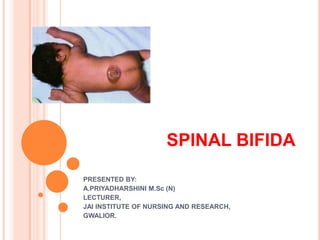
Spina bifida is a birth disorder that involves the incomplete development of the spine. In the first month of pregnancy, a special set of cells forms the “neural tube;” the top of the tube becomes the brain, and the remainder becomes the spinal cord and structures around it. In spina bifida, the neural tube doesn’t close completely and some of the bones of the spine do not close in the back. This can result in an opening anywhere along the spine and may cause damage to the spinal cord and nerves.There are four types of spina bifida: occulta, closed neural tube defects, meningocele, and myelomeningocele. The symptoms of spina bifida vary from person to person, depending on the type and level of involvement. Most cases are mild and do not require special treatment. The more serious cases involve nerve damage.
Occulta is the mildest and most common form in which one or more bones of the spinal column (vertebrae) are malformed. The name “occulta,” which means “hidden,” indicates that a layer of skin covers the opening in the bones of the spine. It usually shows no symptoms and is often found by accident on an x-ray or similar test.
Closed neural tube defects are a diverse group of disorders in which the spine may have malformations of fat, bone, or the membranes (the meninges) that cover the spinal cord. Many of these neural tube defects require surgery in childhood. People with this type of spina bifida may have weakness of the legs and trouble with bowel and bladder control. These issues may change or progress as children grow. It is important to have close communication with doctors to minimize these changes as much as possible.
Meningocele occurs when the meninges protrude through the spine and cause a sac of spinal fluid on the back. This fluid is typically only around the brain and spine, but a problem with the bony covering over the spine allows it to poke out. The malformation contains no nerves and may or may not be covered by a layer of skin. Individuals with meningocele may have minor symptoms.Myelomeningocele is the most severe form of spina bifida. A portion of the spinal cord or nerves are exposed in a sac through an opening in the spine that may or may not be covered by the meninges. The opening can be closed surgically while the baby is in utero or shortly after the baby is born. Most people with myelomeningocele experience changes in brain structure, leg weakness, and bladder and bowel dysfunction.
Myelomeningocele is often called a "snowflake condition" because no two people with the condition are the same. Typically, if the opening in the spine is lower down the back, the person will experience less symptoms. People with myelomeningocele require close follow-up with physicians throughout their childhood and lifespan to maximize their function and prevent complications like kidney failure.Complications of spina bifida may include:
Abnormal sensation or paralysis, which mostly occurs with closed neural tube defects and myelomenin
Neural tube defects are birth defects of the brain, spine, or spinal cord. They happen in the first month of pregnancy, often before a woman even knows that she is pregnant. The two most common neural tube defects are spina bifida and anencephaly.
All about Spina Bifida:
Definition
7 Types
Causes
Health Status
Incidence and epidemiology
Prevention
Social impact
Management
Detection
Diagnosis
Treatment
Antibiotics
Careful observation
Physical therapy
Spina bifida is a birth defect that occurs when the spine and spinal cord don't form properly. It falls under the broader category of NTD (Neural Tube Defects).
Neural tube defects are the most common congenital abnormality in India which can be easily prevented with due information and better nursing practices. Neural Tube Defects can be prevented with intake of folic acid.
My name is Dahianara Moran and I believe being the parent of a child with Spina Bifida is like having a sixth sense, something like a Super Mom. We learn quickly how to defend our little ones from the awkward moments, but most important, we come to enjoy the little things, value our moments, and commit to change the way the world defines “Disability”.
2. DEFINITION:
Spina bifida (Latin: "split spine") is a
developmental congenital disorder caused by the
incomplete closing of the embryonic neural tube.
Some vertebrae overlying the spinal cord are not
fully formed and remain unfused and open.
If the opening is large enough, this allows a portion
of the spinal cord to protrude through the opening in
the bones.
There may or may not be a fluid-filled sac
surrounding the spinal cord.
3. INCIDENCE:
Spina bifida is one of the most common birth
defects, with an average worldwide incidence of
one to two cases per 1000 births, but certain
populations have a significantly greater risk.
Myelomeningocele is the most significant and
common form, and this leads to disability in most
affected individuals.
This condition is more likely to appear in females;
the cause for this is unknown.
4. CAUSES:
Maternal diabetes
Family history
Obesity
Increased body temperature from fever or external
sources such as hot tubs and electric blankets may
increase the chances of delivery of a baby with a spina
bifida.
Medications such as some anticonvulsants.
Pregnant women taking Valproic acid have an increased
risk of having children with spina bifida
Genetic basis.
Folic acid deficiency
5. EMBRYOLOGY:
Spina bifida is caused by the failure of the neural
tube to close during the first month
of embryonic development (often before the mother
knows she is pregnant).
Under normal circumstances, the closure of the
neural tube occurs around the 23rd (rostral closure)
and 27th (caudal closure) day after fertilization.
6.
7. TYPES:
Spina bifida malformations fall into three categories:
spina bifida occulta
spina bifida cystica with meningocele
spina bifida cystica with myelomeningocele.
(The most common location of the malformations is
the lumbar and sacral areas)
8.
9. SPINA BIFIDA OCCULTA:
Occulta is Latin for "hidden". This is the mildest
form of spina bifida.
In occulta, the outer part of some of the vertebrae is
not completely closed.
The splits in the vertebrae are so small that the
spinal cord does not protrude.
The skin at the site of the lesion may be normal, or
it may have some hair growing from it; there may
be a dimple in the skin, or a birthmark.
The incidence of spina bifida occulta is
approximately 10% of the population, and most
people are diagnosed incidentally from spinal X-
rays
10. MENINGOCELE:
The least common form of spina bifida is a posterior
meningocele (or meningeal cyst). In this form, the
vertebrae develop normally, but the meninges are
forced into the gaps between the vertebrae.
11.
12. MYELOMENINGOCELE:
This type of spina bifida often results in the most severe
complications.
In individuals with myelomeningocele, the unfused
portion of the spinal column allows the spinal cord to
protrude through an opening.
The meningeal membranes that cover the spinal cord
form a sac enclosing the spinal elements.
Spina bifida with myeloschisis is the most severe form of
myelomeningocele. In this type, the involved area is
represented by a flattened, plate-like mass of nervous
tissue with no overlying membrane.
The exposure of these nerves and tissues make the
baby more prone to life-threatening infections such
as meningitis.
13.
14. CONTD…
The protruded portion of the spinal cord and the
nerves that originate at that level of the cord are
damaged or not properly developed.
As a result, there is usually some degree
of paralysis and loss of sensation below the level of
the spinal cord defect.
15. CLINICAL MANIFESTATIONS:
Physical Signs:
Orthopedic abnormalities (i.e., club foot, hip
dislocation)
Bladder and bowel control problems, including
incontinence, urinary tract infections, and poor renal
function.
Pressure sores and skin irritations
Abnormal eye movement
68% of children with spina bifida have
an allergy to latex
Paralysis
16. CONTD…
Scoliosis
Back pain
Partial or complete lack of sensation
Weakness of the hips, legs, or feet of a newborn
Other symptoms may include:
Hair at the back part of the pelvis called the sacral area
Dimpling of the sacral area
Difficulty swallowing, which can lead to choking.
Hoarseness.
Breath-holding and problems breathing during sleep.
Below-average intelligence.
17. NEUROLOGICAL COMPLICATIONS:
Many individuals with spina bifida have an
associated abnormality of the cerebellum, called
the Arnold Chiari II malformation. In affected
individuals, the back portion of the brain is
displaced from the back of the skull down into the
upper neck.
18. EXECUTIVE FUNCTION:
Specific areas of difficulty in some individuals
include planning, organizing, initiating, and working
memory. Problem-solving, abstraction, and visual
planning may also be impaired.
Children with spina bifida and shunted
hydrocephalus have higher rates of ADHD.
19. ACADEMIC SKILLS:
Individuals with spina bifida may struggle
academically, especially in the subjects
of mathematics and reading. In one study, 60% of
children with spina bifida were diagnosed with a
learning disability.
20. SOCIAL COMPLICATIONS:
Compared to typically developing children, youths
with spina bifida may have fewer friends and spend
less time with peers.
21. DIAGNOSTIC EVALUATION:
Pregnancy screening:
Neural tube defects can usually be detected during
pregnancy by testing the mother's blood (AFP
screening) or a detailed fetal ultrasound.
Increased levels of maternal serum alpha-
fetoprotein (MSAFP) should be followed up by two
tests - an ultrasound of the fetal spine
and amniocentesis of the mother's amniotic fluid (to
test for alpha-fetoprotein and acetylcholinesterase).
22. PREVENTION:
Dietary supplementation with folic acid has been
shown to be helpful in reducing the incidence of
spina bifida. Sources of folic acid include whole
grains, fortified breakfast cereals, dried beans, leaf
vegetables and fruits.
It is recommended that any woman considering
becoming pregnant take 0.4 mg of folic acid a day.
Pregnant women need 1 mg per day.
23. TREATMENT:
There is no known cure for nerve damage caused
by spina bifida.
The spinal cord and its nerve roots are put back
inside the spine and covered with meninges.
In addition, a shunt may be surgically installed to
provide a continuous drain for the excess
cerebrospinal fluid produced in the brain, as
happens with hydrocephalus.
Shunts most commonly drain into the abdomen or
chest wall.
24. CONTD…
Monitor growth and development of bones,
muscles, and joints.
Treat and evaluate nervous system issues, such as
seizure disorders.
Physical therapy
Speech therapy
25. IMMEDIATE CARE:
Place the child in prone position.
Cover the affected area with sterile gauze piece
dipped in normal saline.
Maintain hydration.
Monitor for associated defects.
26. LIFE LONG TREATMENT:
Catheters
Braces
High fiber diet
Antibiotics may be used to treat or prevent
infections such as meningitis or urinary tract
infections.
27. COMPLICATIONS:
Difficult delivery with problems resulting from a
traumatic birth, including cerebral palsy and
decreased oxygen to the brain
Frequent urinary tract infections
Hydrocephalus
Loss of bowel or bladder control
Meningitis
Permanent weakness or paralysis of legs