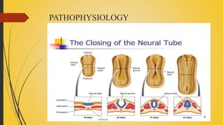
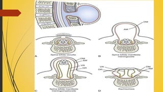
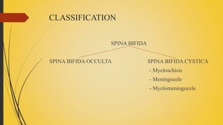
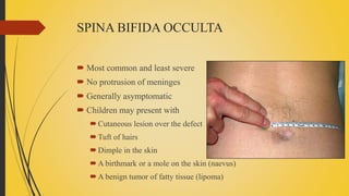
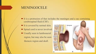
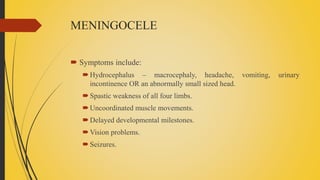
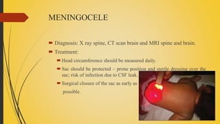
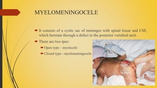
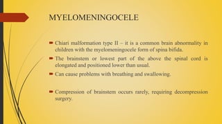
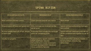
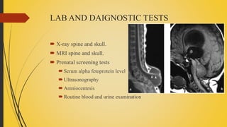
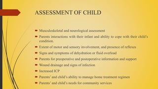
SPINA BIFIDA
INTRODUCTION
 It is a type of neural tube defect.
 A birth defect where there is incomplete closing of the neural arches
and meningeal layers around the spinal cord.
 It is a developmental congenital anomaly.
PATHOPHYSIOLOGY
Ectoderm (outermost layer) of fertilized egg develop a ridge, that
eventually become neural tube.
This neural tube forms spinal cord, brain and meninges.
Spina bifida occurs when a portion of neural tube fail to close properly
PATHOPHYSIOLOGY
CLASSIFICATION
SPINA BIFIDA
SPINA BIFIDA OCCULTA SPINA BIFIDA CYSTICA
- Myeloschisis
- Meningocele
- Myelomeningocele
ETIOLOGY
 Specific cause is unknown
 Various risk factors
Folic acid deficiency
Uncontrolled diabetes
Obesity
Hyperthermia during pregnancy
Medications that interfere with folate metabolism
SPINA BIFIDA OCCULTA
 Most common and least severe
 No protrusion of meninges
 Generally asymptomatic
 Children may present with
Cutaneous lesion over the defect
Tuft of hairs
Dimple in the skin
A birthmark or a mole on the skin (naevus)
A benign tumor of fatty tissue (lipoma)
SPINA BIFIDA OCCULTA
 After 6-8 years of age, children may present with
Progressive deformity of foot
Change in micturition pattern
Alteration in gait
Trophic ulcers over the foot and toes
SPINA BIFIDA OCCULTA
 Diagnosis: Myelogram, CT scan and MRI.
 Treatment:
Progressive disorder needs surgical correction.
Laminectomy and excision of intra-spinal lesion.
MENINGOCELE
 It is a protrusion of that includes the meninges and a sac containing
cerebrospinal fluid (CSF).
 It is covered by normal skin.
 Spinal cord is never involved.
 Usually seen in lumbosacral
region, but may also be seen
thoracic region and skull.
MENINGOCELE
 Symptoms include:
Hydrocephalus – macrocephaly, headache, vomiting, urinary
incontinence OR an abnormally small sized head.
Spastic weakness of all four limbs.
Uncoordinated muscle movements.
Delayed developmental milestones.
Vision problems.
Seizures.
MENINGOCELE
 Diagnosis: X ray spine, CT scan brain and MRI spine and brain.
 Treatment:
Head circumference should be measured daily.
Sac should be protected – prone position and sterile dressing over the
sac; risk of infection due to CSF leak.
Surgical closure of the sac as early as
possible.
MYELOMENINGOCELE
 It consists of a cystic sac of meninges with spinal tissue and CSF,
which herniate through a defect in the posterior vertebral arch.
 There are two tpes:
Open type – myelocele
Closed type - myelomeningocele
MYELOMENINGOCELE
 The child may present with
Flaccid paralysis
Absence of sensation
Drop reflex
Postural abnormalities eg. Clubfoot
Hydrocephalus is usually associated
Musculoskeletal deformity
Contracture of joints, scoliosis and kyphosis
Rupture of sac and infection of CNS
MYELOMENINGOCELE
 Chiari malformation type II – it is a common brain abnormality in
children with the myelomeningocele form of spina bifida.
 The brainstem or lowest part of the above the spinal cord is
elongated and positioned lower than usual.
 Can cause problems with breathing and swallowing.
 Compression of brainstem occurs rarely, requiring decompression
surgery.
MYELOMENINGOCELE
 Diagnosis: Primarily by clinical manifestations
X-ray spine, CT brain and MRI spine, complete neurological
assessment.
Routine blood examination and urine routine.
Prenatal diagnosis – amniocentesis and estimation of Alpha
fetoprotein.
Monitor head circumference daily.
MYELOMENINGOCELE
 Treatment: Surgical correction of the defect
Correction of the musculoskeletal deformities.
Regulation of bowel and bladder function.
Adequate nutrition.
Prevention of rupture of sac by proper positioning of child and sterile
dressing of the sac.
LAB AND DAIGNOSTIC TESTS
 X-ray spine and skull.
 MRI spine and skull.
 Prenatal screening tests
Serum alpha fetoprotein level
Ultrasonography
Amniocentesis
Routine blood and urine examination
ASSESSMENT OF CHILD
 Musculoskeletal and neurological assessment
 Parents interactions with their infant and ability to cope with their child’s
condition.
 Extent of motor and sensory involvement, and presence of reflexes
 Signs and symptoms of dehydration or fluid overload
 Parents for preoperative and postoperative information and support
 Wound drainage and signs of infection
 Increased ICP
 Parents’ and child’s ability to manage home treatment regimen
 Parents’ and child’s needs for community services
NON-SURGICAL INTERVENTIONS
 Preoperatively:
Motivation of parents regarding expression of grief over loss of perfect
child. Eg, - guilt, self-blame, anger etc.
Emotional support to parents
Monitoring of infant’s vital signs and neurologic status
Promote optimal hydration and nutritional status
Maintain integrity of defect, prevent further injury
Prepare parents and infants for surgery
NON-SURGICAL INTERVENTIONS
 Post-operatively:
Maintenance of nutrition and fluid intake
Look for signs and symptoms of infection
Promote healing of surgical site; sterile dressings to be enforced
Monitor vital signs and neurologic status
Emotional support to parents
NON-SURGICAL INTERVENTIONS
 Home care:
Long term management of bowel and bladder training
Provide info about techniques of facilitate mobility and independence
Info about skin care and injury prevention
Educate parents about normal growth and development and also
deviations from normal.
Instruct parents and child to avoid contact with latex and natural rubber
THANK YOU

Spina bifida

  • 1.
  • 2.
    INTRODUCTION  It isa type of neural tube defect.  A birth defect where there is incomplete closing of the neural arches and meningeal layers around the spinal cord.  It is a developmental congenital anomaly.
  • 3.
    PATHOPHYSIOLOGY Ectoderm (outermost layer)of fertilized egg develop a ridge, that eventually become neural tube. This neural tube forms spinal cord, brain and meninges. Spina bifida occurs when a portion of neural tube fail to close properly
  • 4.
  • 6.
    CLASSIFICATION SPINA BIFIDA SPINA BIFIDAOCCULTA SPINA BIFIDA CYSTICA - Myeloschisis - Meningocele - Myelomeningocele
  • 7.
    ETIOLOGY  Specific causeis unknown  Various risk factors Folic acid deficiency Uncontrolled diabetes Obesity Hyperthermia during pregnancy Medications that interfere with folate metabolism
  • 8.
    SPINA BIFIDA OCCULTA Most common and least severe  No protrusion of meninges  Generally asymptomatic  Children may present with Cutaneous lesion over the defect Tuft of hairs Dimple in the skin A birthmark or a mole on the skin (naevus) A benign tumor of fatty tissue (lipoma)
  • 9.
    SPINA BIFIDA OCCULTA After 6-8 years of age, children may present with Progressive deformity of foot Change in micturition pattern Alteration in gait Trophic ulcers over the foot and toes
  • 10.
    SPINA BIFIDA OCCULTA Diagnosis: Myelogram, CT scan and MRI.  Treatment: Progressive disorder needs surgical correction. Laminectomy and excision of intra-spinal lesion.
  • 11.
    MENINGOCELE  It isa protrusion of that includes the meninges and a sac containing cerebrospinal fluid (CSF).  It is covered by normal skin.  Spinal cord is never involved.  Usually seen in lumbosacral region, but may also be seen thoracic region and skull.
  • 12.
    MENINGOCELE  Symptoms include: Hydrocephalus– macrocephaly, headache, vomiting, urinary incontinence OR an abnormally small sized head. Spastic weakness of all four limbs. Uncoordinated muscle movements. Delayed developmental milestones. Vision problems. Seizures.
  • 13.
    MENINGOCELE  Diagnosis: Xray spine, CT scan brain and MRI spine and brain.  Treatment: Head circumference should be measured daily. Sac should be protected – prone position and sterile dressing over the sac; risk of infection due to CSF leak. Surgical closure of the sac as early as possible.
  • 14.
    MYELOMENINGOCELE  It consistsof a cystic sac of meninges with spinal tissue and CSF, which herniate through a defect in the posterior vertebral arch.  There are two tpes: Open type – myelocele Closed type - myelomeningocele
  • 15.
    MYELOMENINGOCELE  The childmay present with Flaccid paralysis Absence of sensation Drop reflex Postural abnormalities eg. Clubfoot Hydrocephalus is usually associated Musculoskeletal deformity Contracture of joints, scoliosis and kyphosis Rupture of sac and infection of CNS
  • 16.
    MYELOMENINGOCELE  Chiari malformationtype II – it is a common brain abnormality in children with the myelomeningocele form of spina bifida.  The brainstem or lowest part of the above the spinal cord is elongated and positioned lower than usual.  Can cause problems with breathing and swallowing.  Compression of brainstem occurs rarely, requiring decompression surgery.
  • 17.
    MYELOMENINGOCELE  Diagnosis: Primarilyby clinical manifestations X-ray spine, CT brain and MRI spine, complete neurological assessment. Routine blood examination and urine routine. Prenatal diagnosis – amniocentesis and estimation of Alpha fetoprotein. Monitor head circumference daily.
  • 18.
    MYELOMENINGOCELE  Treatment: Surgicalcorrection of the defect Correction of the musculoskeletal deformities. Regulation of bowel and bladder function. Adequate nutrition. Prevention of rupture of sac by proper positioning of child and sterile dressing of the sac.
  • 20.
    LAB AND DAIGNOSTICTESTS  X-ray spine and skull.  MRI spine and skull.  Prenatal screening tests Serum alpha fetoprotein level Ultrasonography Amniocentesis Routine blood and urine examination
  • 21.
    ASSESSMENT OF CHILD Musculoskeletal and neurological assessment  Parents interactions with their infant and ability to cope with their child’s condition.  Extent of motor and sensory involvement, and presence of reflexes  Signs and symptoms of dehydration or fluid overload  Parents for preoperative and postoperative information and support  Wound drainage and signs of infection  Increased ICP  Parents’ and child’s ability to manage home treatment regimen  Parents’ and child’s needs for community services
  • 22.
    NON-SURGICAL INTERVENTIONS  Preoperatively: Motivationof parents regarding expression of grief over loss of perfect child. Eg, - guilt, self-blame, anger etc. Emotional support to parents Monitoring of infant’s vital signs and neurologic status Promote optimal hydration and nutritional status Maintain integrity of defect, prevent further injury Prepare parents and infants for surgery
  • 23.
    NON-SURGICAL INTERVENTIONS  Post-operatively: Maintenanceof nutrition and fluid intake Look for signs and symptoms of infection Promote healing of surgical site; sterile dressings to be enforced Monitor vital signs and neurologic status Emotional support to parents
  • 24.
    NON-SURGICAL INTERVENTIONS  Homecare: Long term management of bowel and bladder training Provide info about techniques of facilitate mobility and independence Info about skin care and injury prevention Educate parents about normal growth and development and also deviations from normal. Instruct parents and child to avoid contact with latex and natural rubber
  • 25.